
Spio unidentata Chlebovitsch, 1959
|
publication ID |
https://doi.org/ 10.5281/zenodo.202084 |
|
DOI |
https://doi.org/10.5281/zenodo.6182988 |
|
persistent identifier |
https://treatment.plazi.org/id/9C0687AD-FF91-C35C-FF59-FC8FFC11D465 |
|
treatment provided by |
Plazi |
|
scientific name |
Spio unidentata Chlebovitsch, 1959 |
| status |
|
Spio unidentata Chlebovitsch, 1959 View in CoL
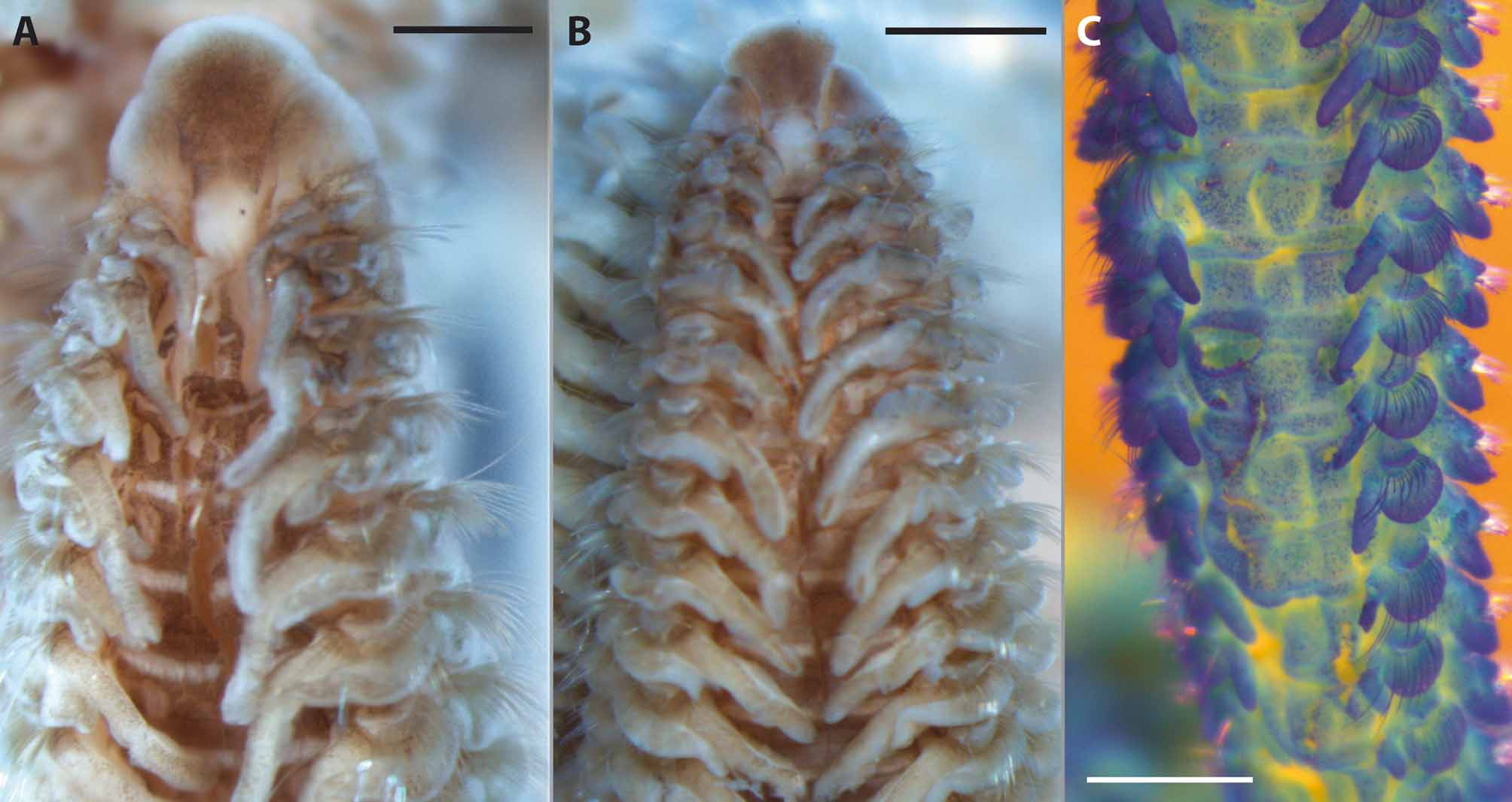
(Figs 6–7)
Spio unidentata Chlebovitsch, 1959: 172 View in CoL –173, Fig. 4.
Type material. Holotype: Kuril Islands, Shikotan Island, Krabovaya Bight, 26 Nov 1954 ( ZIN 3/5337); Paratypes: Kuril Islands, Shikotan Island, Krabovaya Bight, 27 Apr 1955, 1 specimen ( ZIN 1/5335), 12 Dec 1954, 2 specimens ( ZIN 4/5338); Kuril Islands, Shikotan Island, Voshodnaya Bight, 27 Aug 1955, 3 specimens ( ZIN 5/5339).
Diagnosis. Large, dark brown pigmented species; anterior margin of prostomium broadly rounded; branchiae on first chaetiger slightly shorter than those on chaetiger 2; notopodial postchaetal lamellae almost completely fused with branchiae, only distally separated; 5–7 unidentate hooded hooks from chaetiger 36–39.
Description. Large species. Complete specimen with 179 chaetigers about 55 mm long, 2.1 mm wide, without palps (ZIN 4/5338); holotype incomplete, with 152 chaetigers, about 50 mm long, 3.6 mm wide.
Anterior margin of prostomium usually broadly rounded, expanded at anterolateral margin, anterior margin usually projecting over the peristomium (Figs 6A, 7A, B); posterior part of prostomium possibly with distinct elevation beginning in front of posterior pair of eyes and terminating on chaetiger 1–2 (observation not reliable since damage in this area cannot be ruled out); two pairs of black eyes, arranged in trapezoid; anterior pair oval or rounded, widely spaced, posterior pair rounded, closely spaced; anterior pair of eyes usually positioned in the heavily pigmented anterior part of prostomium, therefore hardly visible (Figs 6A, 7A); prostomium distinctly separated from peristomium by a furrow (Figs 6A, 7A, B).
Alcohol-preserved specimens heavily pigmented dorsally, therefore, white band-like nuchal organs and metameric dorsal ciliated organs distinctly visible against dark-brown background (Fig. 6A); nuchal organs up to chaetiger 3; white bands of nuchal organs curved slightly outward in the range of the first transverse ciliary band on chaetiger 2 (Figs 6A, 7A); metameric dorsal ciliated organs, longitudinal, most likely double-paired from the beginning though on most anterior chaetigers only paired metameric dorsal organs found (Figs 6A, 7A); present from between branchiae 3 and 4, up to chaetiger 26–29; unpigmented transverse line between branchiae (in the position of transverse ciliary bands) against heavily pigmented dorsum present (Figs 6A, 7A, B).
Branchiae from chaetiger 1, continuing to very last chaetigers, very last branchiae rudimentary; length of branchiae of first 4–7 chaetigers about 4/5 as long as branchiae of the next consecutive chaetiger and narrower (Figs 6A, 7B); branchiae reaching midline dorsally and touching on chaetigers 1 to about 90; branchiae with broad base, usually also broadly rounded distally (Figs 6B–F, 7A–C); cilia on inner and outer margin not visible.
First notopodium shifted slightly dorsally ( Figs 7 View FIGURE 7 A, B). Notopodial postchaetal lamellae on chaetiger 1 oval (Fig. 6B); elongated with rounded tip on following anterior and middle chaetigers (Figs 6C–E); becoming rounded on posterior chaetigers (Fig. 6F); notopodial postchaetal lamellae almost completely fused with branchiae, only distally separated from branchiae, particularly apparent in posterior chaetigers (Figs 6B–F, 7C). Neuropodial postchaetal lamellae on chaetiger 1 rectangular, elongated towards ventrum on following chaetigers; neuropodial lamellae on chaetigers 10–15 almost twice as long as length of fascicle chaetal region, shorter thereafter (Figs 6C– F). Notopodial postchaetal lamellae on anterior chaetigers about twice as long (ventral-dorsal axis) as neuropodial lamellae, somewhat shorter on middle chaetigers, distinctly shorter than neuropodial lamellae on posterior chaetigers (Figs 6B–F). Noto- and neuropodial prechaetal lamellae on middle and posterior chaetigers well developed FIGURE 6. Spio unidentata Chlebovitsch, 1959 : A. Anterior end, dorsal view, heavily pigmented; specimen damaged dorsally. B–F. Parapodia from chaetigers 1, 6, 17, 47, 150 (=29th last). First parapodium (B) with pigment on parapodial lobes, branchia and laterally between rami. G. Neuropodial capillary from 47th chaetiger, anterior row. H. Hook from 150th chaetiger. I. Neuropodial capillary from 150th chaetiger. J. Smooth notopodial capillary from 47th chaetiger. Scale: A 1 mm, B–F 0.1 mm, G–J 10 µm. All paratype, ZIN 4/5338.
Notopodial chaetae all smooth capillaries: capillaries of anterior and middle chaetigers arranged in two rows, anterior row with shorter capillaries with narrow but distinct sheaths; posterior row with longer capillaries with narrow sheaths; superior fascicle with 3–7 long capillaries present; capillaries of posterior chaetigers not arranged in distinct rows, smooth, of different lengths (Fig. 6J). Neuropodia with rows of smooth capillaries, hooded hooks and inferior fascicle of chaetae in hook-bearing chaetigers; anterior neuropodia with capillaries arranged in two rows, anterior row with shorter capillaries with narrow sheaths, posterior row with longer capillaries with narrow sheaths; from about chaetiger 36–39 anterior row with capillaries with indistinct or no sheath, thinner than in anterior chaetigers, and becoming increasingly thinner towards the end of the body (Figs 6G, I); posterior row of capillaries replaced by single row of 5–7 unidentate hooded hooks from chaetiger 36–39 (Fig. 6H); 3–4 smooth alimbate capillaries in inferiormost position in hook-bearing chaetigers of the middle body region; with a few stout and finely granulated sabre chaetae in posterior hook-bearing chaetigers (Fig 6F).
Pygidium with four anal cirri (single complete specimen available for our study with three remaining cirri, fourth cirrus most likely lost), all of about same size; anus terminal.
Pigmentation. Anterior part, including prostomium, peristomium, branchiae and postchaetal lamellae pigmented dark-brown, only posterior part of prostomium, areas of nuchal organs, metameric dorsal ciliated organs and transverse ciliary bands without brown pigment (Figs 6A, 7A, B).
Methyl green staining pattern. Pre- and postchaetal lamellae of noto- and neuropodia, branchiae, and anal cirri coloured deep blue after application of methyl green; specimens slightly stained ventrally and dorsally ( Fig. 7 View FIGURE 7 C).
Biology. All specimens were found in sandy substrates, under boulders and between rhizomes of Zostera nana , in the upper sublittoral in depth between 5–6 m and in the intertidal ( Chlebovitsch 1959, Buzhinskaya 1990).
Geographical distribution. Known only from the Kuril Islands, Shikotan and Jankich Islands, northwest Pacific ( Chlebovitsch 1959, Buzhinskaya 1990).
Remarks. Spio unidentata is unique among all Spio species in having unidentate hooded hooks, a late start of these hooks on chaetiger 36–39, and a dark brown pigmentation of the anterior part of the body. This species is one of the largest Spio species. Contrary to the original description, we found 5–7 neuropodial hooks from chaetigers 36–39 rather than 6–9 hooks from chaetiger 32–39 ( Chlebovitsch 1959).
| ZIN |
Russian Academy of Sciences, Zoological Institute, Zoological Museum |
No known copyright restrictions apply. See Agosti, D., Egloff, W., 2009. Taxonomic information exchange and copyright: the Plazi approach. BMC Research Notes 2009, 2:53 for further explanation.