
Microspio kussakini Chlebovitsch, 1959
|
publication ID |
https://doi.org/ 10.5281/zenodo.202084 |
|
DOI |
https://doi.org/10.5281/zenodo.6182992 |
|
persistent identifier |
https://treatment.plazi.org/id/9C0687AD-FF94-C358-FF59-FDA7FBEBD2A1 |
|
treatment provided by |
Plazi |
|
scientific name |
Microspio kussakini Chlebovitsch, 1959 |
| status |
|
Microspio kussakini Chlebovitsch, 1959 View in CoL
(Figs 8–9)
Microspio kussakini Chlebovitsch, 1959: 173 View in CoL –175, Fig. 5 View FIGURE 5 .
Type material. Holotype: Kuril Islands, Kunashir Island, Izmena Bight, intertidal, 22 Aug 1951 ( ZIN 1/5317); Paratype: Kuril Islands, Kunashir Island, Izmena Bight, intertidal, 0 4 Jul 1951, 1 specimen ( ZIN 2/5318).
Diagnosis. Prostomium narrow, usually with bluntly rounded anterior part, slightly expanded at anterolateral margin, posterior part distinctly elevated, extended into a caruncle; branchiae on chaetiger 2 only slightly shorter than those on chaetiger 3; notopodium on chaetiger 1 with capillaries; 7–10 bidentate neuropodial hooded hooks with small teeth from chaetiger 18.
Description. Moderate-sized species. Holotype complete without palps, posterior end including pygidium with regenerated anal cirri;with 42 complete and 7 regenerated chaetigers, 1.1 mm wide, about 12 mm long. Paratype incomplete, with 47 chaetigers, 0.9 mm wide, 13 mm long.
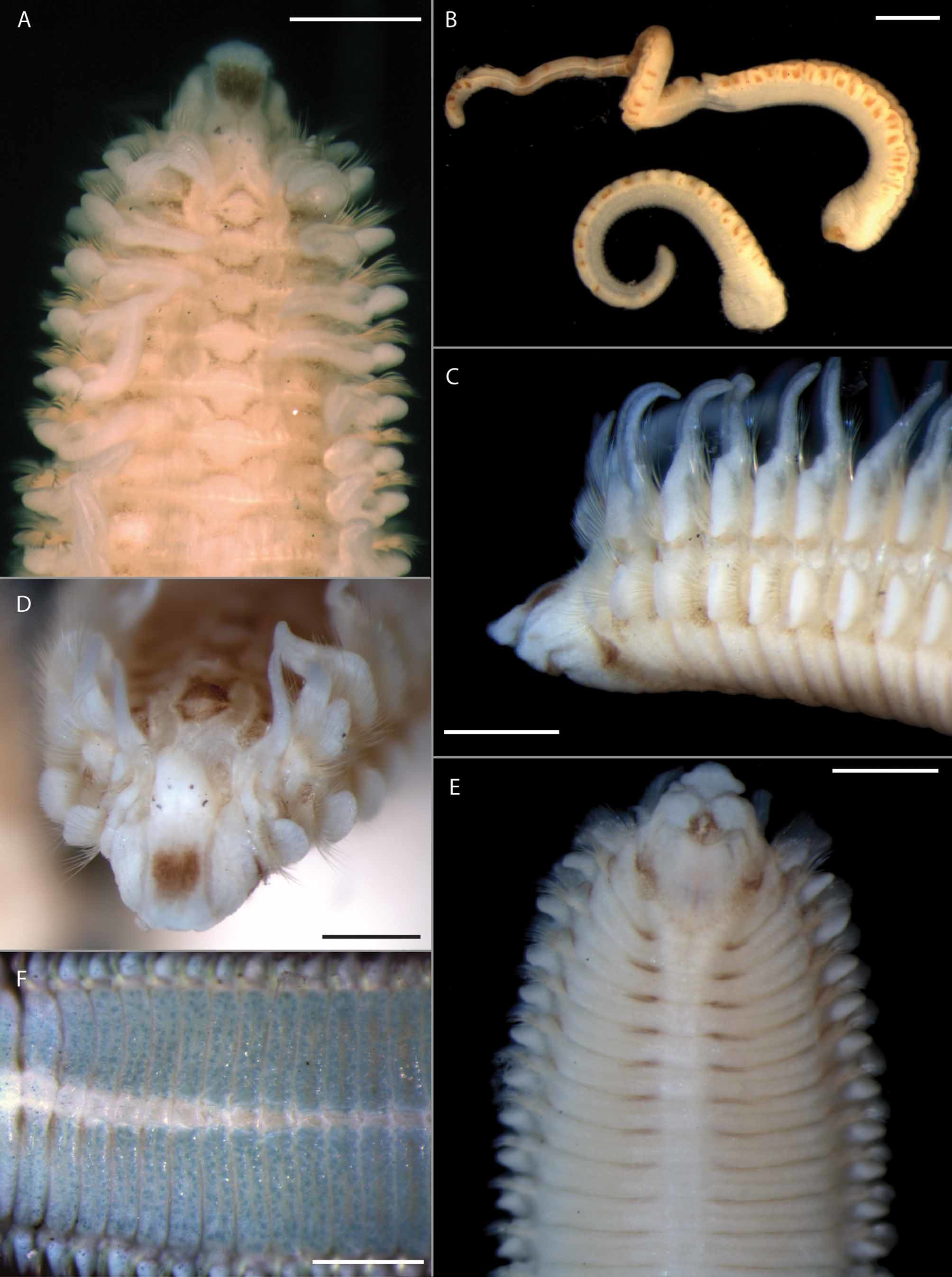
Prostomium narrow and projecting over the peristomium; anterior margin of prostomium usually bluntly rounded, slightly expanded at anterolateral margin, becoming narrower posteriorly (Figs 8A, 9A); posterior end short, extending to chaetiger 1, tapered, distinctly elevated from about the posterior pair of eyes (Figs 8A, 9A); two pairs of dark brown eyes, arranged in trapezoid, anterior pair larger, crescent-shaped, widely spaced, posterior pair smaller, rounded, closely spaced; prostomium distinctly separated from peristomium by a considerable furrow (Figs 8A, 9A).
Nuchal organs indistinct, even with methyl green; extending to chaetiger 2, U-shaped area recognizable but interpretation of arrangement of formerly present ciliated bands impossible (Figs 8A, 9A); metameric dorsal ciliated organs and transverse ciliary bands not visible.
Branchiae from chaetiger 2, present on all fully developed chaetigers in holotype (regenerated posterior part not included) and on all chaetigers in incomplete paratype ( Fig. 9 View FIGURE 9 B); first pair of branchiae shorter and narrower than those on chaetiger 3, thereafter all branchiae of about same length (Figs 8A, 9A); nearly reaching midline dorsally on some anterior chaetigers (chaetiger 2–12 in paratype and 2–3 and again 16–18 in holotype); branchiae with narrow base, rounded distally, cilia on inner and outer margin not visible.
First notopodium shifted slightly dorsally, notochaetae present (Fig. 8A). Notopodial postchaetal lamellae on anterior chaetigers rectangular, longer (dorsal–ventral axis) than neuropodial postchaetal lamellae; on posterior chaetigers oval, shorter than neuropodial lamellae (Fig. 8C); notopodial postchaetal lamellae on anterior chaetigers fused with branchiae, separated from branchiae in posterior chaetigers. Neuropodial postchaetal lamellae on anterior chaetigers rounded, oval on posterior chaetigers; not distinctly enlarged on posterior chaetigers, but longer than notopodial lamellae. Prechaetal lamellae indistinct (Figs 8B, C).
Large number of chaetae broken off in type material. Notopodial chaetae all capillaries without distinct sheath (Fig. 8D); anterior chaetigers with both smooth and distinctly granulated capillaries; middle and posterior chaetigers with smooth notochaetae only; additional superior fascicle of long capillaries present; arrangement of chaetae in rows not observable (probably due to bad condition of the types). Neuropodia with rows of alimbate capillaries and hooded hooks as well as an inferior fascicle of chaetae in the position of sabre chaetae; capillaries of anterior FIGURE 8. Microspio kussakini Chlebovitsch, 1959 : A. Anterior end, dorsal view; specimen lacking first parapodium on left side. B. Parapodium from chaetiger 15. C. Same from 46th chaetiger. D–G. Long notochaeta, inferior neuropodial chaeta, neuropodial hook, thin neuropodial capillary (alternating capillary); all from parapodium 46. Scale: A 0.5 mm, B-C 0.1 mm, D-G 10 µm. A Holotype ZIN 1/5317, all others paratype ZIN N2/5318.
neuropodia probably arranged in two rows, stout and distinctly granulated capillaries (anterior row?) as well as smooth and less stout capillaries (posterior row?) present, all neuropodial capillaries in anterior chaetigers of about same length; 7–10 bidentate hooded hooks in a single row entirely replace both neuropodial capillary rows from chaetiger 18–23 (Fig. 8F); hooded hooks indistinctly narrowed subdistally; main fang and apical tooth small, forming an obtuse angle to shaft; hooks accompanied by or even alternating with very thin alimbate capillaries (Fig. 8G); accompanying capillaries of hook-bearing chaetigers much shorter and thinner than capillaries of anterior chaetigers; few inferior capillaries in the position of sabre chaetae probably present from chaetiger 1; in hook-bearing chaetigers with about three stout sabre chaetae, either smooth or distally granulated, without sheaths (Fig. 8E).
Regenerated pygidium in holotype with four rounded anal cirri of about equal size; dorsal pair more widely spaced than ventral pair; anus terminal ( Fig. 9 View FIGURE 9 B).
Pigmentation. Body light brown without distinct pattern.
Methyl green staining pattern. Inconspicuous; parapodia including postchaetal lamellae and branchiae slightly stained; scattered stained spots dorsally, especially before and after transversal ciliary bands ( Figs 9 View FIGURE 9 A, B).
Biology. Both specimens were found in sandy substrates in brackish water together with the estuarine species Hediste japonica ( Izuka, 1908) , suggesting an estuarine distribution of M. kussakini ( Chlebovitsch 1959) .
Geographical distribution. Known only from the type locality Kunashir Island, Kuril Islands, northwest Pacific Ocean.
Remarks. This species is only known from the type locality. Even though only one complete specimen and one anterior fragment are available, important diagnostic characters could be observed. Microspio kussakini is characterized by the beginning of 7–10 neuropodial hooks with small teeth from chaetiger 18–23, a bluntly rounded anterior margin of the prostomium and the presence of capillaries on the first notopodium. Microspio maori Blake, 1984 is morphologically close to M. kussakini . In M. maori the prostomium is expanded anteriorly, tapering to an entire tip, sometimes appearing conical, and 9–12 hooks with large teeth are present from chaetiger 17–19. Moreover, M. maori possesses two ventral and two ventrolateral anal cirri whereas two ventral and two dorsal anal cirri are present in M. kussakini . The neuropodial hooks first appear on chaetiger 15 in M. moorei ( Gravier, 1911) , but capillaries on chaetiger 1 are absent in this species. Among all other Microspio species neuropodial hooks first appear from chaetiger 9–11.
The two available specimens of M. kussakini are light brown coloured. A special pattern of pigmentation has not been observed in this study and was also not mentioned in the original description. The condition of the specimens in the collection of the Zoological Institute, Saint Petersburg, does not enable a detailed description of other important characters such as the shape of the nuchal organs, the beginning and end of the metameric dorsal ciliated organs, the potential presence of white dots on the ventrum and the number of posterior abranchiate chaetigers. The species description should be amended as soon as new material becomes available.

| ZIN |
Russian Academy of Sciences, Zoological Institute, Zoological Museum |
No known copyright restrictions apply. See Agosti, D., Egloff, W., 2009. Taxonomic information exchange and copyright: the Plazi approach. BMC Research Notes 2009, 2:53 for further explanation.