
Spio picta Zachs, 1933
|
publication ID |
https://doi.org/ 10.5281/zenodo.202084 |
|
DOI |
https://doi.org/10.5281/zenodo.6182986 |
|
persistent identifier |
https://treatment.plazi.org/id/9C0687AD-FF9C-C35E-FF59-F93DFB12D1BA |
|
treatment provided by |
Plazi |
|
scientific name |
Spio picta Zachs, 1933 |
| status |
|
Spio picta Zachs, 1933 View in CoL
( Figs 3–5 View FIGURE 3 View FIGURE 5 )
Spio filicornis picta Zachs, 1933: 129 View in CoL .
Spio picta View in CoL . Buzhinkaya 1985: 134 –135, Fig. 12.
Type material. Lectotype: Sea of Japan, Peter the Great Bay, Stn. 182 ( ZIN 1/25818); Paralectotype: Sea of Japan, Peter the Great Bay, Stn. 116, 1 specimen ( ZIN 2/25819).
Non-type material. Sea of Okhotsk, South Sakhalin, Aniva Bay, Lagoon Busse, littoral, 20 Jul 1947, 1 specimen ( ZIN 4/46812); Sea of Okhotsk, South Sakhalin, Aniva Bay, near village Tobuti, littoral, 19 Jul 1947, 1 specimen ( ZIN 3/46811); Sea of Japan, Tatar Strait, Vostok Bay, depth 4 m, 25 Aug 1980, 1 specimen ( ZIN 5/46983); Sea of Japan, Tatar Strait, Chikhachev Bay, depth 1 m, 29 Aug 1982, 31 specimens ( ZIN 6/47041).
Diagnosis. Prostomium comparatively broad, with bluntly rounded anterior part and parallel lateral margins; branchiae on chaetiger 1 little shorter but distinctly narrower than second pair; notopodial postchaetal lamellae on chaetiger 1 very small, completely fused with branchiae on anterior and medium chaetigers; 8–13 tridentate neuropodial hooded hooks starting from chaetigers 13–19.
Description. Lectotype and paralectotype anterior fragments, in poor condition. One small complete specimen with 58 chaetigers (7.3 mm in length, 0.9 mm in width) without palps, 35 anterior fragments and several detached palps available (longest anterior fragment with 85 chaetigers, width 2.2 mm; maximum width 2.7 mm; maximum length of anterior fragment about 30 mm).
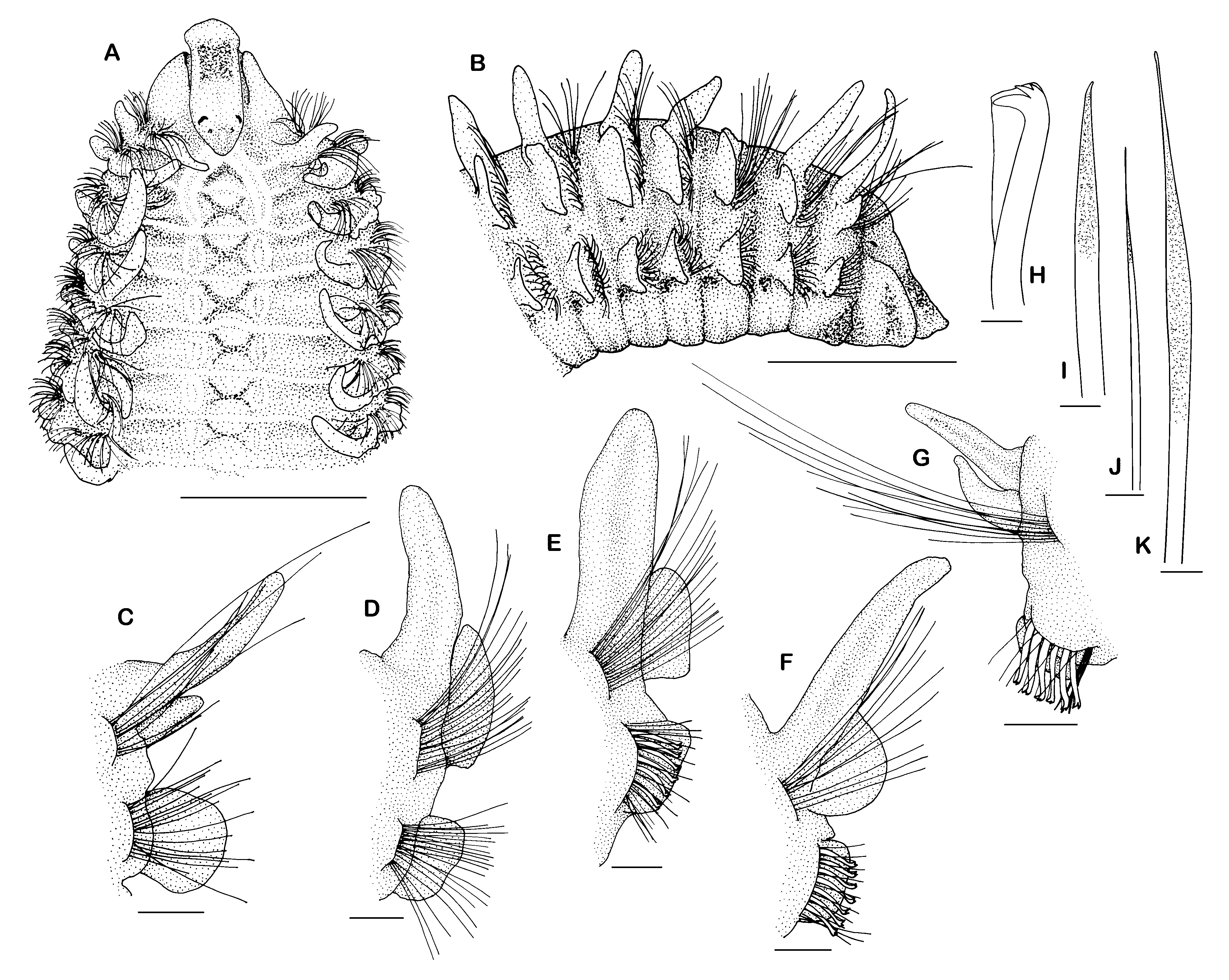
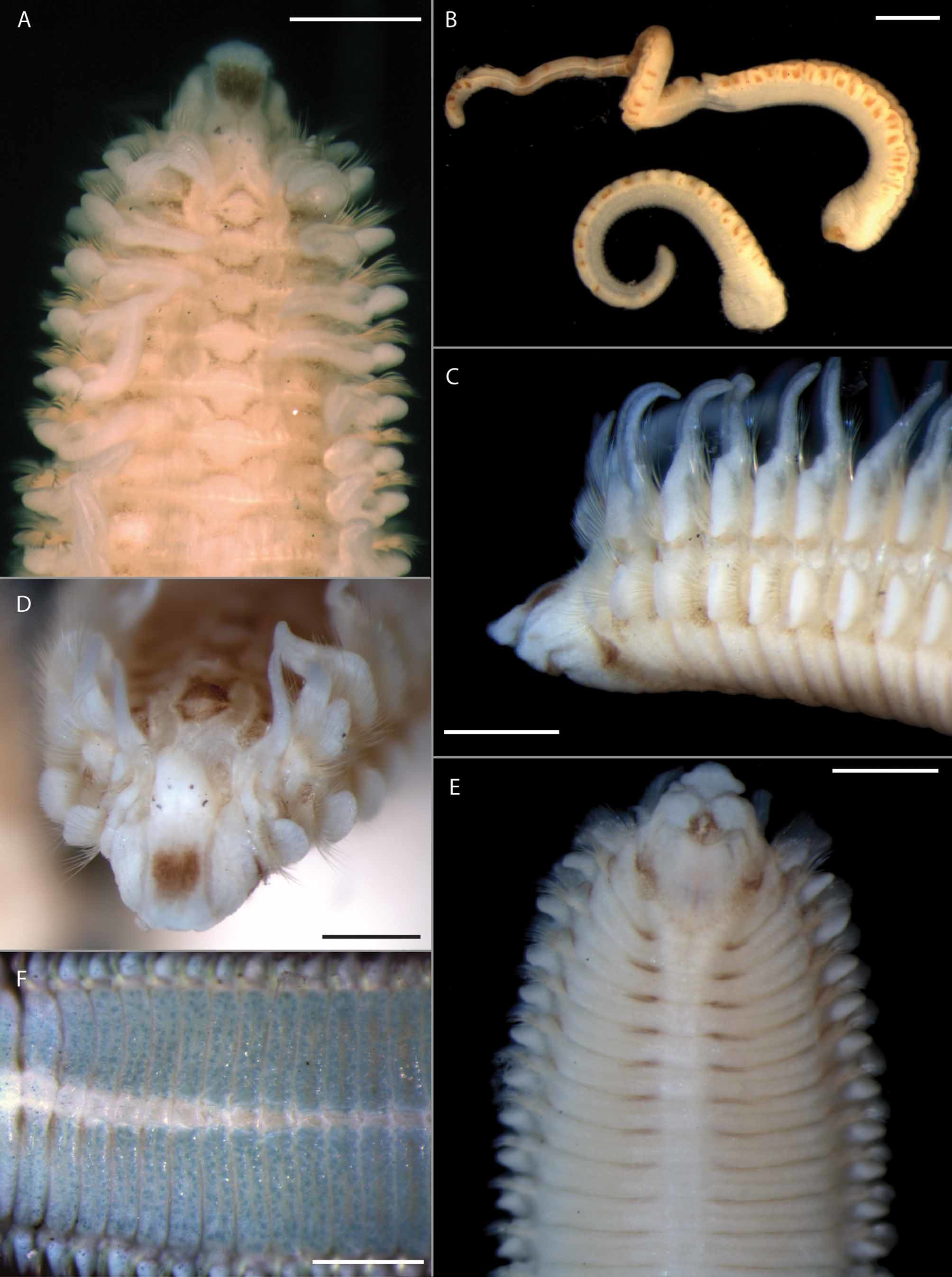
Prostomium comparatively broad ( Figs 3 View FIGURE 3 A, 4A, 5A), often with median longitudinal groove; anterior part of prostomium bluntly rounded or truncate, only slightly expanded at anterolateral margin, often projecting over the peristomium, lateral margins run parallel ( Figs 3 View FIGURE 3 A, 4A, 5A, D); posterior end short, extending to chaetiger 1, tapered, slightly elevated, with indistinct papilla ( Figs 3 View FIGURE 3 A, 4A, 5A, D); usually two pairs of black eyes, arranged in trapezoid or rectangle, anterior pair crescent-shaped or rounded, posterior pair smaller and rounded ( Figs 3 View FIGURE 3 A, 5A, D); sometimes light brown pigment spots between anterior and posterior eyes, easily mistaken as a third pair of eyes ( Fig. 5 View FIGURE 5 D); prostomium distinctly separated from peristomium by a furrow ( Figs 3 View FIGURE 3 A, 5A, D).
Nuchal organs with interrupted median and continuous lateral ciliary bands; median bands interrupted by short transverse ciliary band of chaetiger 2; median and lateral bands up to chaetiger 3, curved slightly outwards in the range of the first transverse ciliary band on chaetiger 2 ( Figs 3 View FIGURE 3 A, 4A, 5A, D); metameric dorsal ciliated organs double-paired, present from between branchiae 3 and 4, maximally up to chaetiger 13 ( Figs 3 View FIGURE 3 A, 4A); transverse ciliary bands distinct ( Figs 3 View FIGURE 3 A, 4A).
FIGURE 4. Spio picta Zachs, 1933 : A. Anterior end, dorsal view. B. Neuropodium at 35th chaetiger (from posterior). C. Ventral view at chaetiger 22 with irregular pattern of pores. D. Three types of neuropodial chaetae in chaetiger 15 (observed with light microscope). Scale: A 100 µm, B–D 10 µm. All ZIN 6/47041.
Branchiae from chaetiger 1, continuing to almost the end of body, only last 5 chaetigers without branchiae (observed on the single complete small specimen); first pair of branchiae usually about two-thirds of length and about one-third of diameter of second pair (Figs 4A, 5C, D); second pair of branchiae usually somewhat narrower and shorter than third pair of branchiae; branchiae do not reach midline dorsally; last 5 pairs of branchiae distinctly shorter than notopodial postchaetal lamellae (observed on the single complete small specimen); branchiae with narrow base, tapering distally, cilia on inner and outer margin not visible ( Figs 3 View FIGURE 3 C–G).
First notopodium shifted slightly dorsally (Fig. 4A). Notopodial postchaetal lamellae on chaetiger 1 very small ( Figs 3 View FIGURE 3 A, 4A); elongated on following chaetigers, completely fused with branchiae from chaetiger 3 or 4 up to about chaetiger 40 ( Figs 3 View FIGURE 3 D–F), thereafter becoming detached and completely separated on posterior chaetigers ( Fig. 3 View FIGURE 3 G); neuropodial postchaetal lamellae on first or first two chaetigers wider (proximal–distal axis) than long (ventral–dorsal axis), thereafter longer than wide ( Figs 3 View FIGURE 3 C–G). No distinct prechaetal lamellae.
Notopodial chaetae all capillaries with narrow sheath; capillaries of anterior chaetigers arranged in two rows: chaetae of anterior row shorter than in posterior row, few of them granulated; chaetae of posterior row longer than in anterior row, lacking granulations; additional superior fascicle of thin capillaries without granulations present; capillaries of middle and posterior chaetigers not clearly arranged in rows, thin, non-granulated, of different length within a fascicle. Neuropodia with rows of capillaries and hooded hooks as well as an inferior fascicle of capillaries in anterior chaetigers and sabre chaetae in hook-bearing chaetigers; capillaries of anterior neuropodia arranged in two rows, anterior row with granulated capillaries with narrow sheaths, becoming thinner in posterior chaetigers ( Fig. 3 View FIGURE 3 J, K), posterior row with non-granulated capillaries with narrow sheaths, posterior row replaced by single row of 8–13 tridentate hooded hooks from chaetiger 13–19 ( Figs 3 View FIGURE 3 H, 4B, D), hooks not narrowed subdistally, with a comparatively long hood, main fang and apical teeth well developed forming a right angle to shaft (Fig. 4D); on posterior chaetigers additional very thin alimbate capillaries with fine granulations present between the hooks or behind the row of hooks (Figs 4B, D); inferior fascicle with 3–5 capillaries without granulations from chaetiger 1 or 2, replaced by usually 3 (2–4) stout, distally granulated sabre chaetae from middle chaetigers ( Figs 3 View FIGURE 3 I, 4B).
Pygidium unknown.
Pigmentation. Conspicuously pigmented species ( Figs 3 View FIGURE 3 A, 5A–E). Light to dark-brown pigment spot on anterior part of prostomium ( Figs 3 View FIGURE 3 A, 5A, D); spot of varying shape, rounded, elongate, sometimes H–, M– or W– shaped. Rounded or oval dorsomedian areas from chaetiger 2 to about chaetiger 8–10 almost completely surrounded by brown pigment, particularly apparent from chaetiger 2 to chaetiger 6; semicircular pigmented areas of consecutive chaetigers abut, resulting in X–shaped appearance of pigment pattern dorsally ( Figs 3 View FIGURE 3 A, 5A). Mouth opening surrounded by light brown pigment ( Fig. 5 View FIGURE 5 E). Border of peristomium/chaetiger 1 with light brown pigment lateroventrally ( Figs 3 View FIGURE 3 B, 5C, E). Transverse brown pigment stripes on ventral segmental borders of chaetigers 1/2 to about chaetigers 9/10 adjacent to unpigmented ventral median longitudinal line, distinct up to between chaetigers 7/8 ( Fig. 5 View FIGURE 5 E). Palps with light brown spots along the food groove ( Fig. 5 View FIGURE 5 B). Light brown pigment spots sometimes visible on both sides of the transverse ciliary bands near the base of the branchiae from chaetiger 2 to about chaetiger 6–11, and on the ventral base of the postchaetal neuropodial lamellae from chaetiger 2 to chaetiger 10 at maximum ( Figs 3 View FIGURE 3 B, 5C).
Methyl green staining pattern. Inconspicuous; posterior, part of prostomium, especially prostomial elevation, branchiae and margin of postchaetal lamellae as well as ventrum slightly stained; irregular pattern of tiny blue dots on ventrum of middle chaetigers ( Figs 5 View FIGURE 5 F); tiny blue dots most likely correspond to small openings on ventrum discovered in SEM studies (Fig. 4C).
Biology. All specimens were found in sandy substrates, in shallow subtidal and intertidal areas, also between rhizomes of Zostera spp., at about 30–32 ‰.
Geographical distribution. Known from shores of the northwest Pacific Ocean, Peter the Great Bay, Sea of Japan, and South Sakhalin, Sea of Okhotsk.
Remarks. Zachs (1933) described a subspecies of S. filicornis as S. filicornis picta among others. This description was very brief and published without illustrations. Buzhinskaya (1985) investigated Zachs’ specimens and newly collected specimens, selected a lectotype and a paralectotype, and provided a more detailed description including figures. She elevated S. filicornis picta to species level.
Spio picta is easily distinguished from other Spio species by its unique pattern of pigmentation, the beginning of about 8–13 tridentate hooded hooks on chaetiger 13–19 as well as the complete fusion of notopodial postchaetal lamellae and branchiae on anterior and middle chaetigers. The combination of these characters has not been described previously. Moreover, the irregular pattern of tiny blue dots visible after application of methyl green and most likely corresponding to the pattern of openings on the ventrum seen in SEM (compare Fig. 4C) has not been observed before. Distinct white dots against a bluish background as found in other Spio species have not been observed for S. picta (compare Meißner et al. 2011).
Intraspecific variations in S. picta include the length of the first pair of branchiae, the intensity of pigmentation, the extension of the metameric dorsal ciliated organ and the number and the start of hooded hooks. The first two characters are not size-dependent. The length of the first pair of branchiae varies between half as long as the second pair of branchiae and nearly the same length. Some specimens were only slightly pigmented but the characteristic pattern illustrated above was still observable. A significant correlation between width of specimens and extension of metameric dorsal ciliated organ has not been found though the greatest extension (up to chaetiger 13) has been found in larger (2.2 mm wide) and the shortest extension (up to chaetiger 9) in smaller specimens (1.3 mm wide). The number and the beginning of neuropodial hooded hooks are obviously correlated to size of specimens. In smaller specimens (<1.4 mm wide) hooks first appear on chaetiger 13 whereas in larger specimens (> 2.0 mm wide) they start on chaetiger 19. The number of hooded hooks ranges from 7–9 in smallest (1.0– 1.6 mm wide) to 11–13 in largest specimens (> 2.0 mm wide). This observation implies a loss of anterior hooks in the course of ontogenesis. In the anteriormost hook-bearing chaetigers, hooks are present only in the inferior part of the neuropodial ramus, indicating a loss of hooks beginning with those from the superior part of the ramus.
| ZIN |
Russian Academy of Sciences, Zoological Institute, Zoological Museum |
No known copyright restrictions apply. See Agosti, D., Egloff, W., 2009. Taxonomic information exchange and copyright: the Plazi approach. BMC Research Notes 2009, 2:53 for further explanation.