
Dina samegreloensis Grosser, Barjadze & Shavadze, 2023
|
publication ID |
https://doi.org/10.5852/ejt.2023.891.2275 |
|
publication LSID |
lsid:zoobank.org:pub:63FFD632-0F96-47B3-A42E-EA30DA7F766E |
|
DOI |
https://doi.org/10.5281/zenodo.8377593 |
|
persistent identifier |
https://treatment.plazi.org/id/4E387A11-FFF9-4ACF-99BA-2F1EC712BE79 |
|
taxon LSID |
lsid:zoobank.org:act:4E387A11-FFF9-4ACF-99BA-2F1EC712BE79 |
|
treatment provided by |
Plazi |
|
scientific name |
Dina samegreloensis Grosser, Barjadze & Shavadze |
| status |
sp. nov. |
Dina samegreloensis Grosser, Barjadze & Shavadze sp. nov.
urn:lsid:zoobank.org:act:
Diagnosis
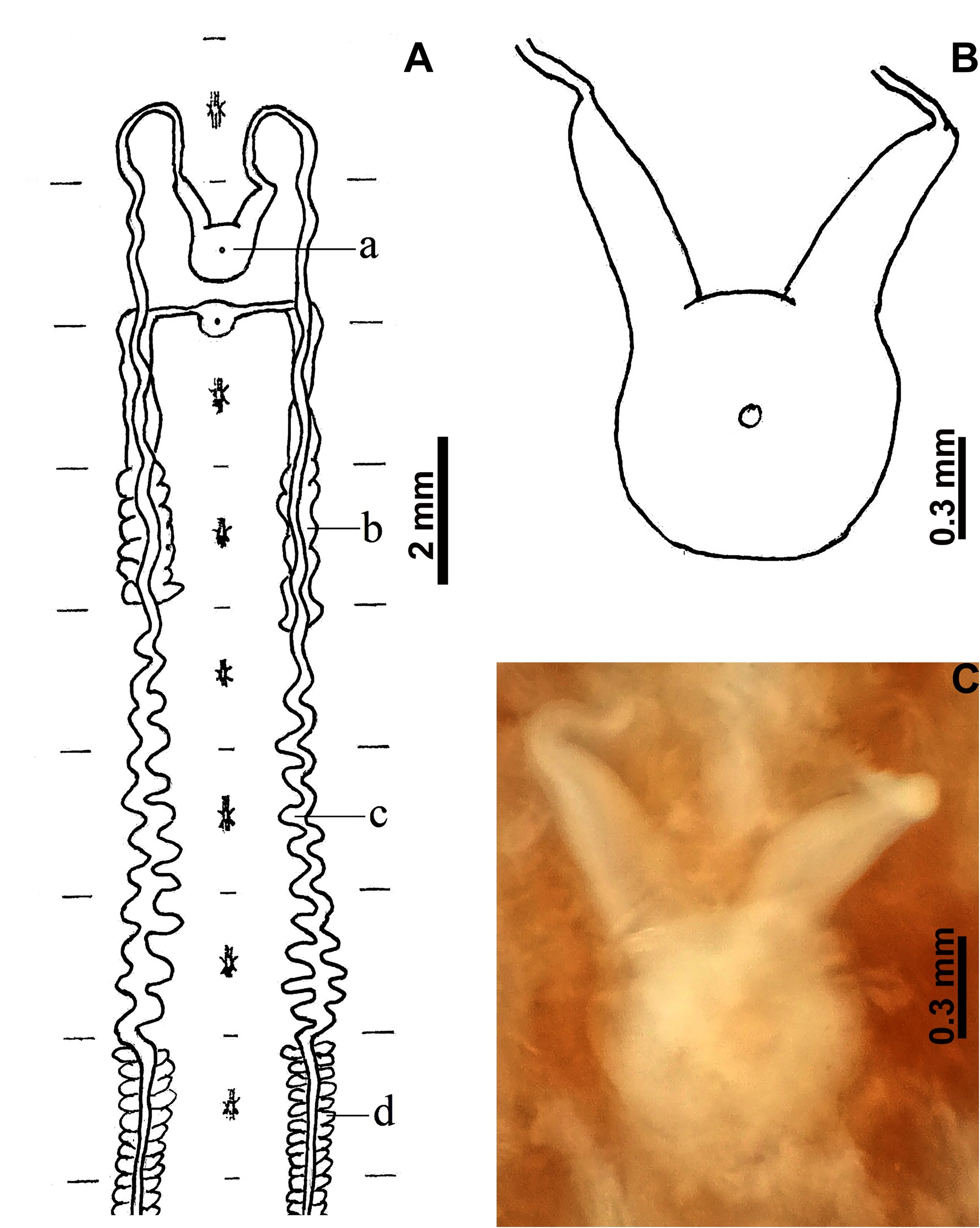
Small-sized erpobdellids with a Dina -like heteronomous annulation.The midbody somites are subdivided into annuli b1, b2, a2, b5 and the broadened annulus b6 ( Fig. 4F View Fig ). The male genital pore is situated in furrow b2/a2 and the female one in furrow b6/b1. The genital pores are separated by three annuli ( Fig. 4G View Fig ). Preserved leeches show numerous small and inconspicuous papillae on the dorsal and ventral sides. The cornua of the genital atrium are short, straight and directed slightly laterally ( Fig. 5B–C View Fig ). The vasa deferentia are very slightly curled up to the third ganglion behind the female genital pore. The ovisacs extend to the end of the second or the beginning of the third somite behind the female gonopore. They are unwinded to the end of the first somite and then coiled to the end ( Fig. 5A View Fig ).
Etymology
Dina samegreloensis sp. nov. is named after the region of Georgia from which the holotype was collected.
Type material
Holotype GEORGIA • body length 32 mm, width 6 mm, caudal sucker width 4 mm; Samegrelo-Zemo Svaneti region, Martvili Municipality, village Pirveli Balda , Motena Cave ; 42°28′35.73″ N, 42°23′28.25″ E; altitude 492 m; 7 Oct. 2021; Sh. Barjadze, L. Shavadze and E. Maghradze leg.; IZISU: AL-T - 00004. GoogleMaps
Description
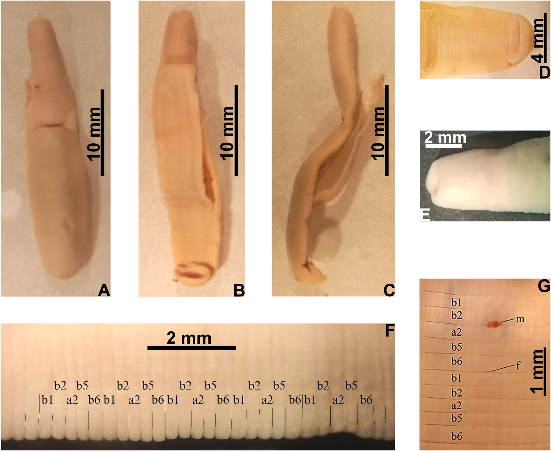
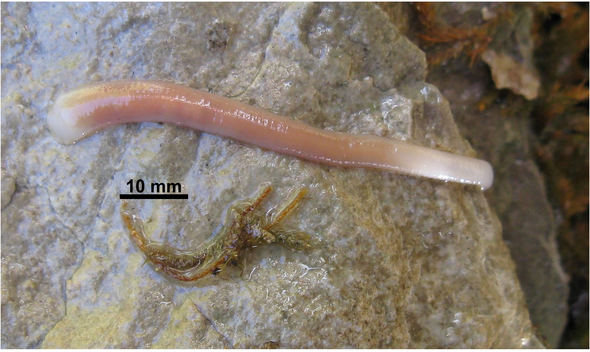
HABITUS. Small-sized erpobdellid. Preserved and contracted individuals reach a body length up to 32 mm and a width up to 6 mm ( holotype, Fig. 4A–B View Fig ). The body dorso-ventrally flattened in the posterior part, the first third (preclitellar and clitellar regions) cylindrical ( Fig. 4C View Fig ). The mouth opening is wide and the upper lip barely noticeably elongated ( Fig. 4E View Fig ). The caudal sucker is slightly wider than half of maximum body width ( Fig. 4D View Fig ). Small papillae numerous on dorsal and ventral surface.
ANNULATION. The annulation is typical of the genus Dina . The midbody somites are quinqueannulate and heteronomousely subdivided by clear furrows into annuli b1, b2, a2, b5 and the clearly broadened annulus b6. Annulus b1 is sometimes also slightly broadened, especially in the posterior part of the body ( Fig. 4F View Fig ). A tendency to split into further annuli is not visible. The male genital pore is situated in furrow b2/a2 and the female one in furrow b6/b1. The genital pores are separated by three annuli ( Fig. 4G View Fig ). The dorsal and ventral sides are roughened by numerouse papillae. The papillae are very small and inconspicuous.
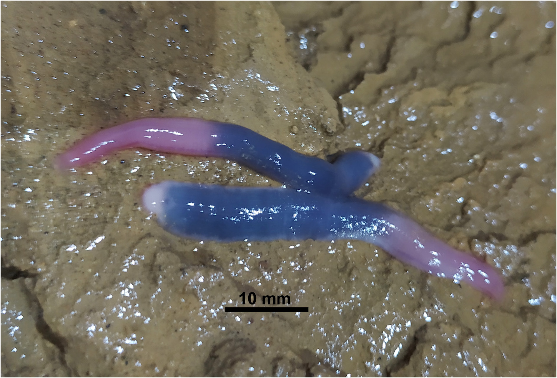
COLOURATION. The colouration of living specimen is pale pink. Preserved specimens are unicolored greyish without any dark patterns ( Fig. 4A–C View Fig ).
EYES. No eyes are visible.
SEXUAL ORGANS. The body of the genital atrium is large. The cornua are short, strong, straight and directed slightly laterally ( Fig. 5B–C View Fig ). The vasa deferentia are clearly offset from the cornua. They run relatively straight and only very slightly curled up to the third ganglion behind the female genital pore, then more coiled up to the end of the fifth somite behind the female genital pore ( Fig. 5A View Fig ). The ovisacs run straight and unwinded to the end of the first somite behind the female genital pore. The right ovisac is strongly coiled and reaches the end of the second somite behind the female gonopore. The left ovisac has a slightly coiled end and reaches the annulus b2 of the third somite behind the female gonopore ( Fig. 5A View Fig ).
Variability
Information on variability is not yet possible. Only the holotype is known.
Habitat
The single individual of this new leech species was found under a stone in the subterranean water stream in the dark zone of Motena Cave.
Distribution
The new species is only known from the type location.
Differential diagnoses
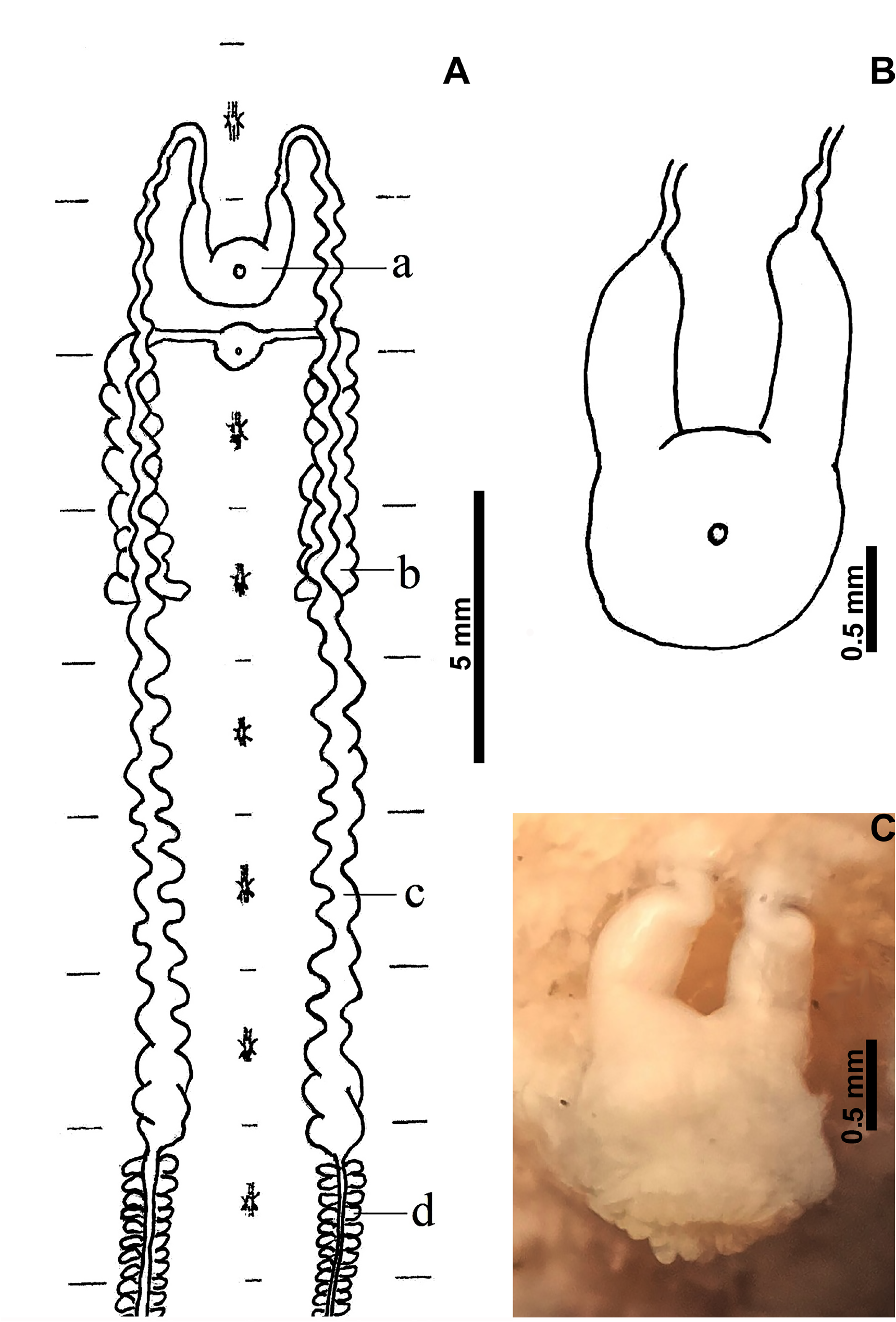
Dina absoloni , a southeastern European cave leech was reported from Georgia by Lukin (1976). This species was not really found in Georgia but was confused with other cave leeches (with D. ratschaensis or an other similar species of Dina , undescribed or here described; see also Discussion). Dina absoloni differs from species of Dina living in Georgian karst caves by the position of the gonopores (the male genital pore is situated in furrow b1/b2 and the female one in furrow b5/b6) and differences in the reproductive system (ovisacs long, reach up to the eight ganglion behind the female gonopore; from the third ganglion onward, the ovisacs extend strong coiled alongside the nervous system to the caudal end; Fig. 7C View Fig ).
The cave dwelling leech Erpobdella borisi Cichocka & Bielecki, 2015 , a species originally described from the Sahoolan Cave in northern Iran ( West Azerbaijan Province) shows a similar gonopore position and also a Dina -like annulation with ring b6 divided into c11, c12. Therefore, the female gonopore is located in furrow b5/c11 ( Cichocka et al. 2015: fig. 4a–b). However, in Dina spp. from the Georgian karst caves, the pore of the male genital organ is located in furrow b2/a2, and the female one in b6/b1. Further diffences are found in the shape of the reproductive system. The ovisacs of E. borisi are long and extend to the seventh ganglion behind the female gonopore ( Cichocka et al. 2015: fig. 5a), while in Dina spp. from Georgian karst caves they are short and extends at most to the third somite behind the female genital pore.
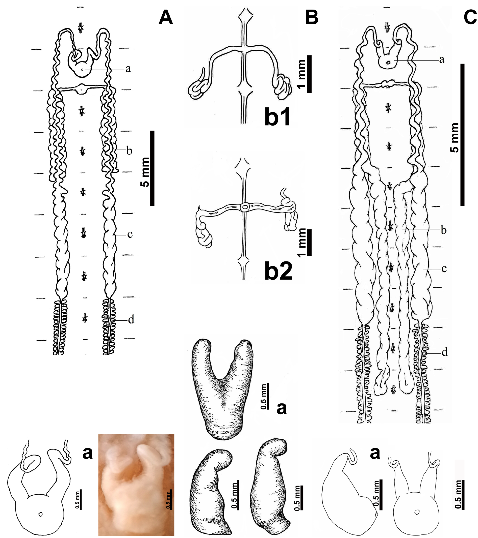
However, with regard to the shape of the vasa deferentia and ovisacs ( Figs 3A View Fig , 5A View Fig , 7A View Fig ) both new species show the greatest similarity with D. ratschaensis . The vasa deferentia of D. samegreloensis sp. nov. are only very slightly curled up to the third ganglion behind the female genital pore. The vasa deferentia of D. imeretiensis sp. nov. and D. ratschaensis are strongly curled along their entire course. The ovisacs of D. imeretiensis and D. ratschaensis are strongly winded along their entire caudal course ( Figs 3A View Fig , 7A View Fig ). In contrast, the ovisacs of D. samegreloensis are only winded in their posterior half ( Fig. 5A View Fig ).
Dina imeretiensis sp. nov. and D. ratschaensis can be clearly separated by the shape of the genital atrium. The cornua of the atrium in D. imeretiensis are nearly parallel and curved ventrally with straight ends ( Fig. 3B–C View Fig ). In D. ratschaensis the cornua are curved first laterally and then sharply to median and only slightly ventrally. The ends of the cornua are strongly kinked ventrally and not clearly offset from the vasa deferentia ( Fig. 7A View Fig ). Further differences occur in the length of the ovisacs. The ovisacs of D. imeretiensis extend up to the second ganglion behind the female genital pore ( Fig. 3A View Fig ). The ovisacs of D. ratschaensis are longer and extend up to the end of the second somite or up to the beginning of the third somite (on annulus b1) behind the female gonopore ( Fig. 7A View Fig ). The colouring of living specimens differs as well. The head, preclitellar region and caudal sucker of D. ratschaensis are whitish, the clitellar and postclitellar regions are light brownish ( Fig. 6 View Fig ). Dina imeretiensis shows a dark bluish postclitellar region, the caudal sucker is whitish and the other body regions are flesh coloured ( Fig. 1 View Fig ).
No known copyright restrictions apply. See Agosti, D., Egloff, W., 2009. Taxonomic information exchange and copyright: the Plazi approach. BMC Research Notes 2009, 2:53 for further explanation.
|
Kingdom |
|
|
Phylum |
|
|
Class |
|
|
Order |
|
|
Family |
|
|
Genus |