
Tityus forcipula ( Gervais, 1843 )
|
publication ID |
https://doi.org/10.11646/zootaxa.5155.2.1 |
|
publication LSID |
lsid:zoobank.org:pub:F23CDF54-115F-4861-8EDB-DAFDF7810586 |
|
DOI |
https://doi.org/10.5281/zenodo.6677778 |
|
persistent identifier |
https://treatment.plazi.org/id/B3264F4B-FF8D-7051-ADB9-FB73FC47F816 |
|
treatment provided by |
Plazi |
|
scientific name |
Tityus forcipula ( Gervais, 1843 ) |
| status |
|
Tityus forcipula ( Gervais, 1843) View in CoL
Figures 1 View FIGURE 1 , 2A–D View FIGURE 2 , 4A, B View FIGURE 4 , 5A–D View FIGURE 5 , 6A–D View FIGURE 6 , 7A–C View FIGURE 7 , 8A–C View FIGURE 8 , 9A, B View FIGURE 9 , 10A, B View FIGURE 10 , 11A–C View FIGURE 11 , 12A, B View FIGURE 12 , 13A, B View FIGURE 13 , 14A, B View FIGURE 14 , 15A, B View FIGURE 15 , 16 View FIGURE 16 ; Tables 2 View TABLE 2 , 4 View TABLE 4 , 5 View TABLE 5 , 6 View TABLE 6 .
References after Fet & Lowe (2000: 245). Tityus forcipula Gervais (1843) View in CoL : 130; Lourenço (1999): 4; Lourenço (2000): 454, 456, 458, 459, figs. 11, 13; Saldarriaga & Otero (2000): 18, table 1; Flórez (2001): 28; Lourenço (2002b): 133; Lourenço (2005): 226; Lourenço (2006): 57, 58, 61; Botero-Trujillo & Fagua (2007): 129, 133, figs. 11, 16, 17; Lourenço (2007): 382; Lourenço & Leguin (2008): 1, 3; Lourenço et al. (2009): 423; Flórez (2011): 764, 765, table 56; González-Sponga (2011): 103; Lourenço & Ythier (2013): 3, figs. 13, 18, table I; Brito & Borges (2015): 2, 7, 9, 14, figs. 1, 4G; Lourenço & Ythier (2017): 29; Ythier (2018): 20; Francke (2019): 24; Miranda et al. (2020): 197, 203, fig. 27; Dupré (2021): 52.
Type material. Holotype: COLOMBIA: adult male, Colombia ( NHM 1846.20 ) . Paratype: unknow sex, unkwon locality ( MNHN) .
Examined material. COLOMBIA: Valle del Cauca department: adult male, Cali, 18 Km highway to the sea, iv.1985 (MUSENUV-Ar 438); GoogleMaps adult female, Cali , Villa Carmelo Pathway , 1500 masl, 13.x.1982 (MUSENUV-Ar 436); GoogleMaps adult female, Cali , Felidia Pathway , vi.1979 (MUSENUV-Ar 434); GoogleMaps adult female, Cali , El Pato Pathway , 2000 masl, iii.1988, G. Salcedo (MUSENUV-Ar 418); GoogleMaps adult female, Cali , El Saladito Pathway , San Antonio Forest , highway to the sea, 3º29’34.22’’N 76º36’49.71’’W, 1813 masl, 21.i.2019, J. Cabra-García (MUSENUV-Ar 2116); GoogleMaps adult female, La Cumbre, Bitaco Pathway, ii.2021, L. Flórez (MUSENUV-Ar 2103); GoogleMaps adult female, La Cumbre, Chicoral Pathway, 28 Km highway to the sea, 1500 masl, iii.1988, C. Sepulveda (MUSENUV-Ar 409); GoogleMaps adult female, Palmira, Panorama Farm, 2100 masl, 9.iii.2005 (MUSENUV-Ar 432); GoogleMaps adult female, Yumbo, Dapa, Bocatoma del Acueducto , 17–18.viii.2016, J. A. Moreno ( IBALCC-RPDR 00256 ); GoogleMaps three adult females and one adult male, Yotoco, Yotoco RNF, 3º52.785’N 76º26.325’W, 1644 masl, 2.xi.2019, D. Garrido & B. Ospina (MUSENUV-Ar 2117); GoogleMaps two adult females, same locality as the previous record, 1.xi.2019 (MUSENUV-Ar 2118); GoogleMaps adult female, Tuluá, 1000 masl, xii.1984 (MUSENUV-Ar 437) GoogleMaps . Risaralda department: three adult males and two adult females, Santuario, San Rafael Plains Natural Regional Park [Parque Natural Regional Planes de San Rafael], 5°7’34”N 76°0’26.4’’W, 2158 masl, 17.x.2012, J. A. Moreno ( MZSP) GoogleMaps .
Diagnosis. Tityus forcipula share a metasoma widening towards segment V in both sexes ( Figures 12A, B View FIGURE 12 , 13A, B View FIGURE 13 ) and a male chela bulkier than female chela ( Figures 7A, B View FIGURE 7 , 8A, B View FIGURE 8 ) with Tityus cuellari , Tityus crassicauda , and Tityus moralensis sp. nov.. However, Tityus crassicauda , Tityus cuellari , and Tityus forcipula share the presence of strongly protruding spinoid granules on the DSM carinae of metasomal segments II–IV ( Figure 14A, B View FIGURE 14 ), whereas, Tityus moralensis , present moderately protruding rounded granules on the DSM carinae of metasomal segments II–IV ( Figure 14C, D View FIGURE 14 ).
On the other hand, Tityus forcipula and Tityus cuellari share the presence of metasomal segment V with DSM carinae composed of moderately to strongly protruding spinoid granules (e.g., Figure 14A, B View FIGURE 14 ), whereas Tityus crassicauda presents a metasomal segment V with DSM carinae composed of slightly protruding granules ( Lourenço & Ythier, 2013: figs 1–4). Finally, Tityus forcipula presents a medium-sized subaculear tubercle with a medium-sized dorsal area between the subaculear tubercle and the aculeus base ( Figure 15A, B View FIGURE 15 ), whereas Tityus cuellari presents a small subaculear tubercle with a small dorsal area between the subaculear tubercle and the aculeus base.
Taxonomic remarks. As mentioned by Brito & Borges (2015), Tityus spinatus was synonymized with Tityus forcipula by Lourenço (1984). However, in recent papers such as Lourenço & Ythier (2013, 2017) T. spinatus is mentioned without any comments about its validity. In fact, Lourenço & Ythier (2013) did not present any formal species revalidation and only provided a single drawing of the metasomal segments II–V + telson of the female holotype. For this reason, we followed Lourenço (1988) and Brito & Borges (2015) and considered Tityus spinatus as a synonym of T. forcipula as proposed by Lourenço (1984), until additional evidence is presented and a formal species revalidation is published.
Description. Based on an adult male (MUSENUV-Ar 438) and an adult female (MUSENUV-Ar 436).
Total length. Male: 53.81 mm and female 61.73 mm (additional measurements in Table 5 View TABLE 5 ).
TABLE 5. (Continued)
Coloration. General pattern (in 70% ethanol) ( Figure 2A–D View FIGURE 2 ) dark reddish-brown. Carapace ( Figure 2A, C View FIGURE 2 ): dark reddish-brown with black variegated pigments; black stripe on anterior margin; lateral and median eyes, surrounded by black variegated pigments. Chelicerae ( Figure 2A, C View FIGURE 2 ): coxa and hand dark yellow, with abundant black pigments over the hand surface; fingers, blackish-brown. Coxosternal region, Legs, Mesosoma , and Pedipalps ( Figure 2A–D View FIGURE 2 ): all reddish-brown, covered with black variegated pigments (except for the coxosternal region and ventral areas of the pedipalp segments). Chela ( Figure 2A–D View FIGURE 2 ) fingers blackish. Metasoma ( Figure 2A–D View FIGURE 2 ): segments dark reddish-brown, progressively darker towards the telson; segments IV–V blackish-brown. Telson ( Figure 2A–D View FIGURE 2 ): blackish-brown; aculeus dark reddish-brown.
Morphology. Body hirsute, covered with abundant microsetae, white colored under UV light. Carapace ( Figure 4A, B View FIGURE 4 ): moderately covered with fine granulation and few coarse granules; anterior margin with deep median notch; anterior median carinae, central lateral, central median, lateral ocular, posterior, posterior median and superciliary carinae, all well-marked, and furrows (anterior median, anterior marginal, central transverse, lateral ocular, supercialiary, posterior transverse, posterior lateral and posterior marginal), all well-marked; ocular tubercle wellmarked, located on the anterior half of carapace; median eyes separated by about 0.6 ocular diameters; with three pairs of lateral eyes and two pairs of lateral micro-ocelli (ADMi and PDMi) (pattern 4A).
Chelicerae ( Figure 4A, B View FIGURE 4 ). Dentition characteristic of the family Buthidae ( Vachon 1963) , densely covered with setae over the internal and ventral surfaces; anterior half of the manus with a transverse row of coarse granules.
Pedipalps. Chela, bulky in male (L/W= 3.71) or slender in female (L/W= 5.31). Trichobothriotaxic pattern Type A, alfa configuration (hand: Eb3:Eb1:Eb2:Esb:Est:Et, fixed finger: eb:esb:est:et:db:dt:it). Femur ( Figure 5A–D View FIGURE 5 ) with five well-marked carinae: VI, DI, DE and VE crenulate, EM serratocrenulate and complete, with intercarinal areas densely covered with fine granulation and few coarse granules. Patella ( Figure 6A–D View FIGURE 6 ) with seven wellmarked carinae: VI, DI, DE and EM, crenulate and complete; VE incomplete- only present on the distal half of the segment (female) or complete (male) and crenulate; DM crenulate and incomplete; IM serratocrenulate and complete, with a short spur near the segment base; with intercarinal areas densely covered with fine granulation. Chela ( tibia) ( Figures 7A–C View FIGURE 7 , 8A–C View FIGURE 8 ) with eight well-marked carinae: VI, VE, D, DS, DMA, IM and ES, crenulate and complete; SA crenulate and incomplete, only present on the anterior half of the hand. Pedipalp movable and fixed fingers each with a basal concavity followed by a well-marked basal lobe (male) ( Figure 8A View FIGURE 8 ) or movable and fixed fingers with obsolete basal concavities and an obsolete basal lobe only present on the movable finger (female) ( Figure 7A View FIGURE 7 ). Oblique rows of granules: Movable finger with 16–16 (male) and 17–17 (female) rows.
Coxosternal region ( Figure 9A, B View FIGURE 9 ). Sternum with posterior depression, outer ridge, and apical button, wellmarked; region covered with abundant coarse and fine granulation, and abundant microsetae; genital operculum longitudinally divided, composed of two sub-triangular plates. Male genital papillae are rounded and with sclerotized tips.
Pectines ( Figure 9A, B View FIGURE 9 ). Pectines are strongly sclerotized and brownish, with white areas (less sclerotized) on the marginal plates ( Figures 2B, D View FIGURE 2 , 9A, B View FIGURE 9 ). Pectinal basal piece shield-like shaped, with a deep anteromedian notch and without glandular areas ( Figure 9A, B View FIGURE 9 ); pectinal tooth counts of 18–17 (male) and 16–18 (female). Intermediate plate, marginal plate, and fulcra densely covered with microsetae ( Figure 9A, B View FIGURE 9 ). Basal middle lamellae, not dilated (male) ( Figure 9A View FIGURE 9 ) or dilated and oval shaped (female) ( Figure 9B View FIGURE 9 ). Female basal middle lamellae with a distal glandular area, white colored ( Figures 2D View FIGURE 2 , 9B View FIGURE 9 ).
Legs. Carinae present; intercarinal areas with fine granulation; ventral telotarsi setae distributed in two rows of ventrosubmedian setae ( type II), acute and stout; telotarsi, counts of ventral macrosetae in the left (L) and right (R) legs on prolateral (pro) and retrolateral (retro) rows from leg I to IV (L (pro/retro) R (pro/retro)): 6/5 6/6; 6/6 5/7; 6/7 6/7; -/- 7/8 (male) and 6/6 6/6; 6/6 7/7; 7/7 6/7; 6/7 7/7 (female). Claws short and symmetrical.
Mesosoma . Tergites I–VI ( Figure 2A, C View FIGURE 2 ), moderately covered with coarse and fine granulation; pre-tergite well defined, with median carina visible on the posterior margin of the post-tergite; tergite VII with DSM and DL carinae complete and crenulate, and median carina composed of a crenulate anteromedian eminence present on the anterior half of the post-tergite. Sternites densely covered with coarse and fine granulation ( Figure 10A, B View FIGURE 10 ); sternites III–VI with a pair of elliptic spiracles on the posterior half, which are progressively larger ( Figure 10A, B View FIGURE 10 ); sternites III–VI with a median longitudinal hyaline suture; sternite V with a large (male) ( Figure 10A View FIGURE 10 ) to medium-size (female) ( Figure 10B View FIGURE 10 ) hyaline subtriangular area on the posterior margin; sternite VI with VSM carinae crenulate, present on posterior half; sternite VII with VSM and VL carinae crenulate, present on posterior 2/3 ( Figure 10A, B View FIGURE 10 ).
Hemispermatophore ( Figure 11A–C View FIGURE 11 ). Thin and sclerotized; pars reflexa, curved but not folded over itself ( Figure 11A, B View FIGURE 11 ); foot narrow and flat ( Figure 11A–C View FIGURE 11 ); pedal flexure inconspicuous ( Figure 11A–C View FIGURE 11 ); body occupying more than 2/3 of the hemispermatophore total length ( Figure 11A–C View FIGURE 11 ). Capsular region with a furrow connecting the internal lobe with the medial area of the region ( Figure 11A View FIGURE 11 ); internal lobe with rounded tip forming an 80º angle ( Figure 11B View FIGURE 11 ); median lobe inconspicuous ( Figure 11B, C View FIGURE 11 ); external lobe thin and not overpassing the internal lobe level and with a translucent area between the middle of the basal lobe and the base of the internal lobe ( Figure 11C View FIGURE 11 ); translucent area narrow but widened basally and distally; basal lobe bifurcate and with a vestigial projection subovate-shaped (in posterior and ventral views) ( Figure 11A, C View FIGURE 11 ); basal lobe with no conspicuous visible surface in lateral view ( Figure 11B View FIGURE 11 ); basal lobe without a “U”-like shaped curvature between its base and the body (in posterior and ventral views) ( Figure 11A, C View FIGURE 11 ).
Metasoma ( Figures 12A, B View FIGURE 12 , 13A, B View FIGURE 13 , 14A, B View FIGURE 14 ). moderately widening towards segment V ( Figures 12A, B View FIGURE 12 , 13A, B View FIGURE 13 ). Segments II–V short and robust (male- L/W ratio: II= 1.29; III= 1.38; IV= 1.39; V= 1.53/ female- L/W ratio: II= 1.32; III= 1.39; IV= 1.46; V= 1.53). Segments I–II ( Figures 12A, B View FIGURE 12 , 13A, B View FIGURE 13 , 14A, B View FIGURE 14 ) with eight complete carinae, parallel and crenulate (paired DL, ML, VL and VSM) and two serratocrenulate (paired DSM); ML of segment II represented by coarse granules on posterior 2/3 and fine granule over the anterior third; intercarinal areas densely covered with fine granulation and moderately covered with coarse granulation; segments III–IV ( Figures 12A, B View FIGURE 12 , 13A, B View FIGURE 13 , 14A, B View FIGURE 14 ) with six complete carinae, parallel and crenulate (paired DL, VL and VSM) and two serratocrenulate (paired DSM), intercarinal areas densely covered with fine granulation and moderately covered with coarse granulation; segment V ( Figures 12A, B View FIGURE 12 , 13A, B View FIGURE 13 , 14A, B View FIGURE 14 ) with five complete carinae and crenulate (VM, paired VL and VSM carinae) and two serratocrenulate (paired DSM), intercarinal areas densely covered with coarse granulation and moderately covered with fine granulation. Segments II–V ( Figure 14A, B View FIGURE 14 ) with DSM carinae, composed of strongly protruding spinoid granules that progressively increase distally, ending in an enlarged distoterminal granule (distoterminal granule not enlarged in segment V).
Metasomal macrosetation. Segments I–IV each with two pairs of VSM macrosetae (2/2): pair VSM1 located in the anterior 1/3, and pair VSM3 located near the posterior margin of the segment, and two pairs of VL macrosetae (2/2): pair VL1 located near the anterior margin, and pair VL2 located in the posterior 2/3 of the segment. Segment V with two pairs of VSM macrosetae (2/2), two pairs of VL macrosetae (2/2), and a single pair of ML macrosetae; pair VSM1 and VL1 located near the anterior margin of the segment; VL2 located on the posterior 2/3, and pair ML1 located dorsolaterally near the posterior margin (behind DSM carinae); anal arch with two pairs of setae on the intercrestal area, one pair of VSM macrosetae (1/1) and one pair of VL macrosetae (1/1).
Telson ( Figure 15A, B View FIGURE 15 ). Covered with abundant microsetae, white colored under UV light ( Figure 15A, B View FIGURE 15 ). Vesicle suboval, not elongated (L/H= 1.75 (male)/ L/W= 1.85 (female)), with dorsal surface smooth and a lateral longitudinal furrow on each side; with VM, paired VSM, VL, and DL carinae, vestigial and moderately marked. Subaculear tubercle small pyramidal, with a rounded apex directed to the middle portion of the aculeus ( Figure 15A, B View FIGURE 15 ); subaculear tubercle with a ventral pair of small rounded granules, pointing to the basal portion of the aculeus; aculeus strongly curved, shorter than vesicle and with a ventral groove.
Variability. Morphometrics. Total length ( including telson): 52.98–62.30 (female) (n= 4, mean= 57.53, standard deviation (SD)= 5.19) and 49.24–53.81 (male) (n= 4, mean= 51.99, SD= 1.95). Chela L/W ratio: 5.15–5.31 (female) (n= 4; mean= 5.02; SD= 0.30) and 3.61–3.71 (male) (n= 4; mean= 3.84; SD= 0.33). Metasomal segment I L/W ratio: 0.92–1.10 (female) (n= 4; mean= 1.02; SD= 0.08) and 1.05–1.15 (male) (n= 4; mean= 1.09; SD= 0.05). Metasomal segment V L/W ratio: 1.42–1.53 (female) (n= 4; mean= 1.48; SD= 0.05) and 1.36–1.53 (male) (n= 4; mean= 1.46; SD= 0.08). Telson vesicle L/H: 1.70–1.86 (female) (n= 4; mean= 1.80; SD= 0.08) and 1.68–1.85 (male) (n= 4; mean= 1.76; SD= 0.07). Counts. Pectinal tooth counts: females 14–18 (n= 8, mode= 16) and males 14–18 (n= 8, mode= 17). Number of movable finger oblique granule rows: females 15–17 (n= 8, mode= 16) and males 16–17 (n= 8, mode= 16). Metasomal macrosetae: The specimens from Risaralda department exhibit three pairs of VSM macrosetae in segments I–IV, instead of two pairs as the other examined specimens.
Natural history. Moreno-González & Hazzi (2012) observed a female of this species feeding upon an adult female of Chactas vanbenedenii ( Gervais, 1843) in Yotoco Forest Reserve, Yotoco, Valle del Cauca Department, Colombia. Seiter et al. (2020) studied life story aspects of T. forcipula and observed that specimens reared in captivity are unable to thrive under high temperatures. Only 10% of the specimens kept at 25–27°C reached adulthood, none reproduced. Among 43 specimens kept between 23–24°C, 21 females and 19 males reached maturity in the fifth instar, but one female and two males required an extra molt to reach maturity. The embryonic development took 208 days (with an average of 12 young per litter) and the postembryonic development took 463 days.
This species has been observed living in sympatry with Chactas vanbenedenii and Tityus parvulus Kraepelin, 1914 across several high-altitude (over 1600 masl) localities in the Cordillera Occidental in Valle del Cauca department, Colombia. The microhabitat that this species seems to prefer is underside rotten logs, inside leaf litter, or under rocks, and rarely it has been observed under tree bark over 1 m above the soil level. A strong association between adult specimens and low vegetation has never been observed, maybe this relationship is due to the particular telotarsi macrosetae structure that this species exhibit (Moreno-González, J. A., pers. obs.).
Distribution ( Figure 16 View FIGURE 16 ). We opted to follow a rather conservative approach. As such, we considered as doubtful records those specimens that we were not able to examine (i.e., Ecuadorian Andes and Central and Oriental Cordilleras from Colombia) ( Figure 16 View FIGURE 16 ). The exact type locality of T. forcipula is not precise, but there is consensus that the male holotype of this species came from Colombia ( Lourenço 1984). Our redescription is based on specimens from the Cordillera Occidental of Cali, Valle del Cauca, Colombia, where Lourenço & Flórez (1990) and Lourenço (1997) identified samples of this species.
Confirmed records. COLOMBIA: Valle del Cauca Department: Cali (Pichinde- Peñas Blancas), Restrepo (road Buga-Buenaventura ), Yotoco ( Yotoco Forest Natural Reserve ), and Yumbo ( Dapa ). New Records. COLOMBIA: Risaralda department: Santuario ( San Rafael Plains Natural Regional Park). Valle del Cauca department: Cali ( El Pato Pathway, El Saladito Pathway ( San Antonio Forest ), Felidia Pathway, Villacarmelo Pathway and 18 Km Highway to the Sea), La Cumbre (Bitaco and Chicoral Pathways), Palmira (Panorama Farm), and Tuluá.
Doubtful records. COLOMBIA: Caldas Department: Manizales . Cundinamarca Department: La Vega ( La Reserva) . Quindío Department: Armenia and Salento. Tolima Department: Ibagué (Cay Pathway). ECUADOR: Azuay province: Cuenca . Cotopaxi Province: Las Pampas. Pichincha Province: Alluriquin and Chiriboga. Santo Domingo de Tsáchilas Province: Santo Domingo de Los Colorados .
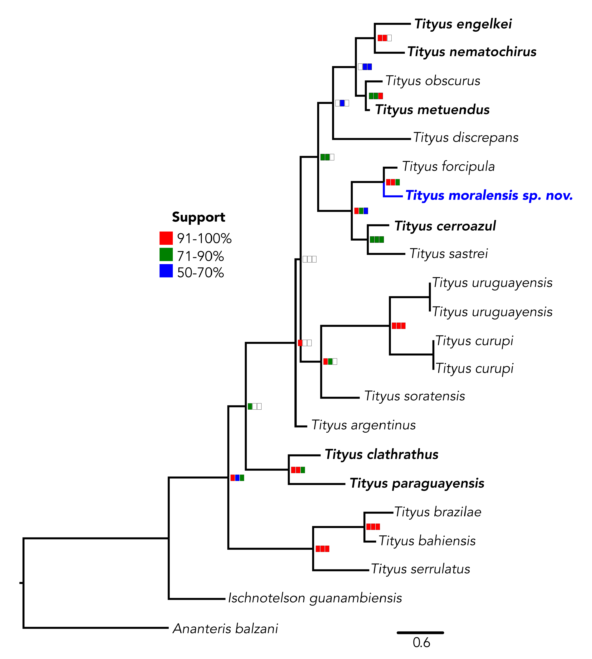
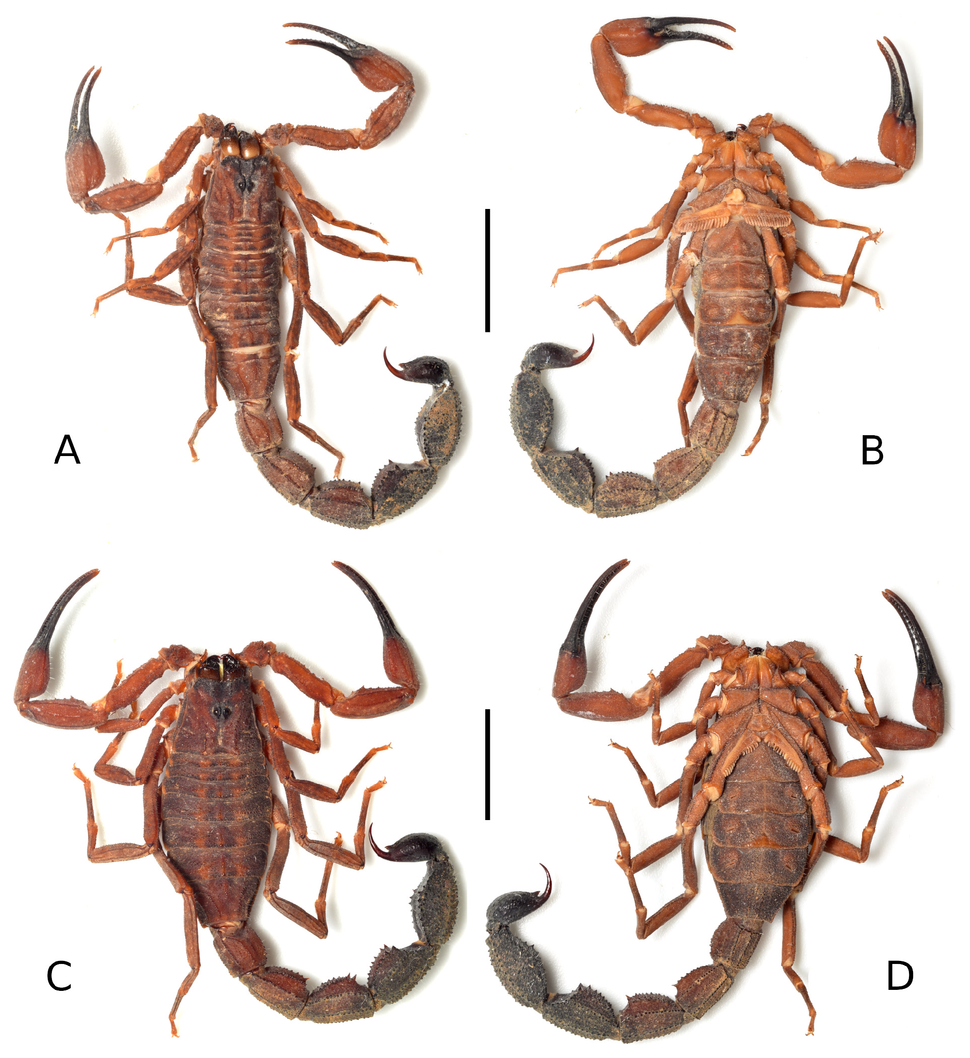

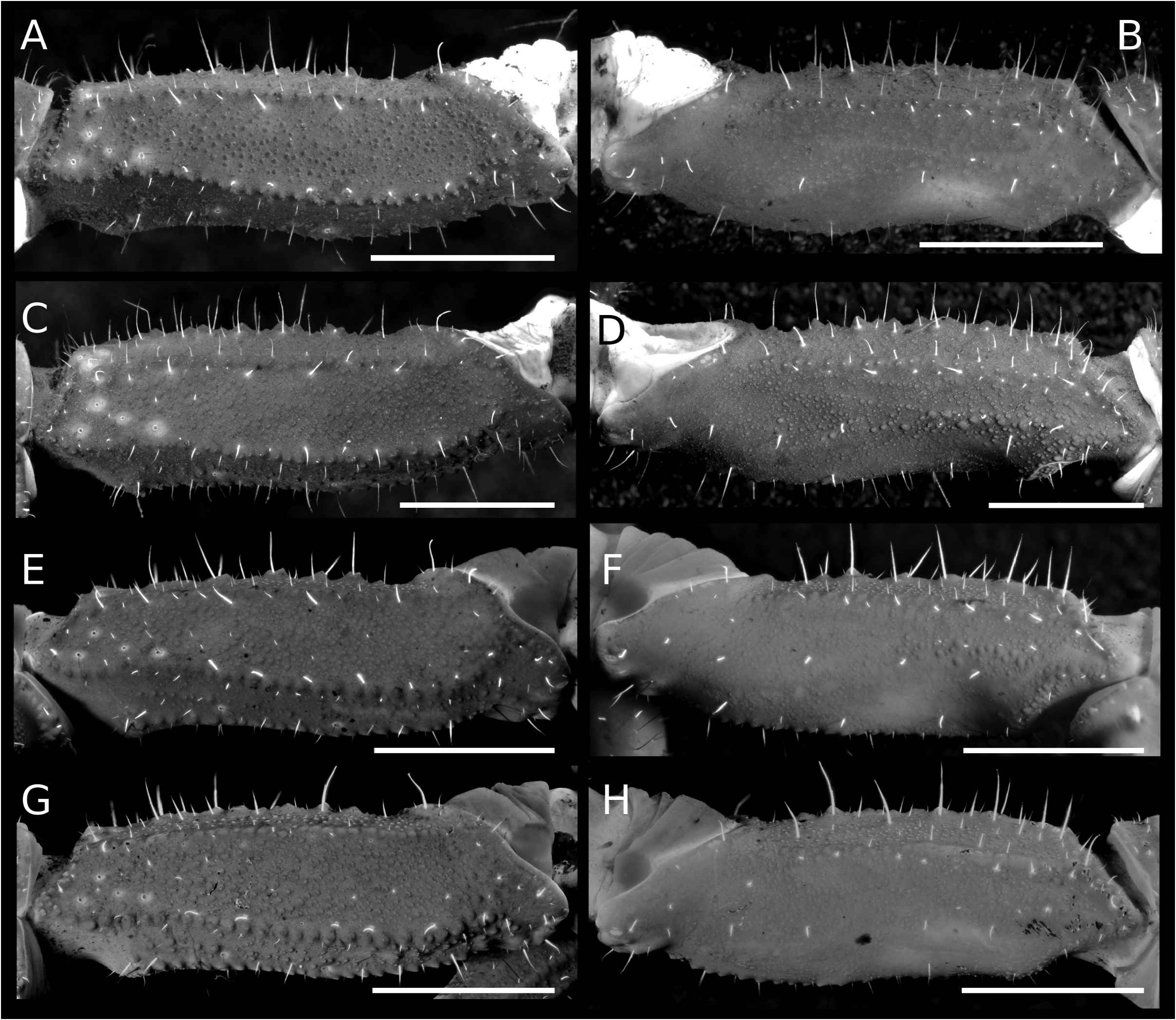

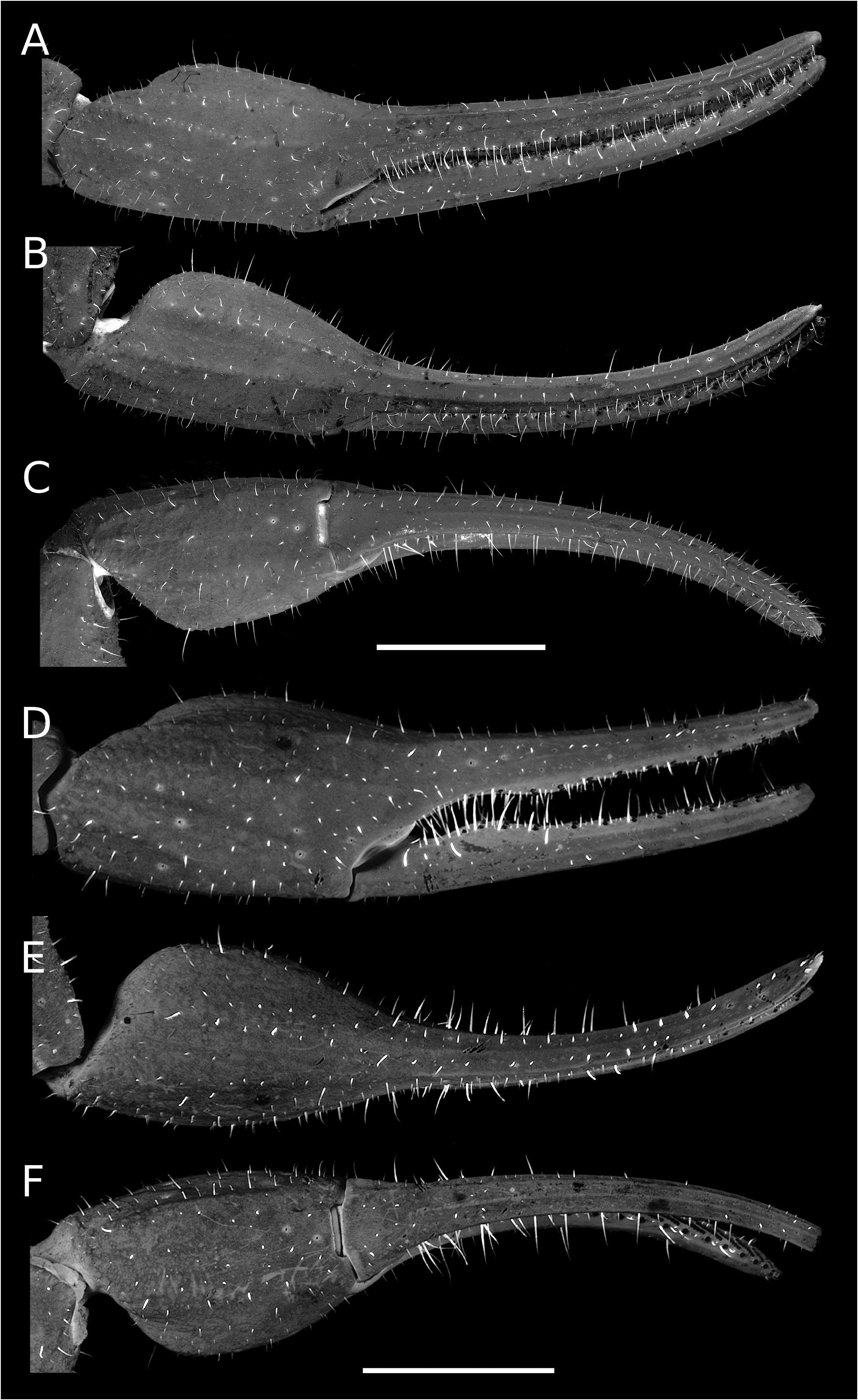
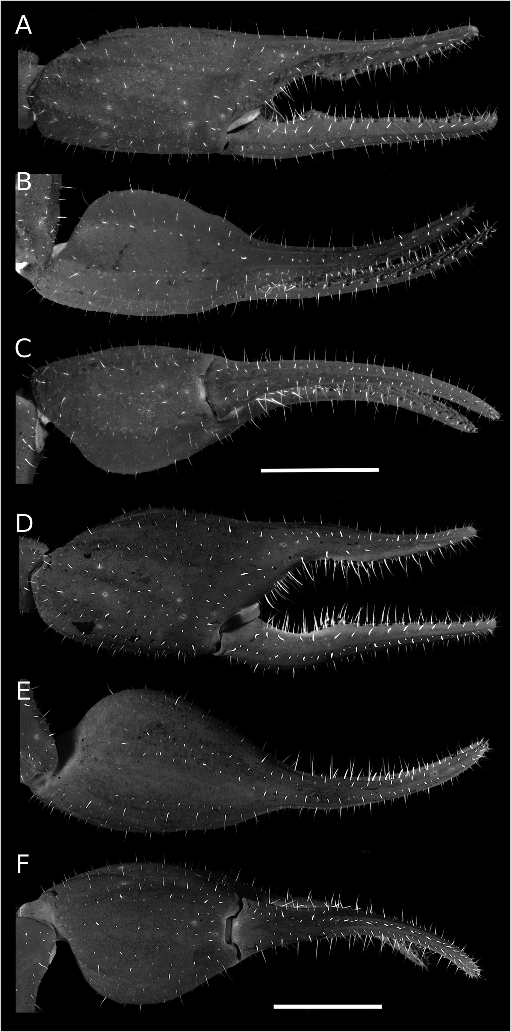

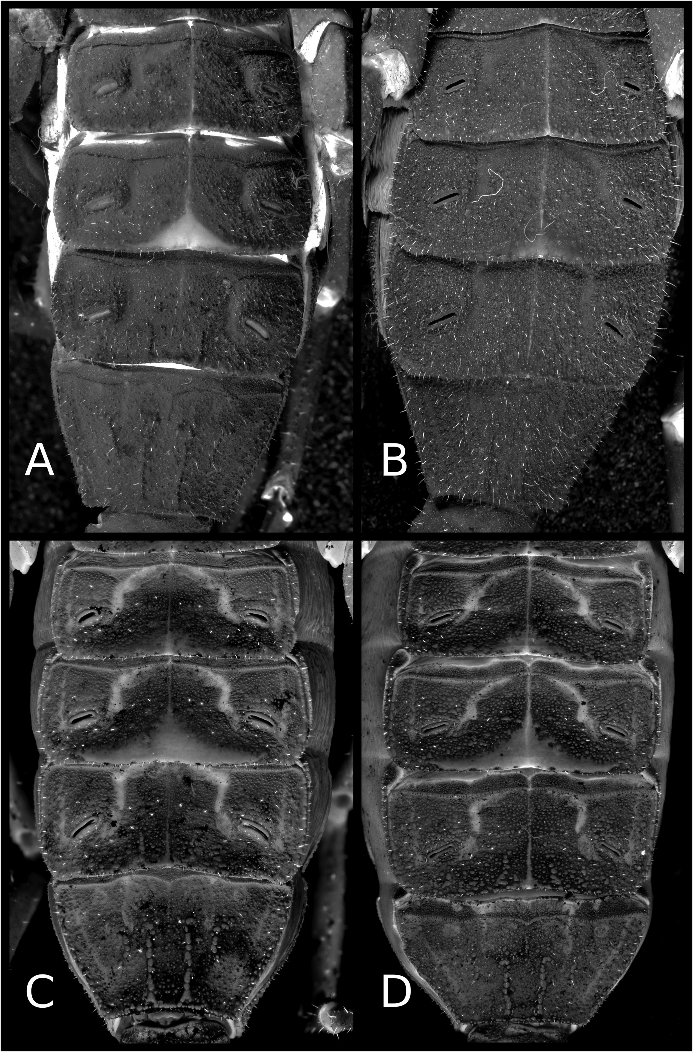
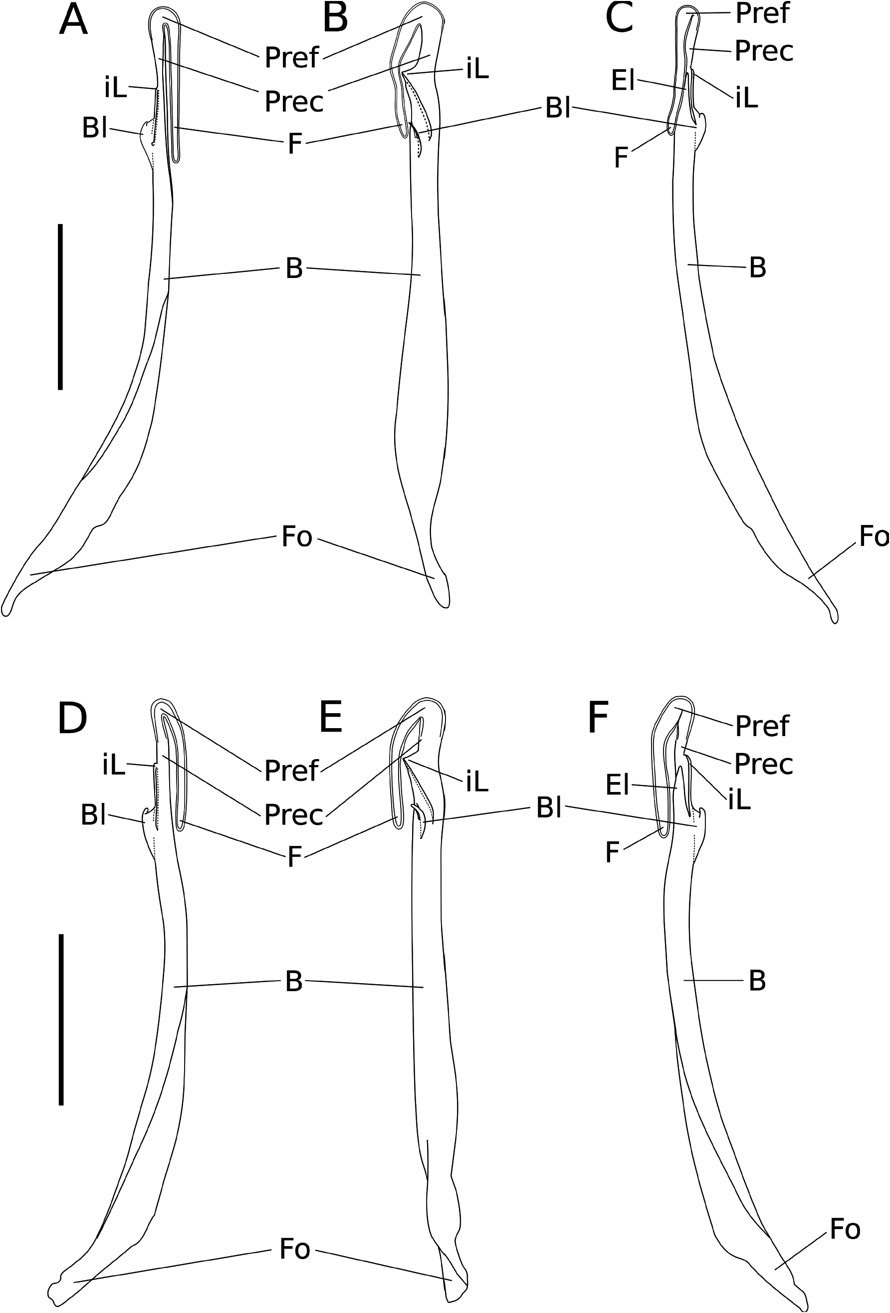

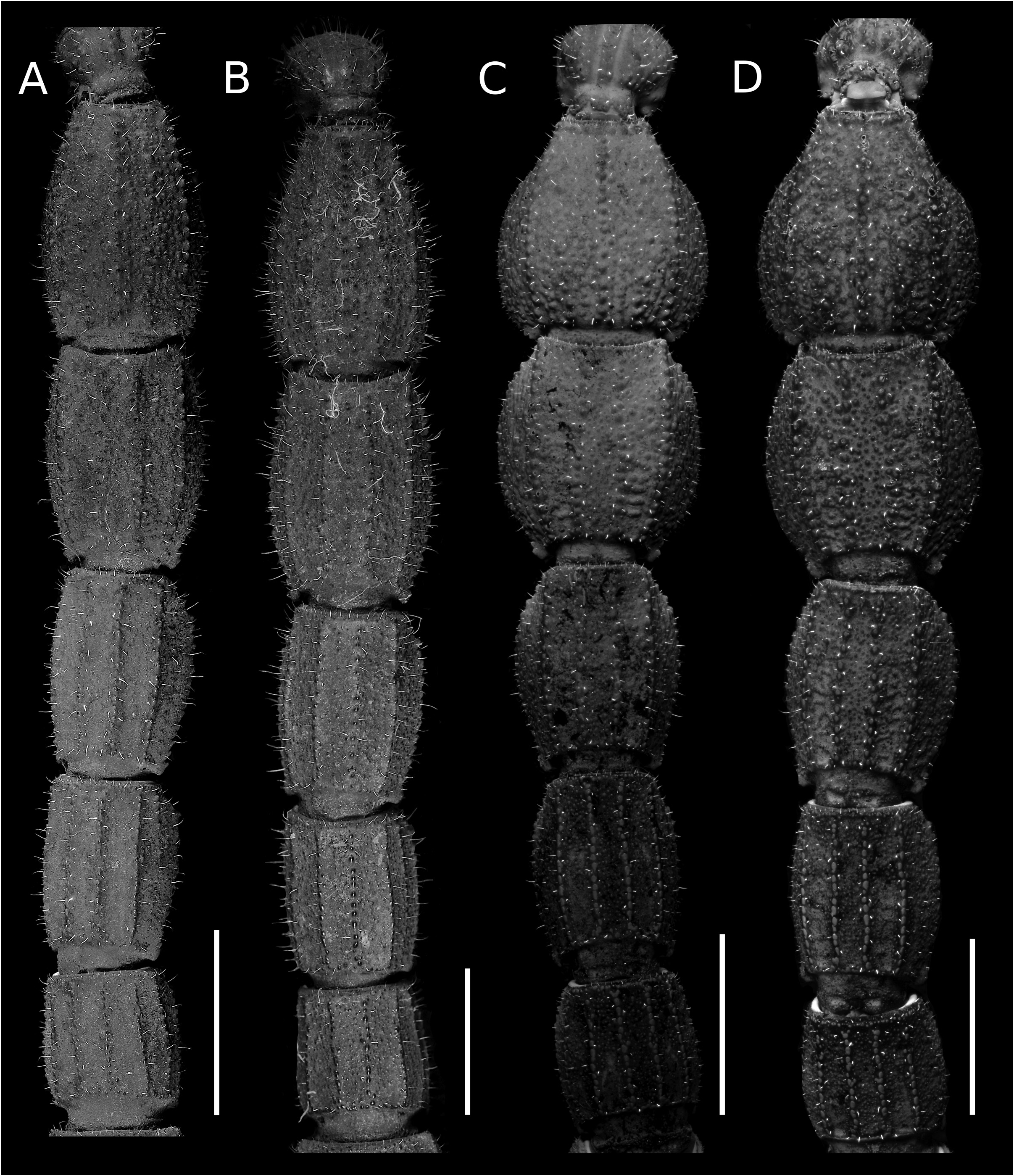
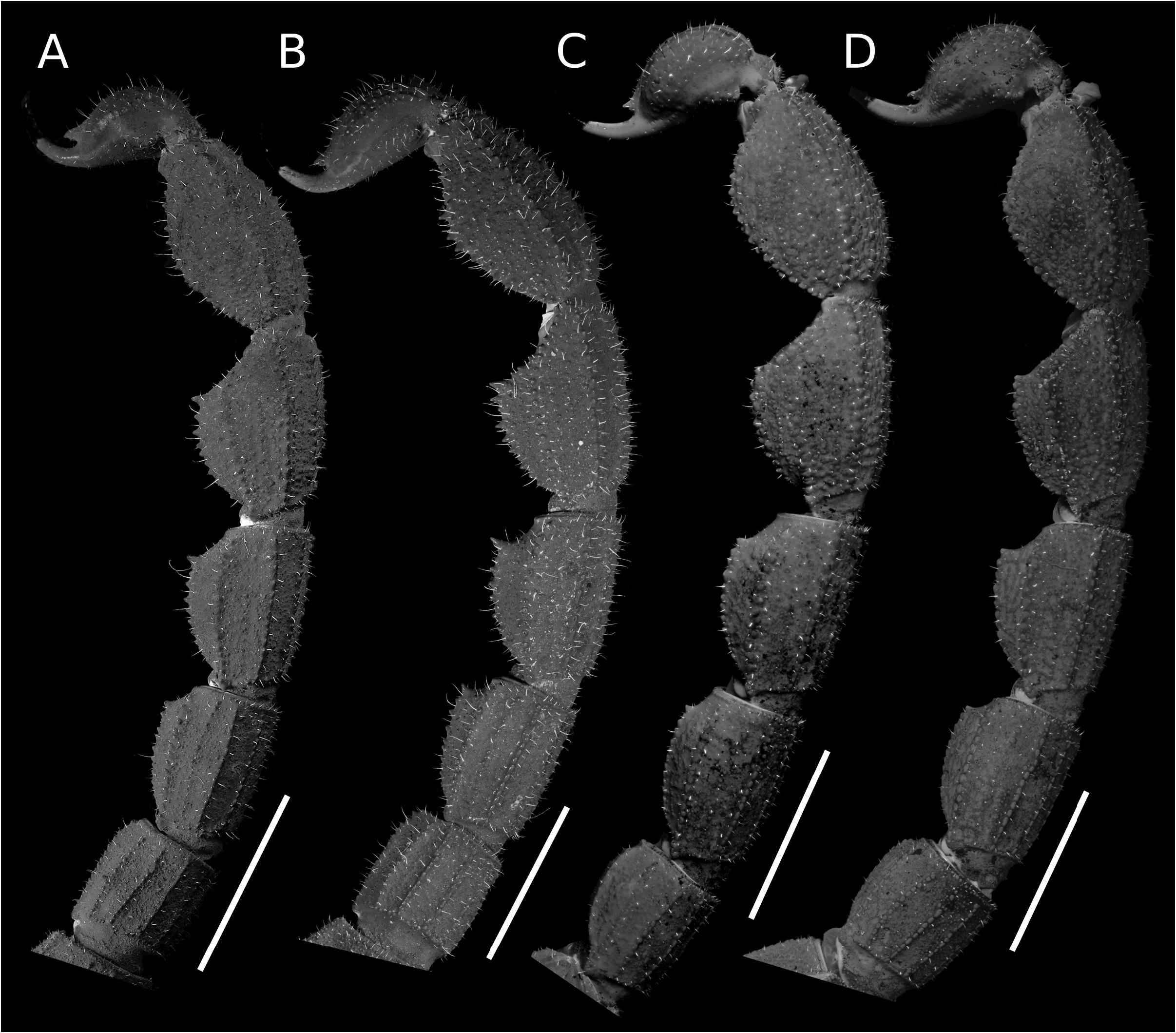

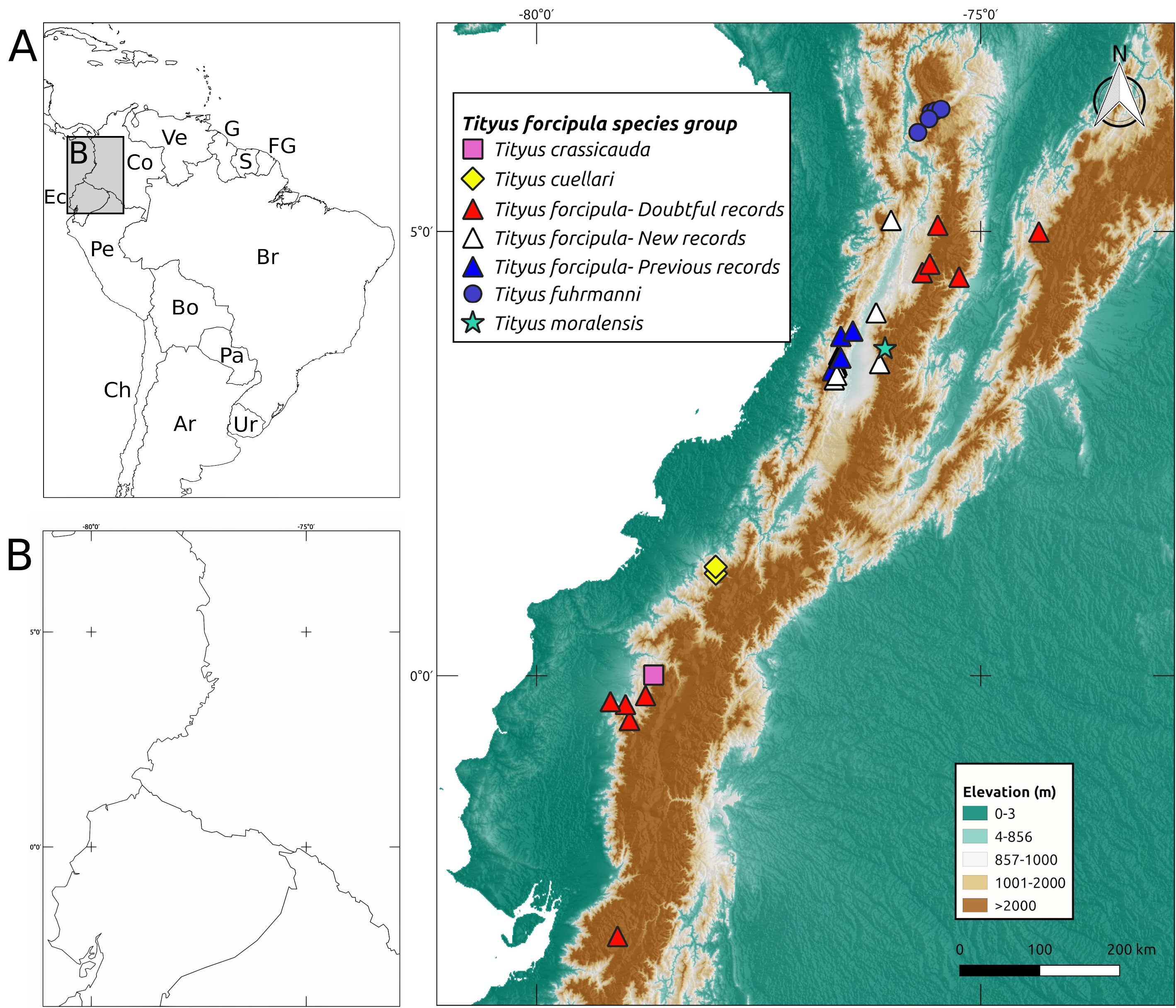
TABLE 5. Measurements (mm) of Tityus forcipula and Tityus moralensis sp. nov. Observation: all specimens, but 1402
| Structure | Measure | Tityus moralensis | Tityus forcipula | |||||||
|---|---|---|---|---|---|---|---|---|---|---|
| 2104 ♂ | 1402 ♀ | 1403 ♀ | 2108 ♀ | 2109 ♀ | 2111 ♀ | 436 ♀ | 2103 ♀ | 438 ♂ | ||
| Body | Total length | 55.03 | 57.99 | 53.38 | 57.93 | 61.55 | 56.25 | 61.73 | 62.30 | 53.81 |
| Carapace | Length | 6.00 | 6.29 | 5.71 | 6.43 | 6.50 | 6.35 | 7.00 | 7.00 | 5.71 |
| Anterior width | 4.14 | 4.57 | 4.00 | 4.71 | 4.65 | 4.75 | 5.00 | 4.86 | 4.14 | |
| Posterior width | 6.29 | 7.00 | 6.57 | 7.29 | 7.90 | 8.00 | 7.71 | 7.86 | 6.29 | |
| Eye diameter | 0.50 | 0.50 | 0.50 | 0.53 | 0.60 | 0.55 | 0.63 | 0.63 | 0.60 | |
| Interocular distance | 0.57 | 0.70 | 0.60 | 0.67 | 0.70 | 0.70 | 0.57 | 0.57 | 0.60 | |
| ocular diad width | 1.23 | 1.43 | 1.30 | 1.43 | 1.40 | 1.40 | 1.57 | 1.53 | 1.37 | |
| Tergite I | Length | 1.00 | 1.07 | 1.07 | 1.13 | 1.50 | 1.05 | 1.13 | 1.27 | 1.00 |
| Tergite II | Length | 1.33 | 1.53 | 1.47 | 1.53 | 1.55 | 1.00 | 1.47 | 1.60 | 1.20 |
| Tergite III | Length | 1.73 | 2.00 | 1.80 | 2.07 | 2.15 | 2.00 | 1.80 | 2.07 | 1.53 |
| Tergite IV | Length | 2.20 | 2.33 | 2.27 | 2.53 | 2.50 | 2.00 | 2.40 | 2.67 | 1.93 |
| Tergite V | Length | 2.33 | 2.73 | 2.53 | 2.80 | 3.00 | 2.50 | 2.80 | 2.80 | 2.13 |
| Tergite VI | Length | 2.47 | 3.00 | 2.80 | 3.00 | 3.60 | 2.50 | 3.07 | 3.07 | 2.27 |
| Tergite VII | Length | 4.27 | 4.73 | 4.33 | 4.73 | 4.75 | 4.25 | 4.87 | 4.93 | 4.13 |
| Mesosoma | Total Length | 15.33 | 17.40 | 16.27 | 17.80 | 19.05 | 15.30 | 17.53 | 18.40 | 14.20 |
| Metasoma I | Length | 4.20 | 4.20 | 4.00 | 4.10 | 4.00 | 4.50 | 4.30 | 4.50 | 4.10 |
| Width | 3.90 | 3.90 | 3.60 | 4.00 | 4.10 | 4.30 | 4.00 | 4.10 | 3.70 | |
| Height | 3.20 | 3.50 | 3.20 | 3.40 | 3.50 | 3.50 | 3.60 | 3.80 | 3.60 | |
| Metasoma II | Length | 5.10 | 5.10 | 4.80 | 5.00 | 5.15 | 4.80 | 5.40 | 5.50 | 4.90 |
| Width | 4.00 | 4.10 | 3.70 | 4.10 | 4.40 | 4.45 | 4.10 | 4.30 | 3.80 | |
| Height | 3.40 | 3.60 | 3.30 | 3.40 | 3.60 | 3.50 | 3.70 | 3.90 | 3.50 | |
| Metasoma III | Length | 5.80 | 5.70 | 5.20 | 5.60 | 6.00 | 6.00 | 6.10 | 6.00 | 5.50 |
| Width | 4.50 | 4.60 | 4.10 | 4.70 | 4.90 | 4.75 | 4.40 | 4.50 | 4.00 | |
| Height | 3.60 | 4.00 | 3.60 | 4.10 | 3.75 | 4.00 | 4.10 | 4.40 | 3.80 | |
| Metasoma IV | Length | 6.30 | 6.30 | 5.50 | 6.40 | 6.20 | 6.75 | 7.00 | 6.90 | 6.10 |
| Width | 5.80 | 5.80 | 5.20 | 5.80 | 6.10 | 5.75 | 4.80 | 5.00 | 4.40 | |
| Height | 4.20 | 4.30 | 3.80 | 4.40 | 4.75 | 4.90 | 4.10 | 4.50 | 4.00 | |
| Metasoma V | Length | 6.50 | 6.60 | 6.20 | 6.60 | 6.90 | 7.00 | 7.50 | 7.40 | 6.90 |
| Width | 6.00 | 6.00 | 5.70 | 5.90 | 6.20 | 6.00 | 4.90 | 4.90 | 4.50 | |
| Height | 4.00 | 3.90 | 3.70 | 4.40 | 4.15 | 4.00 | 4.10 | 3.90 | 3.70 | |
| Metasoma | Length | 27.90 | 27.90 | 25.70 | 27.70 | 28.25 | 29.05 | 30.30 | 30.30 | 27.50 |
......continued on the next page
No known copyright restrictions apply. See Agosti, D., Egloff, W., 2009. Taxonomic information exchange and copyright: the Plazi approach. BMC Research Notes 2009, 2:53 for further explanation.
|
Kingdom |
|
|
Phylum |
|
|
Class |
|
|
Order |
|
|
Family |
|
|
Genus |
|
|
SubGenus |
Tityus |
Tityus forcipula ( Gervais, 1843 )
| Pinto-Da-Rocha, Ricardo & Cabra-García, Jimmy 2022 |
Tityus forcipula
| Gervais 1843 |