
Phallobrycon, Menezes, Naércio A., Ferreira, Katiane M. & Netto-Ferreira, André Luiz, 2009
|
publication ID |
https://doi.org/10.5281/zenodo.275063 |
|
DOI |
https://doi.org/10.5281/zenodo.6216286 |
|
persistent identifier |
https://treatment.plazi.org/id/B62D87F1-FFAC-015B-FF46-8249FAD25503 |
|
treatment provided by |
Plazi |
|
scientific name |
Phallobrycon |
| status |
gen. nov. |
Phallobrycon View in CoL View at ENA , new genus
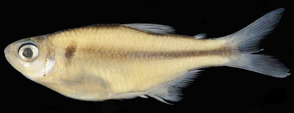
Type species. Phallobrycon adenacanthus , by monotypy and original designation ( Figs. 1 View FIGURE 1 and 2 View FIGURE 2 ).
Etymology. The first part of the name Phallobrycon is from the Greek phallus meaning penis with reference to the urogenital papilla of the male, apparently responsible for the introduction of sperm into the ovary of the female. The word brycon corresponds to the frequently used generic name of small characids.
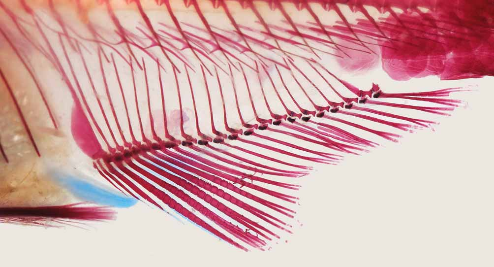
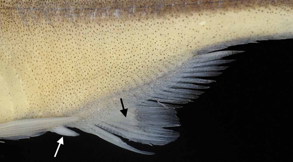
Diagnosis. Among Clade A characids only Phallobrycon and Bryconadenos have the combination of intumescent glandular tissue present on the anterior part of the anal fin and pelvic-fin hooks absent in sexually mature males. In Phallobrycon , however, the glandular tissue is associated with specially developed spines on some anal-fin rays. Two developed spines are present on median unbranched portions of fifth, sixth and seventh anal-fin rays ( Fig. 3 View FIGURE 3 ). These spines are larger and separated from the close-set smaller hooks that appear on distal portions of the anterior anal-fin rays of Phallobrycon , and different from those found on distal portions of several anal-fin rays of many characids. Large spines and small hooks are never found together on any anal-fin ray in Phallobrycon . As opposed to the epithelium clearly divided into a reduced upper part and a more developed lower part forming a cup-shaped structure in Bryconadenos ( Menezes et al., 2009) , a rather simple concentration of glandular tissue on the anterior part of the anal fin is observed in Phallobrycon . Another distinguishing, but not exclusive, character of Phallobrycon is the presence of a developed urogenital papilla in male specimens ( Fig. 4 View FIGURE 4 ).
Discussion. The presence of ii, 8 dorsal-fin rays and four teeth on the inner row of the premaxilla characterizes Phallobrycon as a member of Clade A characids. Gill glands, spermatozeugmata and sperm storage areas in the testis, and presence of spermatozoa within ovarian lumen typical of inseminating Clade A genera, are also present in Phallobrycon ( Figs. 5 View FIGURE 5 , 6 View FIGURE 6 , and 7), but these structures might not be exclusive of members of this clade. Actually gill glands are also present in cheirodontin (Bührnheim & Malabarba, 2007) and aphyocharacin characids (Gonçalves et al., 2005) and was also observed by one of us (KMF) in Bryconamericus exodon , a non-inseminating Clade A species.
Presence of enlarged anal-fin spines in characids was first reported by Weitzman (1977) in Hyphessobrycon diancistrus . Mature males specimens of this species have a remarkably large spine dorsally curved on the posterior margin of fourth unbranched ray and a smaller one on the posterior margin of the unbranched portion of first branched ray (see Weitzman 1977: 353, figs. 2 and 3). Similar spines occupying the same position on the anterior part of the anal fin have been recently described in Hyphessobrycon otrynus by Benine & Lopes (2008: 66, fig. 4), and both Weitzman (1977) and Benine & Lopes (2008) provide information on the occurrence of enlarged anal-fin spines in species of the characid genera Hemigrammus , Tyttobrycon and Moenkhausia . However, the number and location of the spines on the anal fin in Phallobrycon and in the other three characid genera (see Benine & Lopes, 2008) is different, suggesting independent origins. Until a more complete phylogenetic analysis is undertaken, we consider that the presence of enlarged anal-fin spines in Phallobrycon and other characid genera is homoplastic because none of the latter share the features in common between Phallobrycon and Clade A characids.
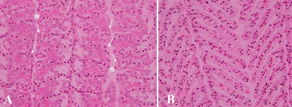
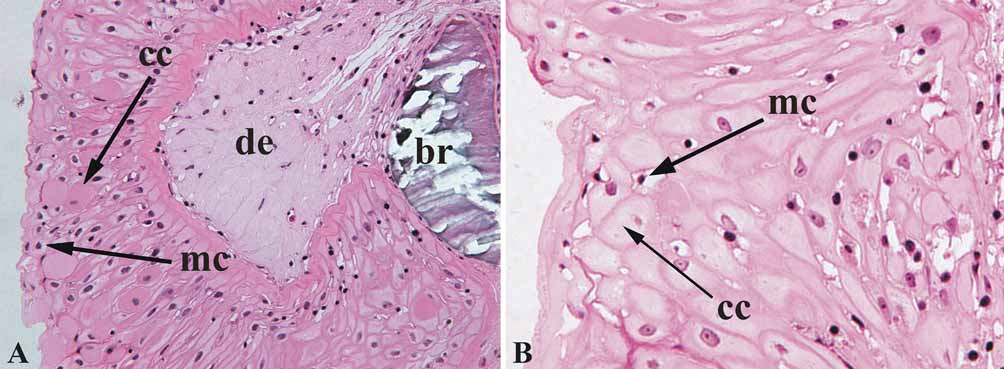
The enlarged anal-fin spines are associated with a mass of white tissue initially identified in H. diancistrus as mucous tissue by Weitzman (1977), and simply as “thick tissue” in H. otrynus by Benine & Lopes (2008). The mass of tissue associated with the enlarged anal-fin spines in Hyphessobrycon diancistrus was later reexamined to reveal the presence of abundant club cells similar to those present in Bryconadenos tanaothoros , in which the cells are arranged into an organ (Weitzman et al., 2005, fig. 21). In a recently discovered and undescribed species of Bryconadenos , the organ is even more developed and elaborate ( Menezes et al., 2009). In Phallobrycon , club cells are also found in the epidermis on the anterior part of the anal fin, not at the surface as proposed by Weitzman et al. (2005), but below a surface layer of mucus cells ( Fig. 8 View FIGURE 8 ). This raises questions about the concentration of glandular tissue in special areas of the characid species indicated by Weitzman et al. (2005: 335). The “thick tissue” of Hyphessobrycon otrynus is probably the result of the thickening of the epithelium caused exclusively by an increase in the layers of club cells. Before its discovery in Phallobrycon , an elongate urogenital papilla presumably used by sexually mature males to transfer sperm to the ovary of the females was only known and described as an intromittent organ in still undescribed inseminating species of Monotocheirodon , also member of clade A characids, by Burns & Weitzman (2006).
In Lophiobrycon weitzmani View in CoL , of the subfamily Glandulocaudinae , which is also inseminating and included in clade A characids, the urogenital papilla is present only in the females (Menezes & Weitzman, in press). In non clade A characids a urogenital papilla is also found in Kolpotocheirodon View in CoL as figured, but not described, in Malabarba & Weitzman (2000: 273 and 274, figs. 2 and 3) and Malabarba, Lima & Weitzman (2004: 323, figs. 6 and 7). According to Flávio C. T. Lima (pers. comm.), the papilla in Kolpotocheirodon figueiredoi View in CoL is morphologically different from that of Phallobrycon View in CoL and larger in females than in the males. Considering that Kolpotocheirodon View in CoL is a member of the subfamily Cheirodontinae , its urogenital papilla most likely developed independently.
Burns & Weitzman (2006) described the histology and anatomy of the intromittent organ present in four populations identified as belonging to Monotocheirodon View in CoL , but not in any of the paratypes of M. pearsoni View in CoL . They also indicate that the four populations of Monotocheirodon View in CoL additionally have sperm with elongate nuclei ( Burns & Weitzman, 2006:530, fig. 1), in contrast with M. pearsoni View in CoL that possess spherical nuclei (aquasperm). Based on this information one might question the inclusion of these populations in Monotocheirodon View in CoL , but if they turn out to be congeneric it might be concluded that the urogenital papilla has evolved in a subset of the genus not including all its representatives. Apart from the developed urogenital papilla, Phallobrycon View in CoL and Monotocheirodon View in CoL share no other exclusive character among clade A characids, and the dentition is quite different in the two genera. Examination of a paratype of Monotocheirodon pearsoni View in CoL (CAS 59792, 29.4 mm SL) by one of us (NAM) revealed the presence of only one row of four mostly pentacuspid premaxillary teeth, seven hexa- to heptacuspid maxillary teeth, and eight mostly pentacuspid dentary teeth gradually decreasing in size. In Phallobrycon View in CoL , on the other hand, there are two rows of four premaxillary teeth, two maxillary teeth with five to six cusps, and a dentary with four anterior pentacuspid and large teeth, followed by two to five smaller tetra or tricuspid teeth. Additionally the two conspicuously enlarged scales on the caudal-fin base typical of M. pearsoni View in CoL , another putative synapomorphy of Monotocheirodon View in CoL , is not present in Phallobrycon View in CoL . Thus the presence of a urogenital papilla in both genera can also be hypothesized as homoplastic.


No known copyright restrictions apply. See Agosti, D., Egloff, W., 2009. Taxonomic information exchange and copyright: the Plazi approach. BMC Research Notes 2009, 2:53 for further explanation.
|
Kingdom |
|
|
Phylum |
|
|
Class |
|
|
Order |
|
|
Family |