
Glyphohesione pattaniensis, Plathong & Plathong & Dean, 2024
|
publication ID |
https://doi.org/ 10.11646/zootaxa.5428.2.4 |
|
publication LSID |
lsid:zoobank.org:pub:ED72D618-B7FA-422A-BAA6-568FA3AB7F7E |
|
DOI |
https://doi.org/10.5281/zenodo.10847455 |
|
persistent identifier |
https://treatment.plazi.org/id/CD47879D-5947-8D2A-FF25-9FC2FED65384 |
|
treatment provided by |
Plazi |
|
scientific name |
Glyphohesione pattaniensis |
| status |
sp. nov. |
Glyphohesione pattaniensis sp. nov.
Figs 3–7 View FIGURE 3 View FIGURE 4 View FIGURE 5 View FIGURE 6 View FIGURE 7
Material examined. Thailand, Southern Gulf of Thailand, Pattani Province. A single incomplete specimen, holotype (PSUZC-POL-0455), chaetigers 21–22 on SEM stub, Sta. PN 03 (7°00'N, 101°20'E), coll. Marine Ecosearch Management Co., Ltd., SCUBA diving, 12 Jul. 2011, sandy mixed with shells fragments, 15 m. GoogleMaps
Diagnosis. Glyphohesione without eyespots. Ventral cirri absent in chaetiger 2. Notopodial spines start on chaetiger 6. Body with small papillae on peristomium and parapodia.
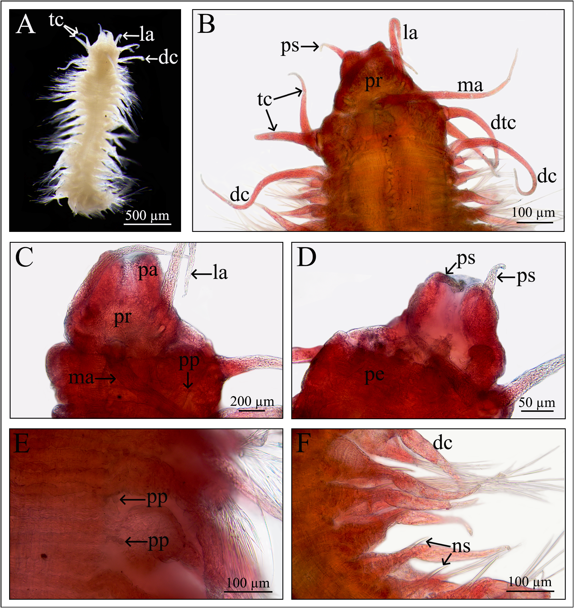
Description. Holotype incomplete, 2.1 mm long 0.5 mm width (at widest point of chaetiger 7, including parapodia), 22 chaetigers ( Fig. 3A View FIGURE 3 ). Body depressed, annulated, with a row of small papillae on peristomium and parapodial bases, mid-ventral groove present; semi-transparent in alcohol, no pigmentation ( Fig. 3A–D View FIGURE 3 ).
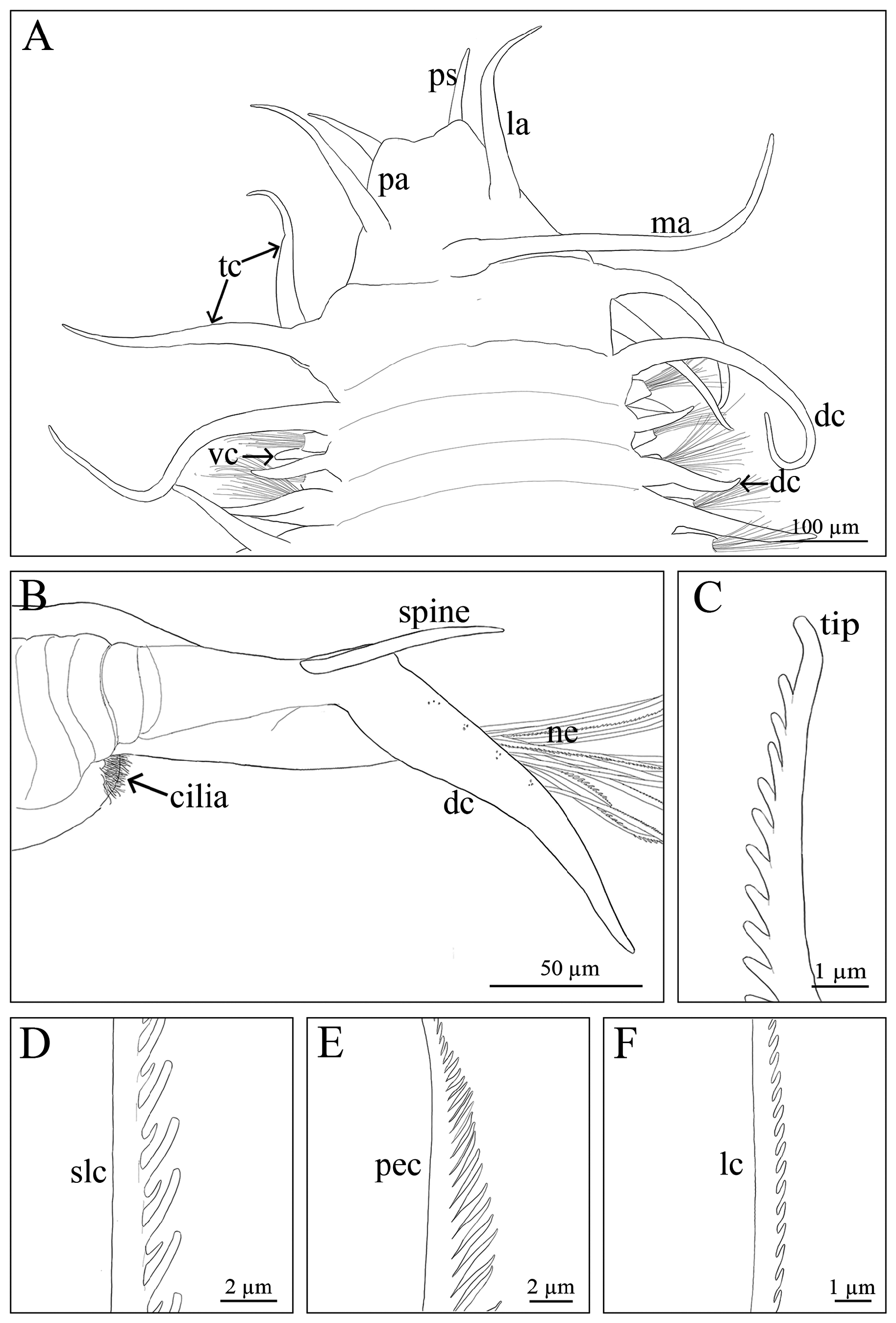
Prostomium bilobed anteriorly, longer than wide (341 µm in length palpostyle and 253 µm wide at the peristomium); three slender cirriform antennae with short ceratophores; median antenna located on the posterior margin of prostomium, about 1.6x longer than lateral antennae.Lateral antennae located at mid-lateral of prostomium; eyespots absent. Palps biarticulate, palpophores large, palpostyles long, slender (0.08 mm), about 0.3x shorter than lateral antennae ( Fig. 3A–C View FIGURE 3 ). Proboscis retracted ( Fig. 3D View FIGURE 3 ).
Peristomium indistinctly separated from prostomium. Two pairs of slender tentacular cirri, slightly shorter than lateral antennae (0.26: 0.28 mm); dorsal tentacular cirri longer than ventral cirri ( Fig. 3B–C View FIGURE 3 ). A transverse row of very small papillae (about 5 µm in diameter) located dorsally about mid-peristomium ( Fig. 3C View FIGURE 3 ).
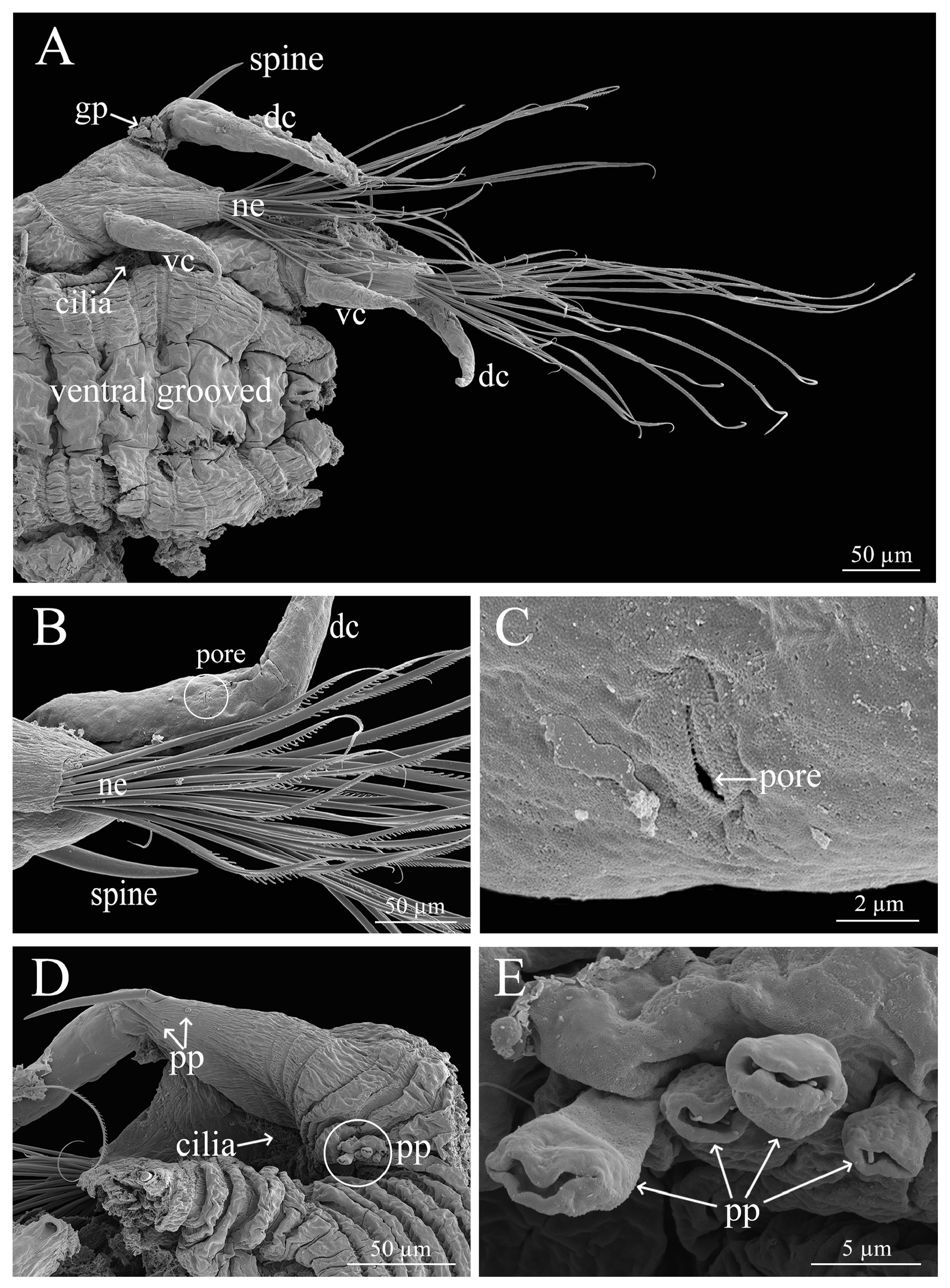
Each chaetiger with rows of 3–5 small epidermal papillae in the posterior dorsolateral margin, and longer than prostomium papillae (up to 7.8 µm in diameter and 12.8 µm long), each row located near parapodial bases ( Figs 3E View FIGURE 3 , 5D–E View FIGURE 5 ) and with 4 axillary ciliated papillae on the parapodia near the aciculum ( Fig. 5D View FIGURE 5 ).
Notopodia with elongated dorsal cirrus; first pair of dorsal cirri longer than those on subsequent chaetigers, about 5.5x longer than dorsal cirri of chaetiger 2 (436:78.5 µm) and 1.7x longer than dorsal tentacular cirri ( Fig. 3A–B View FIGURE 3 ). Ventral cirri shorter than dorsal cirri, ventral cirri of chaetiger 1 shorter than those of other chaetigers, absent on chaetiger 2 ( Fig. 4A View FIGURE 4 ). Dorsum and ventrum of dorsal cirri with rows of pores ( Fig. 5B–C View FIGURE 5 ).
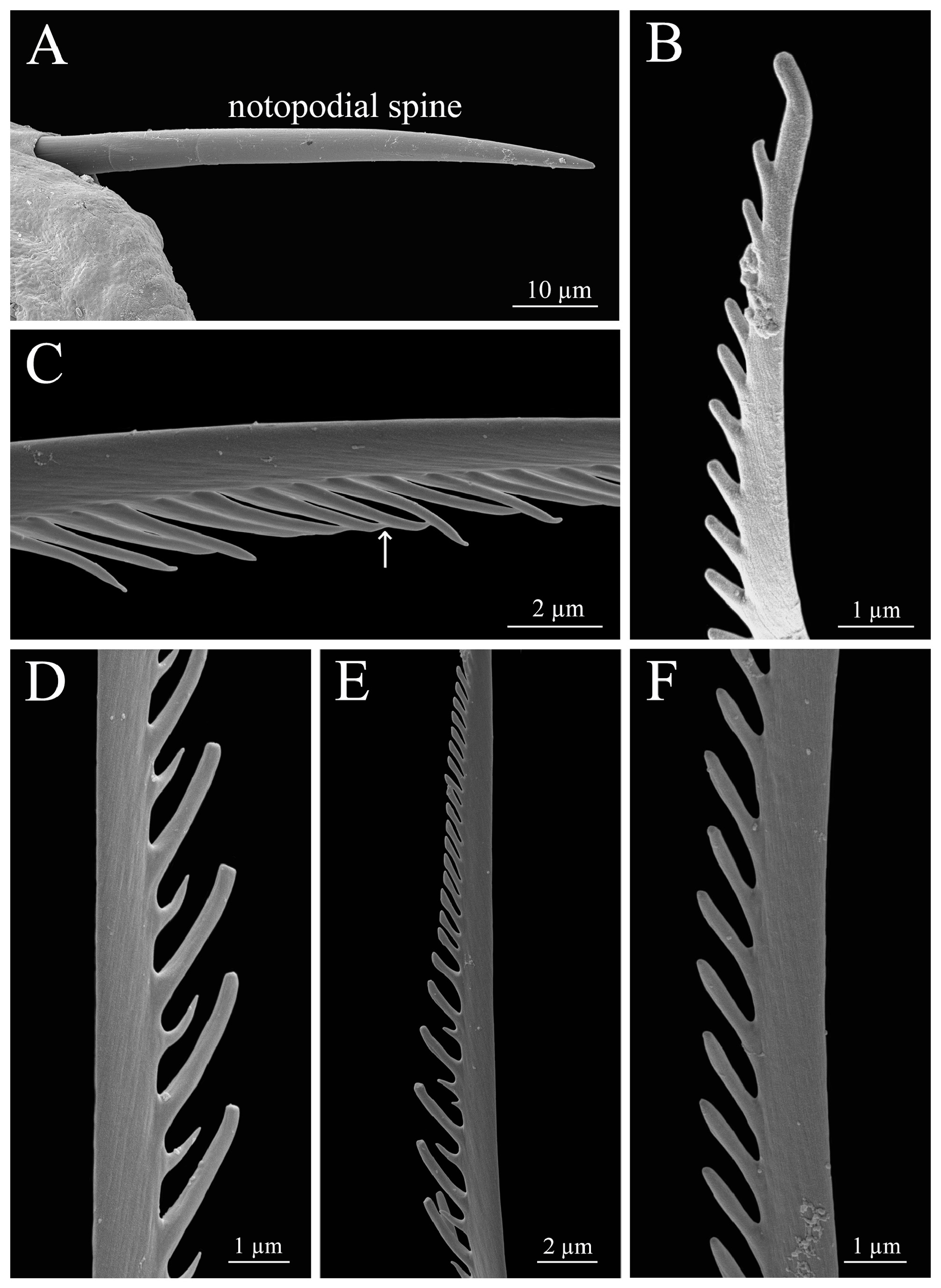
Parapodia biramous. Notopodial lobe reduced; notopodia of chaetigers 1–5 with elongate dorsal cirrus and one notoacicula; from chaetiger 6, notopodia with elongate dorsal cirrus, one notoacicula, and one slightly bent notospine ( Figs 3A–B, F View FIGURE 3 , 5A–B, D View FIGURE 5 , 6A View FIGURE 6 , 7B View FIGURE 7 ).
Neuropodial lobes well developed, conical, truncate ( Fig. 5A–B View FIGURE 5 ); all neurochaetae pectinate with slightly curved tips ( Figs 5B View FIGURE 5 , 6C–F View FIGURE 6 , 7C–F View FIGURE 7 ). Inferior chaetae shorter than superior ones ( Figs 5A, C View FIGURE 5 , 6B, D View FIGURE 6 , 7D View FIGURE 7 ). Neurochaetae numerous, up to 36 chaetae per fascicle ( Figs 4B–C View FIGURE 4 , 5A View FIGURE 5 ).
Oocytes visible in parapodia, very small measure about 11–18 μm in diameter ( Fig. 4D View FIGURE 4 ).
Pygidium unknown.
Etymology. This species is named after Pattani Province, the type locality.
Habitat. Found at 15 m depth of water in sand mixed with shells fragments.
Distribution. Glyphohesione pattaniensis sp. nov. is only known from the Southern Gulf of Thailand.
Remarks. Glyphohesione pattaniensis sp. nov. belongs to the group of Glyphohesione with eyespots absent. This group includes G. campensis , G. klatti and G. longocirrata . Glyphohesione pattaniensis sp. nov. differs from these species by having the lateral antennae located on the mid-lateral of the prostomium. In G. campensis , G. klatti and G. longocirrata the lateral antennae are found on the anterior of the prostomium ( Friedrich 1950; Licher 1994; Ribeiro et al. 2020). Although it most resembles G. klatti by having the first notopodial spines beginning on chaetiger 6, those of G. klatti actually vary from chaetigers 5–8 while those of the new species are only found on chaetiger 6 ( Friedrich 1950). Glyphohesione pattaniensis sp. nov. also differs from G. campensis and G. longocirrata as its notopodial spines start from chaetiger 6 while those of G. campenisis and G. longocirrata present on 5 and 10–15 respectively ( Licher 1994; Ribeiro et al. 2020). Additionally, the new species lacks ventral cirri at chaetiger 2 and small epidermal papillae are present on the prostomium and the parapodia. These characters have not been reported in other species in the genus previously.
Glyphohesione pattaniensis sp. nov. also differs from G. campensis and G. longocirrata by having curved tipped neurochaetae instead of the straight tips of G. campensis ( Ribeiro et al. 2020) and bifid tips seen in G. longocirrata ( Licher 1994) .
Glyphohesione pattaniensis sp. nov. also differs from all known species of the genus in having a greater number (up to 35) of neurochaetae per fascicle. Glyphohesione campensis , G. klatti , G. longocirrata and G. nicoyensis bear fewer neurochaetae with 8, 25, 14 and 18 chaetae per fascicle respectively ( Dean 1998; Friedrich 1950; Licher 1994; Ribeiro et al. 2020).

No known copyright restrictions apply. See Agosti, D., Egloff, W., 2009. Taxonomic information exchange and copyright: the Plazi approach. BMC Research Notes 2009, 2:53 for further explanation.
|
Kingdom |
|
|
Phylum |
|
|
Class |
|
|
Order |
|
|
Family |
|
|
Genus |