
Paucibranchia gemmata ( Mohammad, 1973 ) Molina-Acevedo, 2018
|
publication ID |
https://doi.org/ 10.11646/zootaxa.4480.1.1 |
|
publication LSID |
lsid:zoobank.org:pub:0D3D99EC-107A-4D6B-B19E-52147C6C141E |
|
DOI |
https://doi.org/10.5281/zenodo.5953875 |
|
persistent identifier |
https://treatment.plazi.org/id/CE78C444-FFF0-2147-FF5B-A6DCFCFEFC7F |
|
treatment provided by |
Plazi |
|
scientific name |
Paucibranchia gemmata ( Mohammad, 1973 ) |
| status |
comb. nov. |
Paucibranchia gemmata ( Mohammad, 1973) View in CoL n. comb.
Figures 38–41 View FIGURE 38 View FIGURE 39 View FIGURE 40 View FIGURE 41 , Tables 1–2
Marphysa gemmata Mohammad, 1973:32 View in CoL –34, Figs. 4–5 View FIGURE 4 View FIGURE 5 ; Katsiaras et al. 2014:211 –213, Figs. 8a–d View FIGURE 8 , 9a–d View FIGURE 9 , Tab. 2.
Material examined. Type material: Holotype BNHM 1971.49 View Materials , Al-Dbaiyyah , Kuwait, Arabian Gulf, 9 May 1969, 28°57’ N 48°11’ E, in sand, intertidal, coll. M. B. Mohammad. GoogleMaps
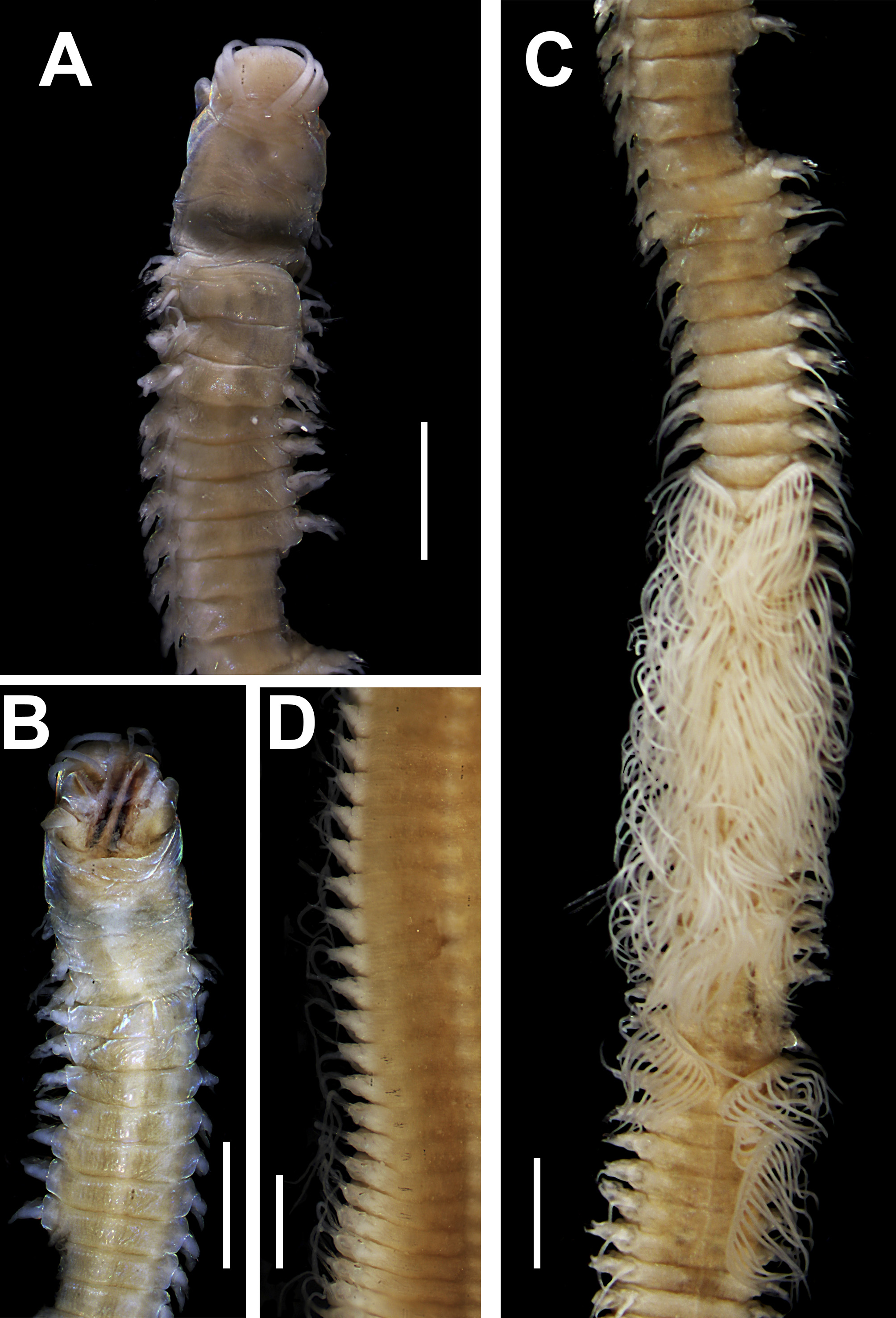
Description. Holotype incomplete, dried, with 160 chaetigers, L10= 5.9 mm, W10= 1 mm, the fragment with TL= 51 mm. Anterior region of body with convex dorsum, and flat ventrum, without groove; body depressed from chaetiger 6, widest at chaetiger 92, tapering after chaetiger 136.
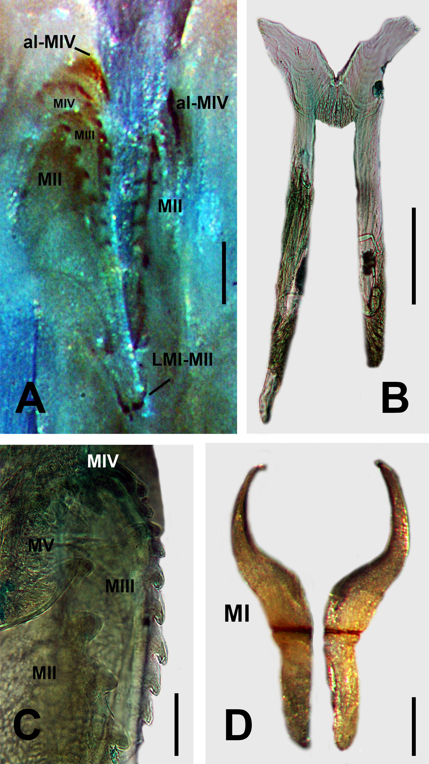
Prostomium in poor condition, entire, 0.8 mm long, 1.2 mm wide, frontally rounded, without median sulcus ( Fig. 38A View FIGURE 38 ), ventral sulcus deep ( Fig. 38B View FIGURE 38 ). Prostomial appendages in a semicircle, median antenna equidistant. Palps reaching first chaetiger; right lateral antennae broken, left lateral antennae reaching second chaetiger; median antennae reaching four chaetiger. Palpophores and ceratophores ring-shaped, short, thick; palpostyles and ceratostyles tapering, thick, without articulation. Eyes absent.
Peristomium wider than prostomium (1 mm long, 1.5 mm wide), first ring two times longer than second ring, separation between rings distinct on all sides ( Fig. 38A–B View FIGURE 38 ). Inferior lip broken.
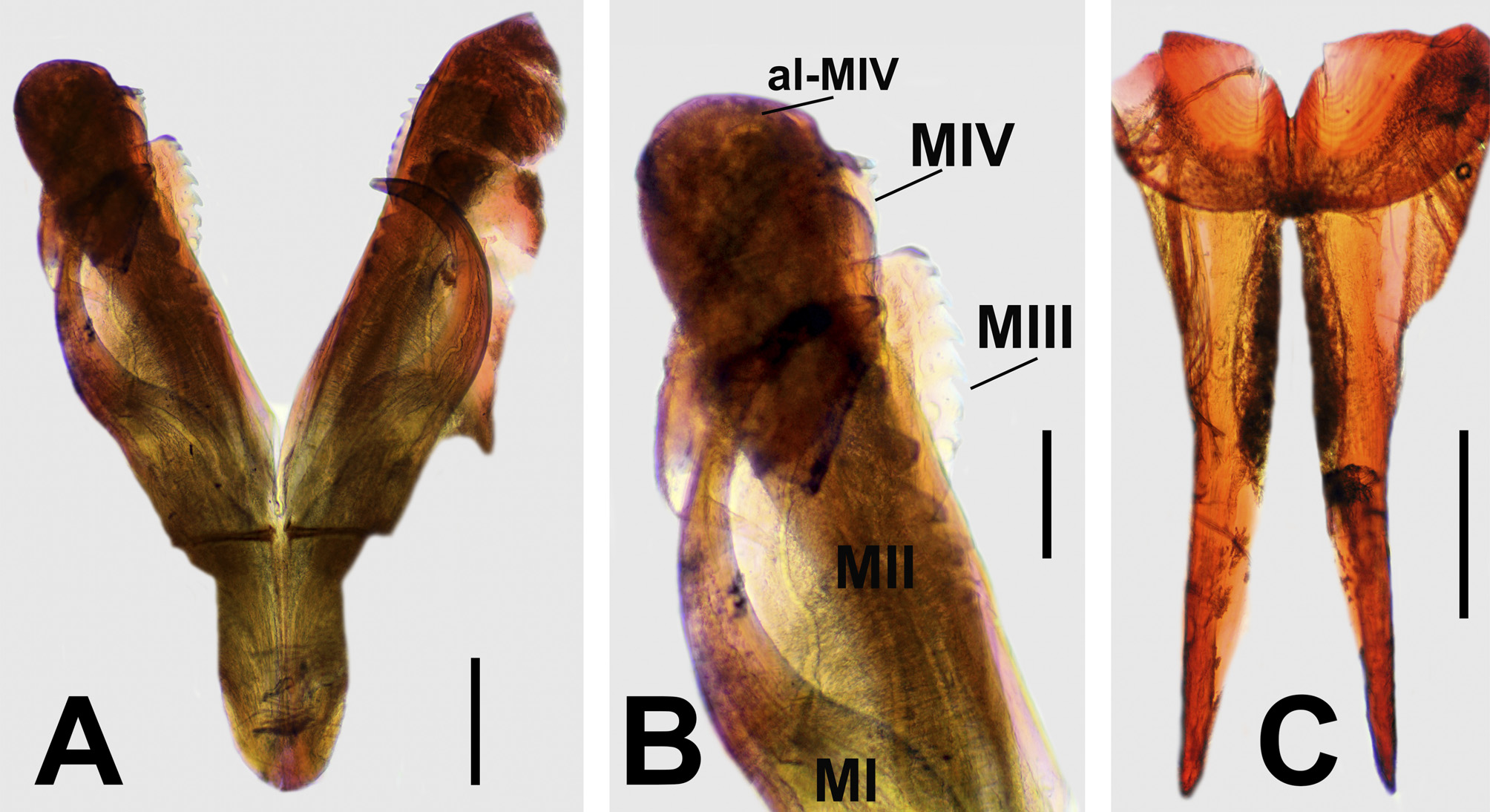
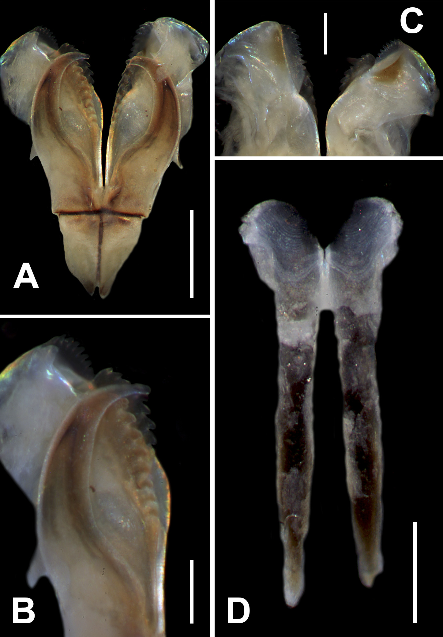
Maxillary apparatus with MF= 1+1, 8+8, 7+0, 7+9, 1+1 ( Fig. 39A View FIGURE 39 ). Maxillary carriers 2.1 times shorter than length of MI. MI forceps-like; closing system 10.5 times shorter than length of MI; ligament between MI and MII slightly sclerotized ( Fig. 39A–B View FIGURE 39 ) . MII wide ; teeth curved, cavity opening oval 4 times shorter than length of MII ; ligament between MII and MIII, and right MIV, slightly sclerotized ( Fig. 39A–B View FIGURE 39 ) . MIII short; with triangular teeth; with attachment lamella slightly sclerotized ( Fig. 39A–B View FIGURE 39 ). Left MIV with small basal teeth; attachment lamella triangular, wide, situated 1/2 along length of posterior edge of maxilla, slightly sclerotized. Right MIV with teeth of similar size; attachment lamella slender, semicircular, better developed in the middle, situated 1/3 along length of posterior edge of maxilla ( Fig. 39A–C View FIGURE 39 ). MV square, with a short rounded tooth ( Fig. 39A–B View FIGURE 39 ). Mandibles dark; cutting plates whitish, with 18 growth rings ( Fig. 39D View FIGURE 39 ).
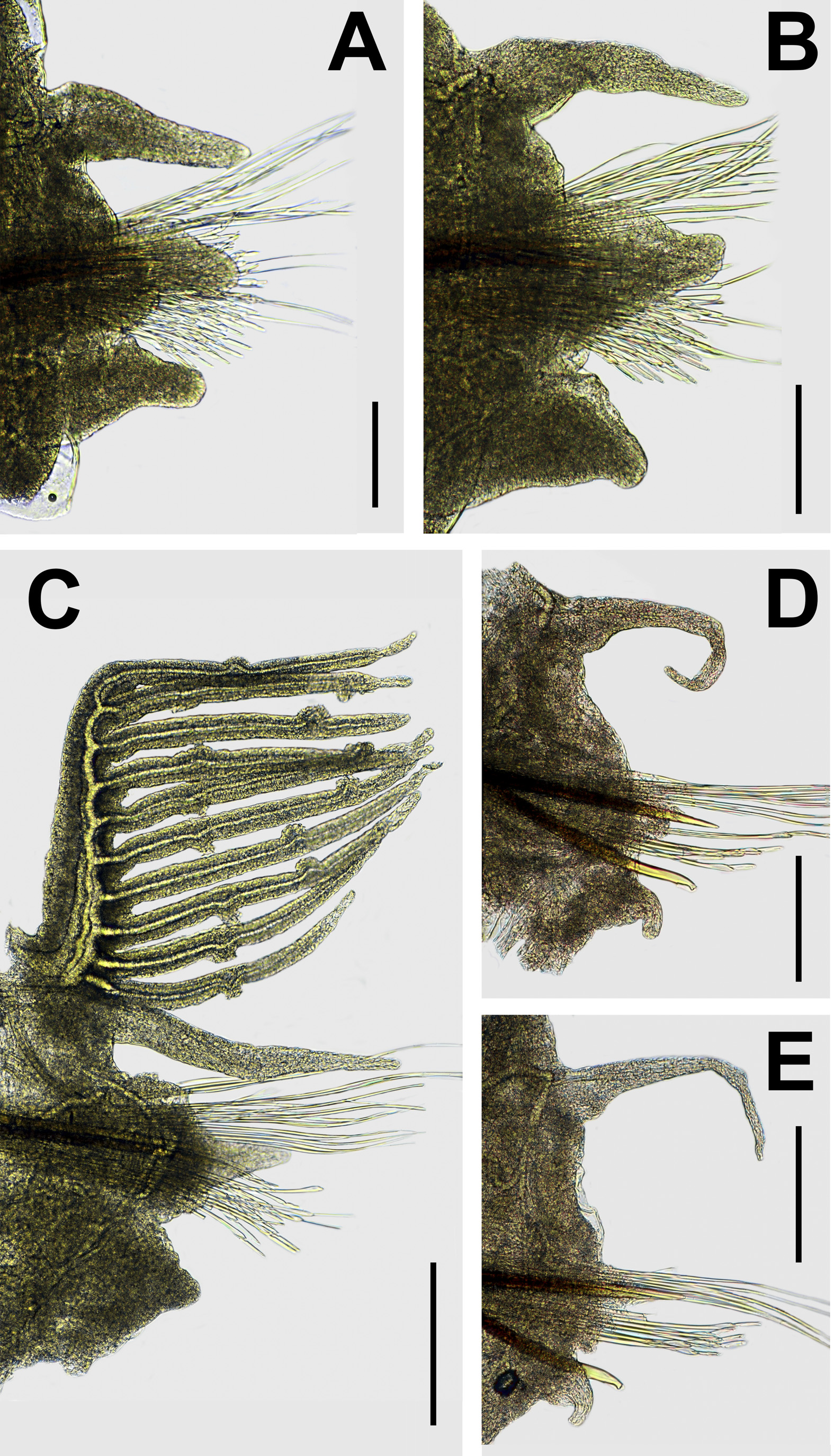
Branchiae pectinate with up to 16 filaments, in chaetigers 22–45 ( Figs. 38C View FIGURE 38 ; 40C View FIGURE 40 ). Number of branchial filaments per chaetiger in order anterior-posterior: 10, 13, 14, 13, 15, 12, 13, 14, 16, 11, 16, 13, 13,?, 11, 15, 9,?, 8, 13, 12, 12, 16. Basal branchial filaments longer than dorsal cirri.
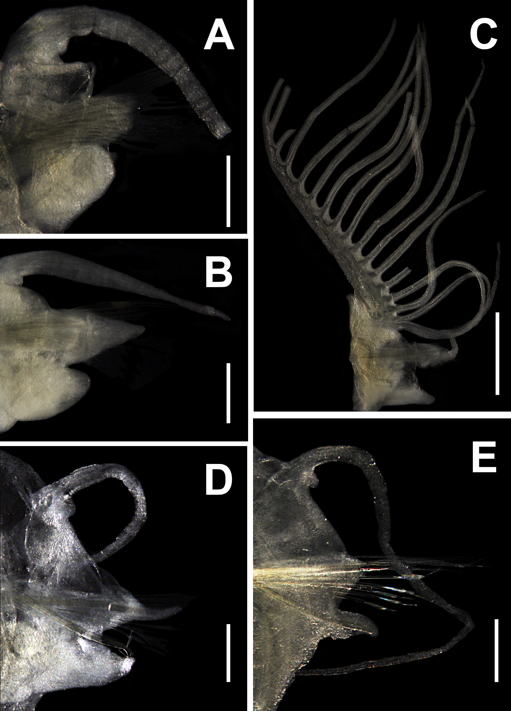
First parapodia smallest; most developed in chaetigers 3–17, following ones becoming gradually smaller. Notopodial cirri conical, increasing in size from chaetiger 3 (Ldc4: 0.60 mm; Ldc16: 0.86 mm) from chaetiger 46, gradually decreasing in width and increasing in length; in posterior region, filiform, two times longer than prebranchial region ones (Ldc105: 1.3 mm); Hayashi & Yamane’s organ present ( Fig. 40A–E View FIGURE 40 ). Prechaetal lobes as a transverse fold in all chaetigers ( Fig. 40A–E View FIGURE 40 ). Chaetal lobes in chaetigers 1–36, rounded, shorter than postchaetal lobes, with aciculae emerging dorsal to midline; from chaetiger 29, triangular, longer than other lobes, with aciculae emerging of midline ( Fig. 40A–E View FIGURE 40 ). Postchaetal lobes well developed in chaetigers 1–94, conical; thinner, elongated in branchial region; decreasing in size in chaetigers 53–94, following ones inconspicuous ( Fig. 40A–E View FIGURE 40 ). Ventral cirri digitiform in chaetigers 1–14; in chaetigers 15–66 with oval swollen base and digitiform tip; from chaetiger 67, conical, gradually reducing in size posteriorly ( Fig. 40A–E View FIGURE 40 ).
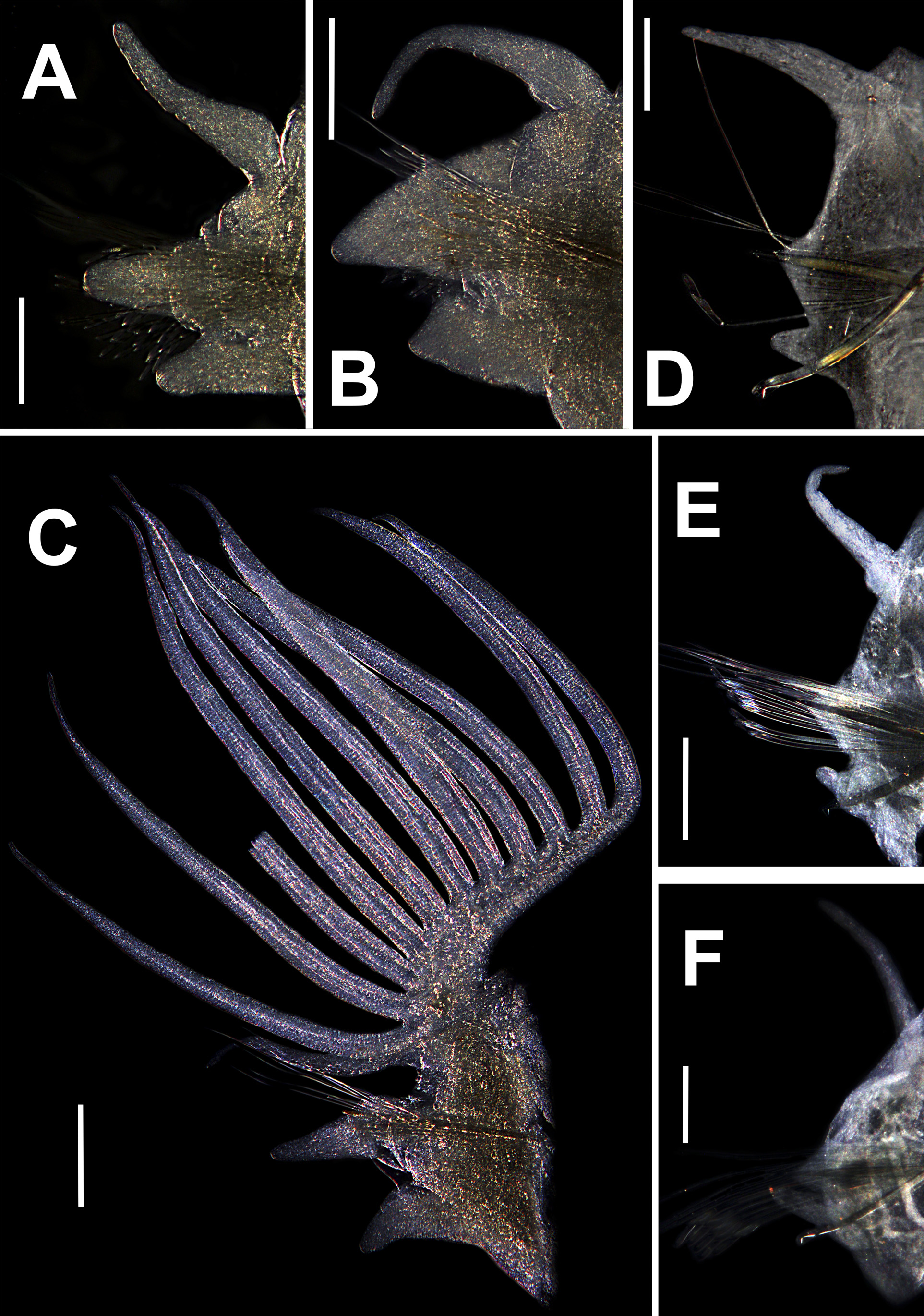
Aciculae blunt; translucent ( Fig. 40A–E View FIGURE 40 ). First 17 chaetigers with 3 aciculae; in chaetigers 18–29 with 2 aciculae; from chaetiger 30, with only one acicula.
Limbate chaetae of two sizes in same chaetiger, larger in anterior region, reduced in number around chaetiger 27. One types of pectinate chaetae; in anterior chaetigers isodonts narrow, with 1–2 pectinate, with up to 3–4 teeth, with transverse distal edge; in median-posterior pectinate not observed. Compound falcigers present in all chaetigers; in anterior region with blade of three sizes (longer 108.5 µm; Fig. 41A View FIGURE 41 , median 90 µm, Fig. 41B View FIGURE 41 ; smaller 80 µm, Fig. 41C View FIGURE 41 ), the smaller more abundant; all with triangular teeth, of similar size, distal tooth directed upward, proximal tooth directed laterally; in media-posterior chaetigers with all blades of similar size, shorter than blades of anterior chaetigers (56 µm, Fig. 41D View FIGURE 41 ), with triangular teeth, distal tooth shorter than proximal, directed upward, proximal tooth directed laterally. Subacicular hooks bidentate, translucent, starting in chaetiger 61L–62R; with one hook per chaetiger; with triangular teeth, distal tooth smaller than proximal, directed upward; proximal tooth directed laterally ( Fig. 41E View FIGURE 41 ).
Distribution. Kuwait, Persian Gulf.
Remarks. The specimen studied here is the only record that has been found of this species. Katsiaras et al. (2014) commented that they did not observe pectinate chaetae in the posterior chaetigers, and this peculiar condition is confirmed here.
Paucibranchia gemmata n. comb. resembles P. adenensis n. comb., P. conferta n. comb., P. patriciae n. sp. and Paucibranchia sp. 2 in the translucent color of subacicular hooks and bidentate form, and the presence of only compound falcigers. However, P. gemmata n. comb. is different because it has dorsal cirri two times longer in the postbranchial region; whereas in all previous species dorsal cirri are of the same size in pre- and postbranchial regions. In addition, P. gemmata n. comb. has falcigers with blades of three sizes in anterior chaetigers; whereas P. adenensis n. comb. and Paucibranchia sp. 2 only have falcigers with blades of two size, and in P. patriciae n. sp. all blades are of similar size. Finally, the aciculae are translucent in P. gemmata n. comb.; whereas in P. conferta n. sp. are reddish on the basal end and translucent distally. The comparison with other Paucibranchia n. gen. species having only compound falcigers present is provided in Table 2.

No known copyright restrictions apply. See Agosti, D., Egloff, W., 2009. Taxonomic information exchange and copyright: the Plazi approach. BMC Research Notes 2009, 2:53 for further explanation.
|
Kingdom |
|
|
Phylum |
|
|
Class |
|
|
Order |
|
|
Family |
|
|
Genus |
Paucibranchia gemmata ( Mohammad, 1973 )
| Molina-Acevedo, Isabel C. 2018 |
Marphysa gemmata
| Mohammad, 1973 :32 |
| Katsiaras et al. 2014 :211 |