
Amaeana occidentalis ( Hartman, 1944 )
|
publication ID |
https://doi.org/10.11646/zootaxa.3994.1.1 |
|
publication LSID |
lsid:zoobank.org:pub:093B124E-58AE-4303-8C07-2D7B27E6AC38 |
|
DOI |
https://doi.org/10.5281/zenodo.6094918 |
|
persistent identifier |
https://treatment.plazi.org/id/DD7687BB-FF9E-FFBF-FF66-FF62D8CEFD74 |
|
treatment provided by |
Plazi |
|
scientific name |
Amaeana occidentalis ( Hartman, 1944 ) |
| status |
|
Amaeana occidentalis ( Hartman, 1944) View in CoL
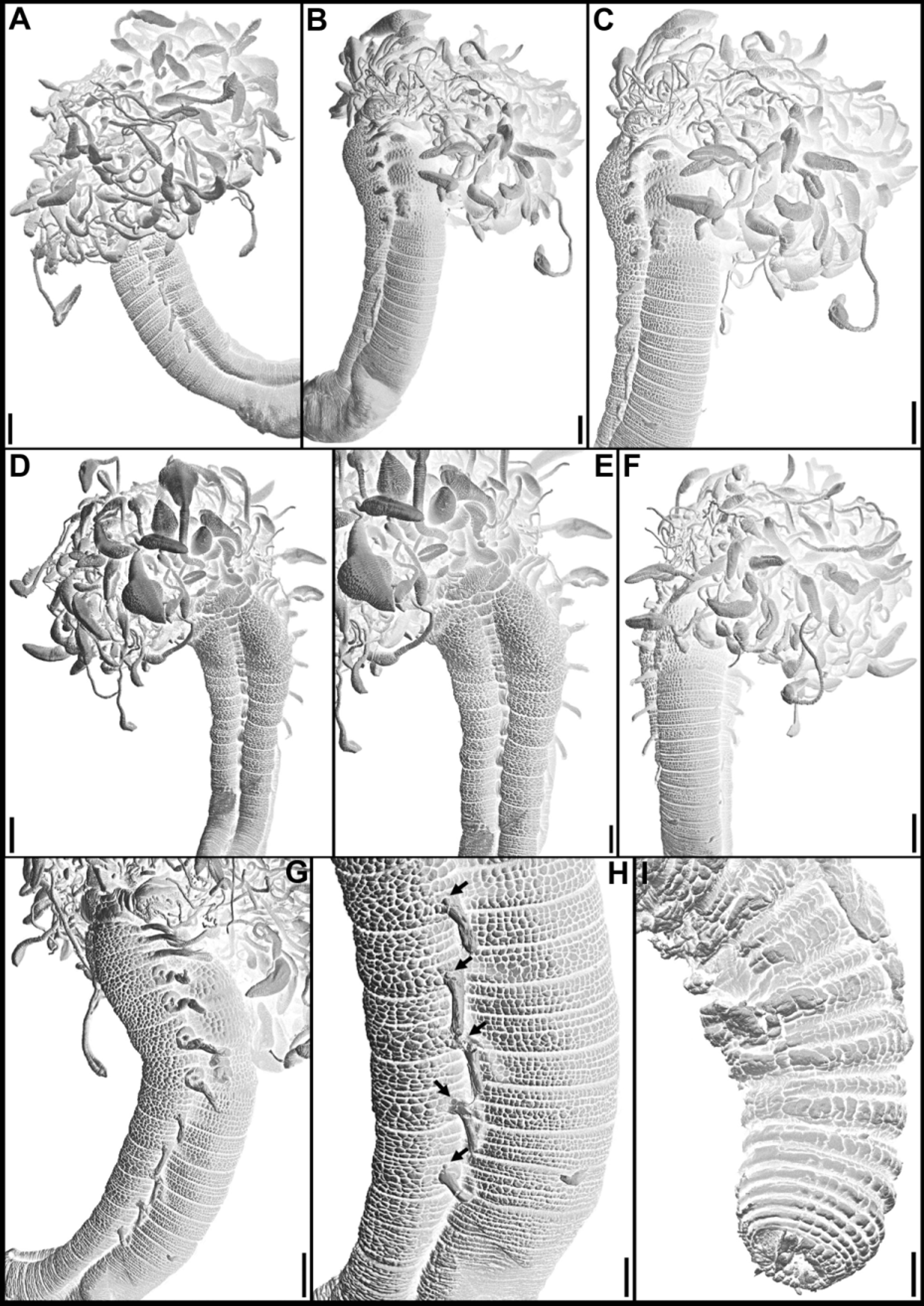
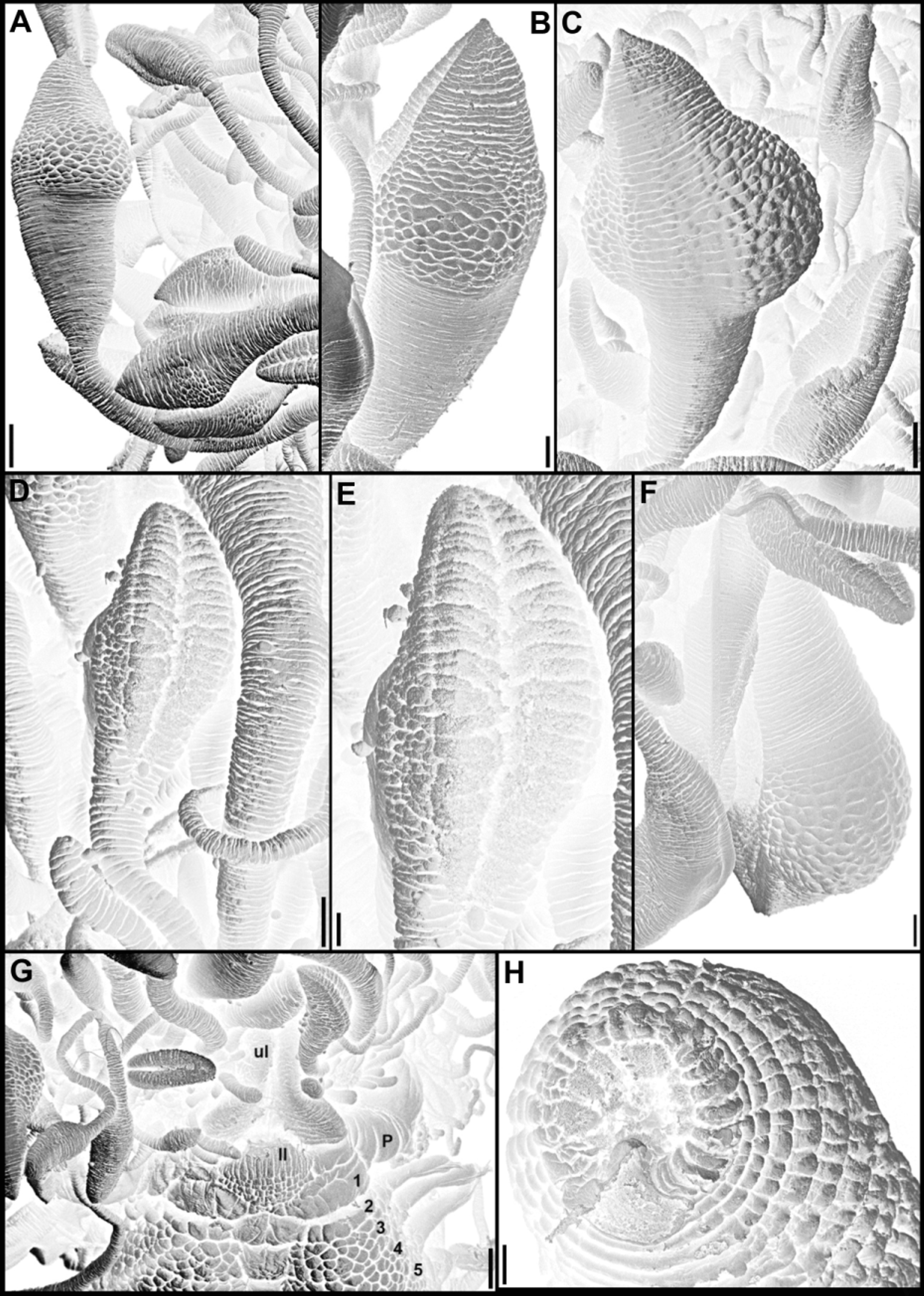
Figures 18 View FIGURE 18 , 19 View FIGURE 19 , 20 View FIGURE 20 , 21 View FIGURE 21 and 22 View FIGURE 22
Amaea occidentalis Hartman 1944: 277 –278, Pl. 26, figs 68, 69.
Amaeana occidentalis View in CoL . Hartman 1959: 495. — Hilbig 2000: 238 –240, Fig. 9.1.
Material examined. Holotype LACM-AHF Poly 230 (coll. USA, California, Marin County, Tomales Bay, Inverness, just north of village, 38°05'54"N, 122°51'07"W, mud flat, soft black mud mixed with shell fragments, by O. Hartman, Jul.1935): incomplete, in good state of preservation, ~ 65 mm long, ~ 4 mm wide. Non-type specimens: LACM-AHF Poly 6517 [coll. USA, California, Monterey County, Moss Landing, Elkhorn Slough, several hundreds north of the current mouth (south mouth before mouth shifted), 36°38'31"N, 121°47'10"W, mudflat, Sta. McGinitie 5, coll. T. H. Bullock, 19.Jun.1939]: 6 specimens, some complete, although several in more than one piece, most of them in good state of preservation, dark brown to purple; slides (from complete specimen, in two pieces, LACM-AHF 6517): notochaetae segment 7, neurochaetae mid-abdominal segment, neurochaetae posterior segment. LACM-AHF Poly 6518 (coll. USA, California, Humboldt County, off Eureka, Eel River South, MBARI- Seep, 40°42'14"N, 124°22'22"W, 39.62 m, sediment near cold seep, Shipek grab, R/V McGAW Sta. Shipek 65, by Lisa Levin, Leslie H. Harris et al., 21.Oct.1998, id L. Harris): complete specimen, in excellent state of preservation, ~ 31 mm long, 2 mm wide, mounted on SEM stub.
Type locality. USA, California, Marin County, Tomales Bay, 38°05'54"N, 122°51'07"W, 58 m.
Description. Holotype incomplete specimen, ~ 65 mm long, ~ 4 mm wide at segment 8, maximum width of body. Life specimens yellow sulphur, becoming purple after preservation (L. Harris personal communication).
Prostomium at base of upper lip, both basal and distal parts developed, basal part as thickened crest, distal part with large, rounded flaring lobes, mid-dorsal process not visible, if present, hidden by large number of buccal tentacles; prostomium covering segment 1 laterally and terminating laterally to lower lip, near mouth ( Figs 18 View FIGURE 18 A–E, G–H; 19A, C; 20B–E, G; 21G). Buccal tentacles of three types, short ones thin, uniformly cylindrical, except for expanded sphaerical to elliptical tips; intermediate tentacles thin, uniformly cylindrical, also expanded at tip; long buccal tentacles progressively widening at tip, towards clearly marked subdistal cylindrical inflation, with short and pointed tip ( Figs 18 View FIGURE 18 A–E, G–H; 19A, C; 20A–G; 21A–G); all types with ciliated groove only conspicuous at tips.
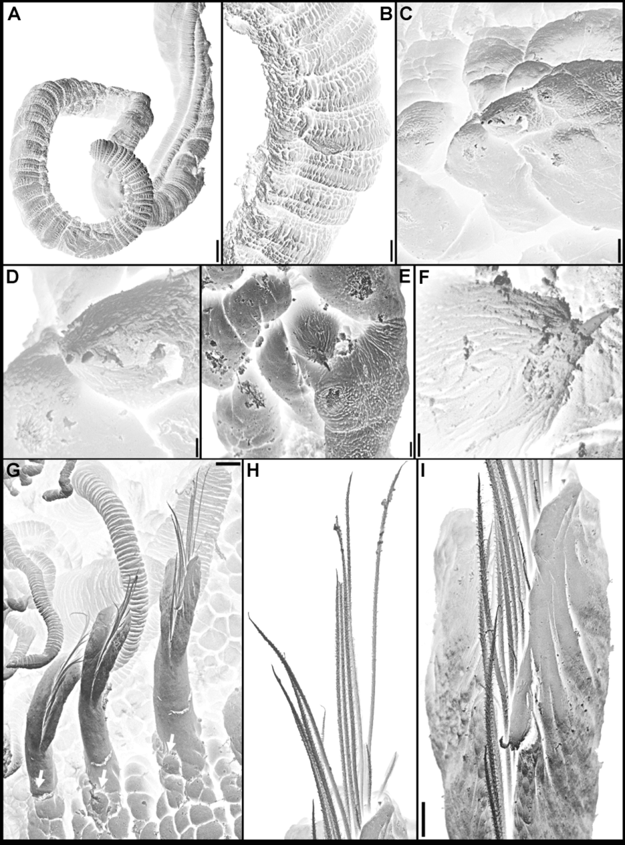
Peristomium restricted to lips, upper lip almost circular; lower lip short, rounded ( Figs 18 View FIGURE 18 A, C, E, G–H; 19A, C; 20B, D–E, G; 21G). Body abruptly broader on segments 3–5, of relatively uniform width until segment 10, slightly tapered on segments 11–14, then more markedly tapering to narrower uniformly cylindrical posterior body, beginning from segment 17–18 and tapering near pygidium ( Figs 18 View FIGURE 18 A–H; 19A, C, F; 20A–I); long achaetous gap between termination of notopodia and beginning of neuropodia, corresponding to segments 15–37, with poorly marked segmentation and fragile, with thin body wall dorsally, distinctly longer than region with notopodia ( Figs 18 View FIGURE 18 A, B–D, F; 19A, F; 20A–B, G–H).
Segments biannulated, segment 1 short, visible dorsally and ventrally; segment 2 narrower and shorter than following segments, with relatively small, pentagonal mid-ventral shield at beginning of mid-ventral groove, extending anteriorly through segment 1 until near ventral edge of lower lip, shield about same length as that of segment 3 and slightly broader ( Figs 18 View FIGURE 18 A–E, G–H; 19A, C; 20D–E; 21G). Ventrum highly glandular, covered with small papillae, arranged in paired ventro-lateral pads on segments 2–14; papillae about same size throughout and slightly more numerous on anterior segments; from after termination of notopodia, paired longitudinal crests bordering mid-ventral groove through posterior body ( Figs 18 View FIGURE 18 A, C, E, H; 19A, C, F; 20A–E, G–H; 22A–B).
Notopodia extending through 12 segments, until segment 14; elongate, cylindrical notopodia, with equal sized lobes and elongate and distally blunt tips ( Figs 18 View FIGURE 18 A–E, G–H; 19A, C; 20A–H; 21G; 22G–I). Acicular, narrowlywinged notochaetae in both rows, wings inconspicuous under higher magnifications of light microscopy, those from anterior row ¼ length of chaetae from posterior row, or less, barely protruding from parapodial lobes ( Figs 21 View FIGURE 21 B, D–E; 22G–I).
Neuropodia present from segment 38, laterally to mid-ventral groove, on outer margins of longitudinal crests. On anterior neuropodia, neurochaetae as up to 12–13 relatively stout, distally tapered spines, slightly expanded subdistally, with oblique tip ( Fig. 19 View FIGURE 19 G); posterior neuropodia with fewer spines, 8–9 ( Fig. 19 View FIGURE 19 H); at least some spines protruding from neuropodia ( Figs 18 View FIGURE 18 A–B, F; 19A, F; 20I; 22A–E).
Nephridial and genital papillae anterior to bases of all notopodia, those of segments 6–9 largest ( Figs 18 View FIGURE 18 B–E, G–H; 19A, C; 20A–H). Pygidium crenulate, with slightly larger ventral papilla ( Figs 19 View FIGURE 19 A, F; 20I; 21H).
Remarks. Amaeana occidentalis is characterised by the great length of the achaetous gap between termination of notopodia and beginning of neuropodia, consisting of more than 20 segments. Also, members of A. occidentalis present the highest numbers of spines per neuropodium, 12–13 on anterior neuropodia, 8–9 on posterior ones, and numbers of pairs of notopodia among all species of Amaeana , 12 pairs, although this latter character is shared with A. accraensis and A. yirrarn ( Table 1).
The only other species with more segments in the achaetous gap is A. antipoda ( Augener 1926) , according to the original description, but members of this species only have 13–15 achaetous segments and have fewer neuropodial spines per neuropodium, about 4–5 spines, and only 11 pairs of notopodia ( Table 1). In addition, as said above, we consider this species as nomem nudum (see Material and methods).
The other species of Amaeana with 12 pairs of notopodia are both easily distinguished from A. occidentalis ( Hartman 1944) , by the number of spines per neuropodium, in addition to the extension of the achaetous gap. The type of A. accraensis has an achaetous gap of 5–6 segments and 8 spines per neuropodium, while in A. yirrarn Hutchings, 1997 , the gap extends for 7–9 segments and there are 1–2 spines per neuropodium ( Table 1). In addition, notochaetae in the posterior row are distally pinnate among specimens of A. accraensis , while in A. occidentalis they are acicular.


No known copyright restrictions apply. See Agosti, D., Egloff, W., 2009. Taxonomic information exchange and copyright: the Plazi approach. BMC Research Notes 2009, 2:53 for further explanation.
|
Kingdom |
|
|
Phylum |
|
|
Class |
|
|
Order |
|
|
SubOrder |
Terebelliformia |
|
Family |
|
|
Genus |
Amaeana occidentalis ( Hartman, 1944 )
| Nogueira, João Miguel De Matos, Carrerette, Orlemir & Hutchings, Pat 2015 |
Amaeana occidentalis
| Hilbig 2000: 238 |
| Hartman 1959: 495 |
Amaea occidentalis
| Hartman 1944: 277 |