
Magelona cf. falcifera Mortimer & Mackie, 2003
|
publication ID |
https://doi.org/10.5281/zenodo.208658 |
|
DOI |
https://doi.org/10.5281/zenodo.5658287 |
|
persistent identifier |
https://treatment.plazi.org/id/EA76A055-FFB5-FF9D-FF0A-D79A0DFDFA1F |
|
treatment provided by |
Plazi |
|
scientific name |
Magelona cf. falcifera Mortimer & Mackie, 2003 |
| status |
|
Magelona cf. falcifera Mortimer & Mackie, 2003 View in CoL
Figures 9–10 View FIGURE 9 View FIGURE 10 , 13 View FIGURE 13 K
Magelona falcifera Mortimer & Mackie, 2003: 167 View in CoL –169, fig. 3?
Material examined. Persian Gulf, IRAN—Stn. 8 ( NMW.Z.2010.037.0008; 11 af, and 1 dissected af), 1998; Stn. 13 ( NMW.Z.2010.037.0009; 1 c, 11 af), 1998; Stn. 14 ( NMW.Z.2010.037.0010; 20 af), 1998; Stn. 24 ( MNCN.16.01/13232; 2 af), 1998; Stn. 8(1) ( NMW.Z.2010.037.0011; 37 af), 2002; Stn. 13(1) (MB29–000189; 23 af), 2002; Stn. B4–10 ( MNCN.16.01/13233, grab A, 15 af; MNCN.16.01/13234, grab B, 22 af; MB29–000190; grab C, 6 af), 2005; Stn. B4–15C ( NMW.Z.2010.037.0012; 1 af), 2005; Stn. F10 ( NMW.Z.2010.037.0013, grab 1, 2 c, 44 af; NMW.Z.2010.037.0014, grab 2, 4 af), 2006; Stn. F20(3) ( NMW.Z.2010.037.0015, 16 af), 2006. QATAR—Stn. E72A ( MNCN.16.01/13235; 1 af), 2005; Stn. G93 ( MNCN.16.01/13236, grab A, 3 af; MB29– 0 0 0 191, grab B, 1 af; MB29–000192, grab C, 1 af), 2005.
Diagnosis. Prostomium width similar to length, subtriangular without prostomial horns. Chaetigers 1–8 with lanceolate postchaetal lamellae, notopodial prechaetal lamellae indistinct and without dorsal superior processes. Chaetiger 9 with broad triangular postchaetal lamellae. All thoracic chaetae capillary. Abdominal chaetigers with lanceolate lateral lamellae and one enlarged hooded hook in anterior abdominal parapodia; other hooded hooks bidentate, those nearest the lateral lamellae more slender. Hooks in 2 groups, vis-à-vis. Lateral pouches absent. Methyl green staining particularly strong between chaetigers 5–9.
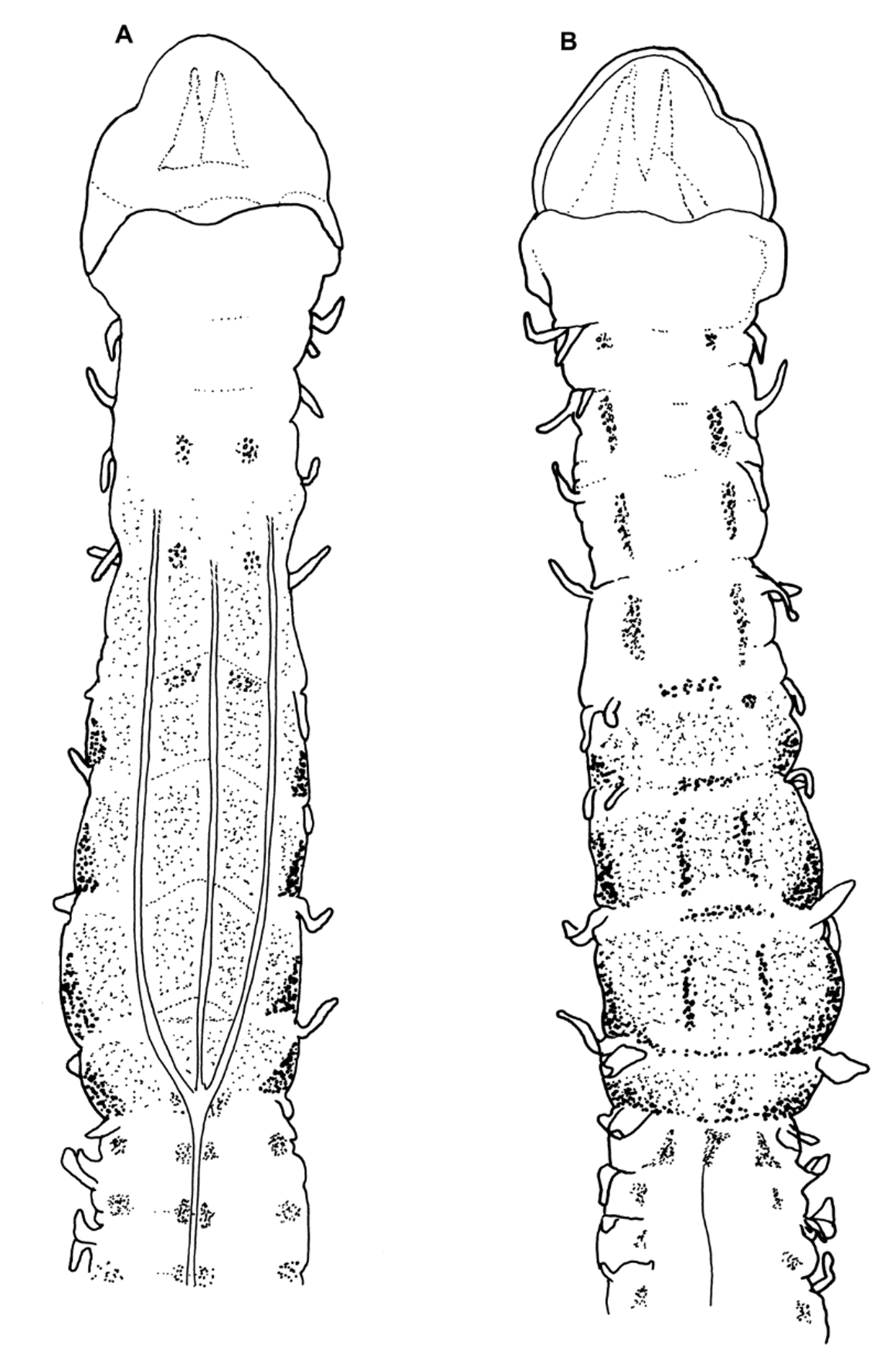
Description. A small slender species; difference between abdomen and thorax not marked ( Figure 9 View FIGURE 9 A). Dimensions of figured complete specimen (NMW.Z.2010.037.0009): prostomium 0.4 mm long, 0.4 mm wide; thorax (including prostomium) 1.8 mm long, 0.45 mm wide (measured at widest point around chaetiger 7); abdomen 0.4 mm wide; total length 17.0 mm for 67 chaetigers. Other complete specimens (2) 5.5–6.7 mm for 45–52 chaetigers; remaining material 12–53 chaetigers, 1.3–13.5 mm.
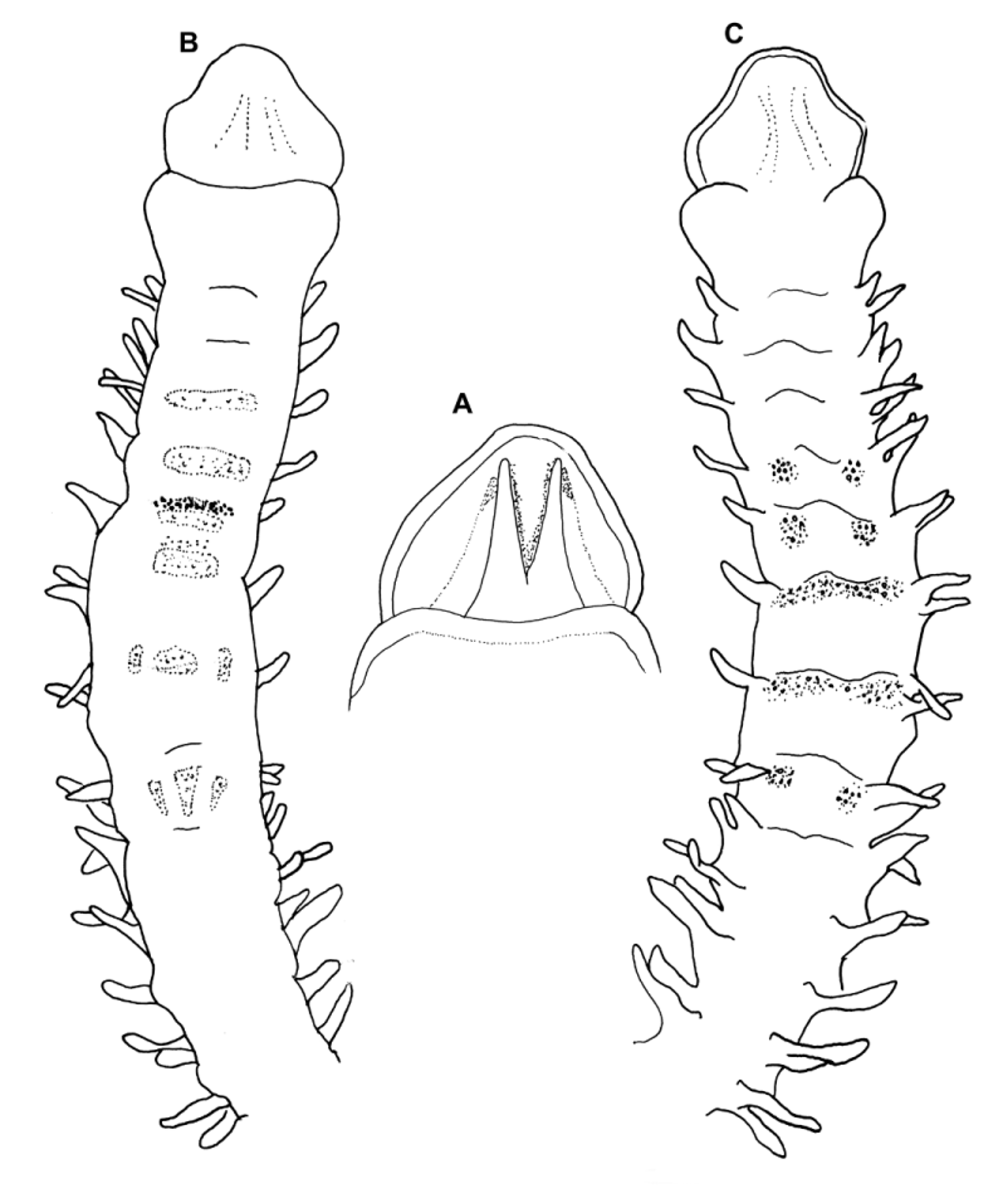
Prostomium as long as, or less than width, subtriangular; anterior margin smooth and rounded, occasionally straight ( Figure 9 View FIGURE 9 B) (Note: prostomium folded and curled in anterior drawing, Figure 9 View FIGURE 9 A). Two pairs of longitudinal dorsal muscular(?) ridges; outer pair shorter and more indistinct, abutting inners for entire length; inner pair separated for majority of length, tips reaching distal prostomial margin. Indistinct (muscular?) areas either side of ridges, visible only under compound microscope. Proboscis everted in many specimens, oval to circular when partially everted, heart - shaped when fully everted. Proboscis ridged inferiorly, superior surface appearing smooth.
Palps present on several specimens, arising ventrolaterally from base of prostomium, reaching at least chaetiger 16–20, non-papillated region reaching chaetiger 3–5. Papillae: long, of similar lengths, digitiform; with 1 row of papillae, either side of inconspicuous ventral groove for length of palp.
Achaetous region behind prostomium roughly one and a half times the size of chaetiger 1; dorsal antero-lateral margins of which are rounded and expand over the base of the prostomium. Chaetigers 1–8 similar; parapodia biramous ( Figures 9 View FIGURE 9 C–G); notopodia with low indistinct prechaetal lamellae. Superior notopodial prechaetal processes (DML) and ventral neuropodial lobes (VNL) absent. Postchaetal lamellae lanceolate, of about equal size in both rami, gradually becoming longer and broader along thorax.
Chaetiger 9: segment slightly narrower and thinner than previous segments. Prechaetal lamellae low; postchaetal lamellae broad triangular, with narrow tips ( Figure 9 View FIGURE 9 H). Chaetae of all thoracic chaetigers simple capillaries.
Abdominal chaetigers with basally constricted lanceolate lateral lamellae of about equal size in both rami (Figures 9I –K), becoming more slender posteriorly. Lamellae do not appear to extend postchaetally. Small triangular processes (DML and VML) present at inner margins of chaetal rows.
A large sickle-shaped hook in both rami of anterior abdominal chaetigers present, no secondary teeth observed ( Figure 9 View FIGURE 9 L); decreasing in size around chaetiger 20 and not apparent after chaetiger 25. A single small slender bidentate hook ( Figure 9 View FIGURE 9 O) is present at the base of the lateral lamellae (and next to the enlarged hooded hooks in the anterior abdomen); appearing to emerge where the lamellae is basally constricted. Remaining hooks ( Figures 9 View FIGURE 9 M–N) bidentate of similar size, hooks in two groups, main fangs vis-à-vis. Approximately four hooks present in anterior abdomen (one slender, one enlarged and two ‘ordinary’ hooks, as seen in figured specimen) increasing medially to approximately 6–8 hooks and posteriorly 4–6. Sporadic tridentate hooks observed in the posterior abdomen.
Eggs observed posteriorly (NMW.Z.2010.037.0008b; dissected af, visible from approximately the 30th chaetiger) packed within the body cavity, approximately 75 μm in diameter. Pygidium small with two slender lateral anal cirri ( Figure 9 View FIGURE 9 P). No pouches observed.
Colour. No living animals observed, preserved colour uniformly cream/white in alcohol. Glandular areas noticeable interparapodially within the abdomen, staining lightly with Rose Bengal in some specimens. Staining with methyl green ( Figure 10 View FIGURE 10 ) shows a diffuse overall stain, particularly strong between chaetigers 5–9. Dorsally, pale green speckled areas are present between chaetigers 1 to 4−5 (also seen as white speckles in unstained material). Darker transverse bands also present, level with parapodia on chaetigers 6–8; two longitudinal lines on chaetigers 7–8 either side of mid - dorsal line, and much darker pigmentation on lateral margins of chaetigers 6–9 (also seen on venter). Staining particularly strong around the parapodia of chaetiger 9; additional stain seen as speckles abdominally. Ventrally, speckled patches (lighter in colour) present medially between chaetigers 3–5 and denser staining medially between chaetigers 5–8. Abdominally, speckled areas interparapodially and ventrally either side of the mid-ventral line.
Habitat. Found at 10 stations from 4 surveys off the coast of Iran, in medium sand, shelly muddy sand, and fine shelly sand, 10–19 m, and 2 stations from one survey off Qatar, in medium sand, 15–18.4 m. Evidence of a sediment tube, present on many specimens.
Distribution. Iran, Qatar (present study), Seychelles ( Mortimer & Mackie 2003).
Remarks. The Persian material in general conforms well with the type material, however, several perceived differences exist. The original description depicts the prostomial shape as subhexagonal, however, having reviewed all material, prostomial shape varies from subtriangular to subhexagonal depending on the degree to which the lateral edges are inverted. Figure 11 View FIGURE 11 A shows the prostomial shape of a dissected paratype, which agrees well with those seen in the Persian material. The figured Persian specimens appear slightly broader than the type material, however, both sets of material are generally of a similar size and breadth. Lastly, there seems to be a variation in the methyl green staining patterns ( Figures 10–11 View FIGURE 10 View FIGURE 11 ). The types of M. falcifera show a diffuse overall stain, with dorsal transverse white bands on chaetigers 6 and 7 and additional white patches on chaetigers 4, 5 and 8. Ventrally, white transverse bands are present between chaetigers 3–6, with additional white patches level with chaetigers 7 and 8, those of the later chaetiger being somewhat triangular. Additional, strong green speckles are often present as a transverse line around chaetigers 4–6 (one transverse line shown on Figure 11 View FIGURE 11 , around chaetiger 5). No abdominal staining observed. The stain dissipated very quickly in the Seychellois material, in contrast to the Persian material that persisted for some time, still evident days after initial staining.
The morphological similarity between the Persian material and the type specimens is strong, with the only major difference appearing to be the variation in staining patterns. However, none of the perceived differences is deemed significant enough to warrant separation of this material at this time.


No known copyright restrictions apply. See Agosti, D., Egloff, W., 2009. Taxonomic information exchange and copyright: the Plazi approach. BMC Research Notes 2009, 2:53 for further explanation.
|
Kingdom |
|
|
Phylum |
|
|
Class |
|
|
Order |
|
|
Family |
|
|
Genus |
Magelona cf. falcifera Mortimer & Mackie, 2003
| Mortimer, Kate, Cassà, Susanna, Martin, Daniel & Gil, João 2012 |
Magelona falcifera
| Mortimer 2003: 167 |