
Thouarella parva Kinoshita, 1908
|
publication ID |
https://doi.org/ 10.11646/zootaxa.3602.1.1 |
|
publication LSID |
lsid:zoobank.org:pub:10304FBF-3969-4EFA-83F1-BB8A5E2B37F3 |
|
persistent identifier |
https://treatment.plazi.org/id/EE36E867-FFDD-FFB2-FF0A-A935FE140CE2 |
|
treatment provided by |
Felipe |
|
scientific name |
Thouarella parva Kinoshita, 1908 |
| status |
|
23. Thouarella parva Kinoshita, 1908 View in CoL
Fig. 40 a,b View FIGURE 40
Thouarella (Diplocalyptra) parva Kinoshita, 1908d: 53–56 View in CoL , figs 1–3; Cairns & Bayer 2009: 28 (list)
Thouarella (Amphilaphis) parva Kükenthal 1915: 149 View in CoL (key); 1919: 410; 1924: 290 (key)
Material unavailable: Holotype, Kodzu Island, Japan, depth unknown. Unfortunately the holotype was not present within the University of Tokyo Museum collection (Dr Ueshima, pers. comm.), and so Kinoshita’s original description of a 73 mm fragment (1908d) is summarised below.
Description
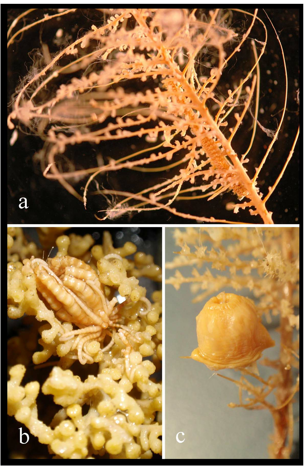
The colony is uniplanar with dichotomous branching. The branchlets diverge from the main stem at around 50˚, 5–13 mm between branchlets. The colony colour is white to light yellow.
The polyps are 1 mm high and upwardly inclined to the branchlets at around 45˚, the widest point being around the marginals. They are slender toward the base ( Fig. 40a View FIGURE 40 ), arranged in pairs, with a density of about 6 per cm, and each has 8 longitudinal rows of body-wall scales. There are 5–6 scales in the abaxial rows, and 5 in the outer and inner lateral rows, reducing to 4 adaxially.
From a lateral view the operculars are completely concealed by the marginals. Very small accessory operculars are present ( Fig. 40 View FIGURE 40 bI). The operculars are lanceolate ( Fig. 40 View FIGURE 40 bII), 160–360 µm high and 90–160 µm wide. The distal half of the outer opercular surface is slightly concave and both the inner and outer surfaces have granules arranged radially in the proximal area. The inner surface is likely to be tuberculate proximally, as is typical for the genus.
The marginals are an irregular triangle-shape ( Fig. 40 View FIGURE 40 bIII) and approximately the same size as the largest opercular (400 µm), although wider proximally.
The body-wall scales are not as tall as the marginals (350–380 µm) and are broader ( Fig. 40 View FIGURE 40 bV, bVI). The outer surface of the body-wall scales has radially arranged granules from the central proximal area whilst the inner scale surface is tuberculate with a smooth band along the distal edge, occasionally with several ridges perpendicular to the distal edge.
The coenenchymal scales are smaller than body-wall scales, and circular to elliptical in shape. The outer surface is sculpted and folded with sparse tubercles on the inner surface. The distal and lateral edges of all sclerites are finely serrate with a coarsely lobate proximal edge.
Distribution
This species is only known from Kodzu Island and Sagami Bay, Japan. The depth from which the sample was taken is unknown.
Remarks
Kinoshita was the first to note accessory operculars in Thouarella (1908d, Fig. 2 View FIGURE 2 , shown here in Fig. 40b View FIGURE 40 ) and T. parva remains one of the few Thouarella species with this kind of sclerite.
The marginals of the polyps of T. parva ( Fig. 40 View FIGURE 40 bIII) appear to have a small keel on their inner surface, and the specimen is thus considered to be Thouarella . However, this is far from clear and the rounded distal edge of what appear to be marginals in Fig. 40b View FIGURE 40 , and lack of a clear keel could indicate that this is actually a species of Plumarella . More material is required to confirm the placement of T. parva within Thouarella .
Comparisons
Dichotomous branching precludes this species from being T. hilgendorfi , T. moseleyi , T. laxa , T. tydemani , or T. grasshoffi from within Group 2. Additionally, these species all have tall, triangular marginals that are absent in T. parva .
Only two other Thouarella species have dichotomous branching: T. coronata and T. biserialis . The polyps of T. coronata diverge at 90˚ to the branch, whereas those of T. parva depart at 45˚. This seems to be a very small difference, however, without more material of T. parva it is impossible to determine the extent of the character differences between these species. There are believed to be specimens of T. parva in Japan (Cairns, pers. comm.), but until this material is examined these species are considered to be distinct.
Polyps of both T. parva and T. biserialis depart from branchlets at approximately 45˚ and all their sclerites appear to be very similar shapes and sizes. The polyps of T. biserialis have marginals with a distinct keel, which may be present on T. parva , but the polyps of T. biserialis are clavate whereas those of T. parva are modestly flared with a relatively tall operculum.

No known copyright restrictions apply. See Agosti, D., Egloff, W., 2009. Taxonomic information exchange and copyright: the Plazi approach. BMC Research Notes 2009, 2:53 for further explanation.
|
Kingdom |
|
|
Phylum |
|
|
Class |
|
|
Order |
|
|
Family |
|
|
Genus |
Thouarella parva Kinoshita, 1908
| TAYLOR, M. L., CAIRNS, S. D., AGNEW, D. J. & ROGERS, A. D. 2013 |
Thouarella (Amphilaphis) parva Kükenthal 1915: 149
| Kukenthal, W. 1915: 149 |
Thouarella (Diplocalyptra) parva
| Cairns, S. D. & Bayer, F. M. 2009: 28 |
| Kinoshita, K. 1908: 56 |