
Polydora carinhosa, Radashevsky, Vasily I., Lana, Paulo C. & Nalesso, Rosebel C., 2006
|
publication ID |
https://doi.org/10.5281/zenodo.174538 |
|
DOI |
https://doi.org/10.5281/zenodo.6256912 |
|
persistent identifier |
https://treatment.plazi.org/id/F72C87EB-FF96-FFAF-057E-FE9988DC3447 |
|
treatment provided by |
Plazi |
|
scientific name |
Polydora carinhosa |
| status |
sp. nov. |
Polydora carinhosa View in CoL sp. nov.
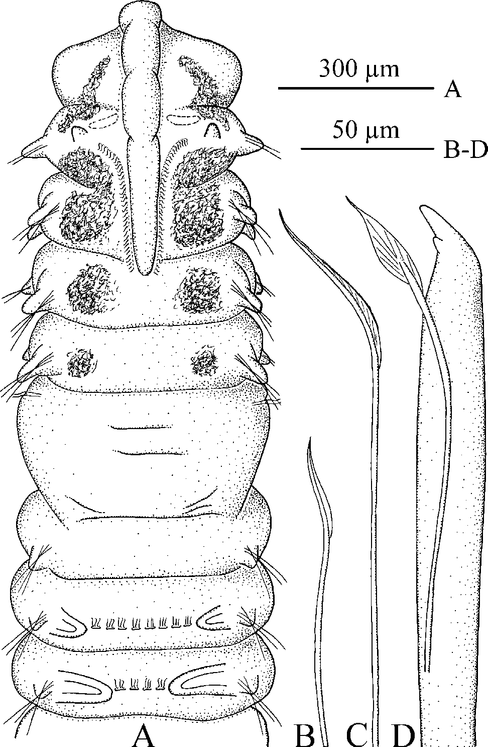
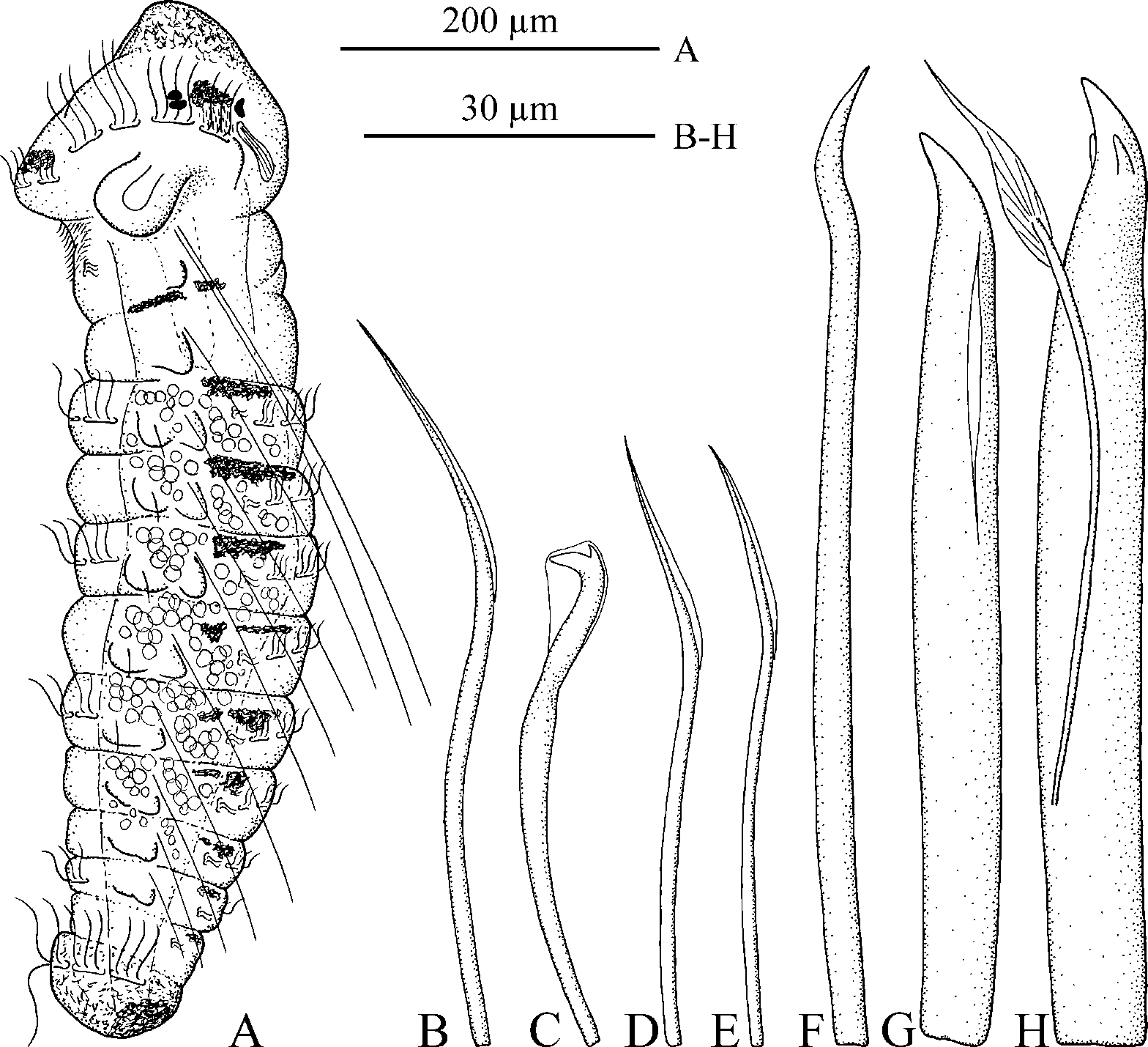
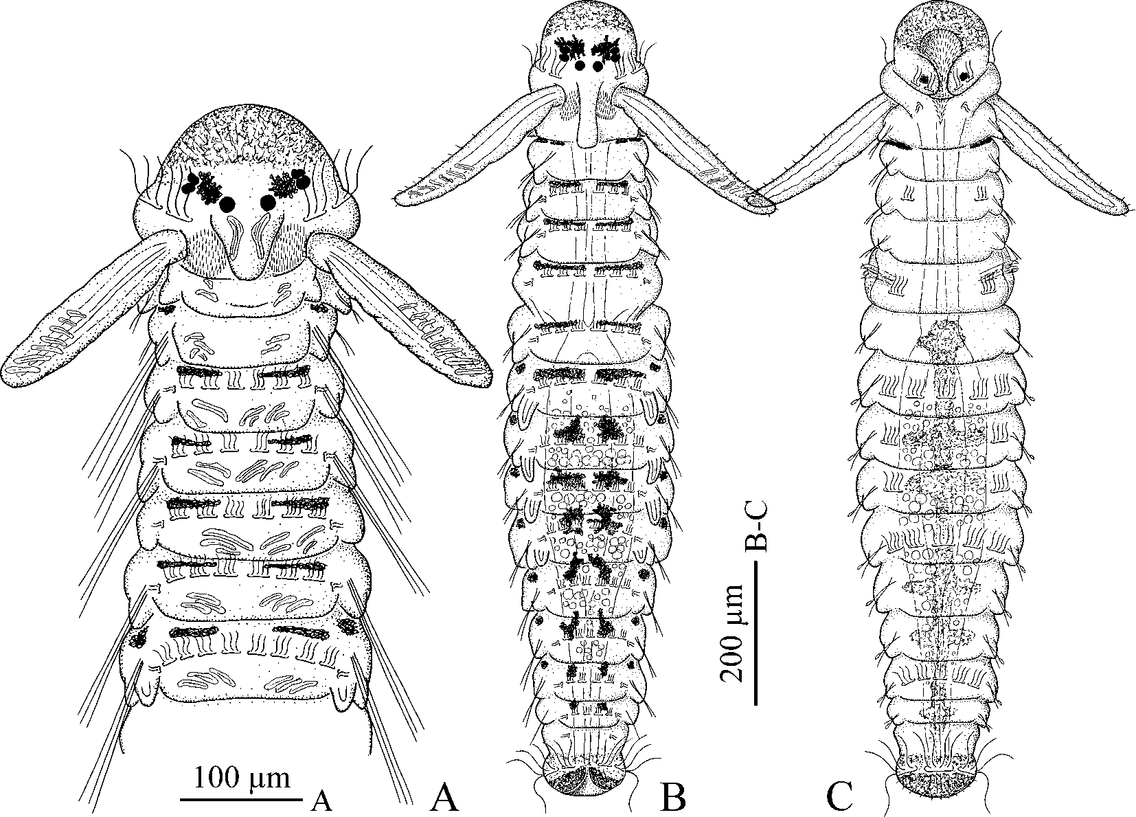
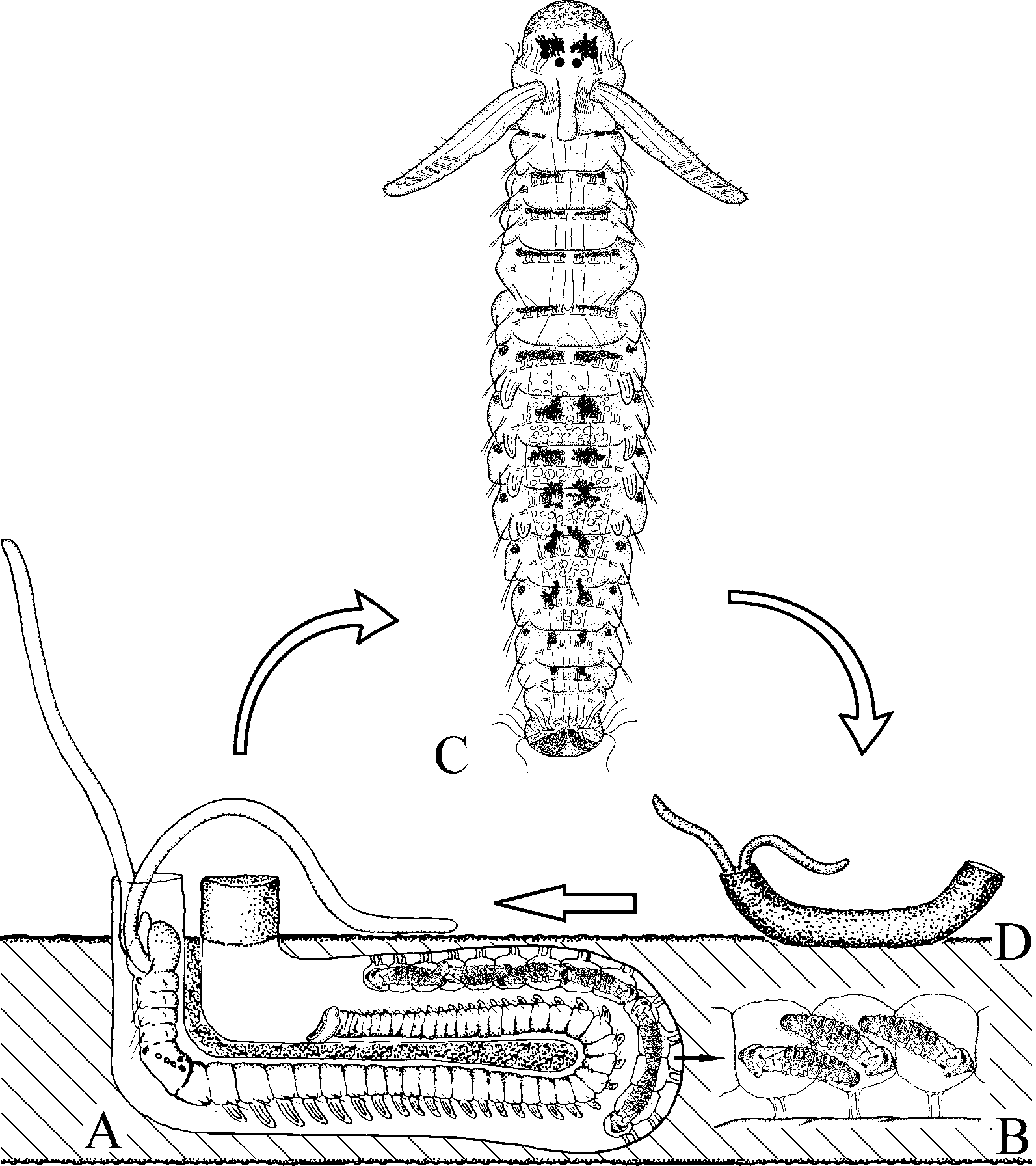
( Figs 10–12 View FIGURE 10 View FIGURE 11 View FIGURE 12 )
Material
Paraná, Paranaguá Bay, 25°28´S, 48°27´W, 2 m, from shells of the oyster C. rhizophorae , 13 Sep 2001, coll. V.I. Radashevsky, MZSP 182 ( holotype). Santa Catarina, Florianópolis, Praia da Ponta de Sambaqui, 27°28.5´S, 48°33.7´W, 1 m, from shells of the cultured oyster C. gigas , coll. Y.M.B. Neptune, 25 Apr 2003, MZSP 181 (1 paratype).
Adult morphology
Holotype complete male, with 103 chaetigers; paratype anterior fragment of female. Prostomium rounded anteriorly. One pair of black eyes present in holotype; eyes absent in paratype. Caruncle extending to middle of chaetiger 3. Occipital antenna absent. Black pigment diffused on dorso-lateral sides of peristomium, in front of palp bases, and on dorsal side of four anterior chaetigers; segmental patches of pigment larger on anterior chaetigers ( Fig. 10 View FIGURE 10 A). Narrow black line present along longitudinal ciliated groove on palps.
Chaetiger 1 with short capillaries in neuropodia and small postchaetal lamellae in both rami, notochaetae absent. Posterior notopodia with packets of needle-like spines besides capillaries. Needles not protruding beyond body surface, beginning from chaetiger 27 in holotype.
Chaetiger 5 greatly modified, with 4 dorsal superior winged capillaries ( Fig. 10 View FIGURE 10 C), 6 major modified spines alternating with bilimbate-tipped companion chaetae and arranged in a slightly curved, diagonal row ( Fig. 10 View FIGURE 10 D), and 6 winged ventral capillaries ( Fig. 10 View FIGURE 10 B); postchaetal lamellae absent. Dorsal superior and ventral capillaries shorter and fewer than those on chaetigers 4 and 6. Major spines falcate, with small lateral accessory tooth.
Hooded hooks in neuropodia from chaetiger 7, bidentate, with constriction on shaft.
Branchiae on chaetigers 7–90, full-sized from chaetiger 11. Nototrochs from chaetiger 7 onwards ( Fig. 10 View FIGURE 10 A).
Pygidium small, cup-shaped.
Holotype male, with sperm first present in chaetiger 27.
Habitat
Polydora carinhosa bores in shells of the oysters C. rhizophorae and C. gigas .
Reproduction
Polydora carinhosa is gonochoristic. Females deposit eggs into capsules which are joined to each other in a string. Each egg capsule is attached by two thin stalks to the inner wall of the burrow. Larvae develop inside the capsules until about the 14-chaetiger stage. The kind of lecithotrophy (endo- or exolecithotrophy) in P. carinhosa is unknown since only two broods with 10- and 13–14-chaetiger larvae were found.
Larval development
Ten-chaetiger larvae ( Fig. 11 View FIGURE 11 A) about 650 µm long, with three pairs of black eyes including two pairs of lateral eyes and one pair of median eyes; lateral eyes positioned close to each other and obscured by ramified melanophores positioned above them. Prostomium and peristomium weakly demarcated. A group of short non-motile apical cilia present on frontal edge of prostomium. Lateral lips of peristomium well developed, forming a voluminous vestibulum lined with short fine cilia. Vestibular ciliation running posteriorly over low ventral peristomial lip as a triangular neurotroch. One pair of small ciliated cells positioned on either side of neurotroch in the middle of chaetiger 1.
Prototroch formed by two bands of long cilia running along lateral sides of peristomium, and two shorter bands of shorter cilia running across lateral lips of peristomium. Short compound cilia positioned along outer edges of vestibulum; these cirri probably sensory, moving slower than simple cilia. Nototrochs from chaetiger 3 onwards. Grasping cilia on either side of each nototroch, beating perpendicular to body axis and holding long bristles along the dorsum when larva is swimming in the capsule. Gastrotrochs on chaetigers 3, 5, 7, and 10. Telotroch interrupted middorsally, forming a gap where long bristles are held when larva is swimming in the capsule.
Fine grains of yellow pigment dispersed on anterior part of prostomium, on ventral side of posterior chaetigers and on pygidium. A pair of ramified melanophores present on prostomium between median and lateral eyes. A pair of small melanophores on ventral side of lateral peristomial lips, just on prototroch level. Black pigment present laterally between chaetigers 1 and 2. Distinct transverse paired melanophores on dorsal side of chaetigers 3–6, in front of nototrochs; from chaetiger 7 those melanophores becoming stellar, ramified. Paired black pigmentation present on dorsal sides of pygidium.
Larval serrated bristles in all notopodia, those on chaetiger 1 longest.
Voluminous vestibulum opening posteriorly into short esophagus which extends to end of chaetiger 2. Buccal bulb absent. A muscular sphincter present between esophagus and stomach. The stomach narrowing posteriorly and weakly separated from the hindgut. Wall of the stomach containing numerous oil drops; its inner surface lined with fine cilia.
Two pairs of protonephridia in chaetigers 1 and 2.
Fully developed, ready to hatch larvae 800–850 µm long for 13–14 chaetigers. Fine granules of yellow pigment dispersed on anterior part of prostomium, on pygidium, and on ventral side from chaetiger 6 ( Fig. 12 View FIGURE 12 C). Two large black patches on dorsal side of pygidium, along edge of gap; no middorsal melanophore.
Short single motile cilia on tips of postchaetal lamellae. Numerous non-motile sensory cilia on frontal edge of prostomium, on palps and on posterior edge of pygidium.
One pair of banana-shaped cells with striated contents positioned posterior to median eyes; large fusiform cells with striated contents positioned inside anterior end of prostomium and in palps; elongated cells of irregular shape present on dorsal and ventral sides of chaetigers, and inside pygidium.
Gastrotrochs on chaetigers 3, 5, 7, 10, and 13. Those on chaetigers 3 and 5 composed of two lateral ciliated cells, those on other chaetigers with five ciliated cells.
Chaetiger 5 with 2 dorsal superior capillaries ( Fig. 11 View FIGURE 11 E), 4 dorsal modified chaetae ( Fig. 11 View FIGURE 11 F–H) and 3 ventral capillaries ( Fig. 11 View FIGURE 11 D). Modified chaetae including first two provisional spines and posterior two chaetae of quasi-adult type. Provisional spines including one heavy falcate spine with longitudinal groove ( Fig. 11 View FIGURE 11 G), and one thinner, awl-like spine with sigmoid distal end ( Fig. 11 View FIGURE 11 F); chaetae of quasi-adult type including heavy falcate spines with two lateral teeth, and bilimbate-tipped companion chaetae ( Fig. 11 View FIGURE 11 H).
Hooded hooks in neuropodia from chaetiger 7, 2– 3 in a series, accompanied by 2–3 winged capillaries ( Fig. 11 View FIGURE 11 B,C).
Lateral organs as small pits 3–5 µm in diameter with stiff, non-motile cilia 10–15 µm long between noto- and neuropodia on all chaetigers.
Glandular pouches in chaetigers 6–11, large in anterior chaetigers and gradually diminishing in size posteriorly, each composed of 1–2 large secretory cells enveloped by thin common membrane but opening to the exterior separately.
Circulatory system developed and functional.
Protonephridia in chaetigers 1 and 2. Metanephridia from chaetiger 7 onwards.
Settlement and metamorphosis
Larvae of P. carinhosa underwent gradual metamorphosis and loss of provisional larval features inside the egg capsules when they grown to 900 µm long for 14 chaetigers. The largest larva without bristles in notopodia was 1035 µm long for 14 chaetigers. The 14-chaetiger larvae hatched and settled after a short planktonic stage ( Fig. 13 View FIGURE 13 ). Adult mode of feeding after settlement was enabled by rapid elongation of the palps, modification of the prostomium, enlargement of the ventral peristomial lip and transformation of the lateral peristomial lips into dorso-lateral ciliary folds. In further development, the prostomium became separated from the peristomium; the caruncle and nuchal ciliated bands elongated posteriorly over chaetiger 3, and nototrochs were lost on anterior chaetigers.
Remarks
Five Polydora View in CoL species, besides P. carinhosa View in CoL , have been described with needle-like spines in posterior notopodia. Those include P. aura Sato-Okoshi, 1998 View in CoL from Japan, P. latispinosa View in CoL from Australia, P. f u s c a Radashevsky & Hsieh, 2000 and P. v i l l o s a Radashevsky & Hsieh, 2000 from Taiwan (see Radashevsky & Hsieh 2000: table 2), and Polydora robi Williams, 2000 View in CoL from Philippines and Indonesia. The spines in P. f u s c a, P. robi View in CoL and P. v i l l o s a are separate and greatly protrude through the cuticle, whereas in P. aura View in CoL , P. carinhosa View in CoL and P. latispinosa View in CoL they are gathered into tight packets and do not protrude through the cuticle. Polydora aura View in CoL , P. f u s c a, P. latispinosa View in CoL and P. ro b i differ from this new species in having an occipital antenna on the caruncle. Also, these four species have no dorsal superior capillaries on chaetiger 5, whereas those chaetae are present in P. carinhosa View in CoL and P. v i l l o s a. Polydora fusca View in CoL inhabits mud tubes on soft bottom; P. v i l l o s a bores in corals, while P. aura View in CoL , P. carinhosa View in CoL , P. latispinosa View in CoL and P. ro b i bore in shells of various mollusks. Polydora carinhosa View in CoL and P. ro b i differ from other needle-bearing species in having an entire prostomium instead of a prostomium with a weak incision on the anterior margin. Polydora robi View in CoL is unique among these species in having a pygidium surrounded by anal papillae, without a cup-shaped or disk-like expansion.
Polydora carinhosa View in CoL is also remarkable in that the larval development is completed entirely in the egg capsules inside the mother’s tube, and larvae settle shortly after hatching. Among Polydora View in CoL species, benthic lecithotrophic development was also described in P. c u r i o s a, P. hoplura View in CoL and P. nuchalis Woodwick, 1953 View in CoL . In P. c u r i o s a, females deposit a few large eggs in each capsule, all of which develop into larvae (endolecithotrophy) ( Radashevsky 1994). In P. hoplura View in CoL and P. nuchalis View in CoL , females deposit many small eggs, few of which develop into larvae engulfing non-developing nurse eggs (adelphophagia or exolecithotrophy) ( Wilson 1928; Woodwick 1953, 1960). The kind of lecithotrophy in P. carinhosa View in CoL is unknown since only two broods with developed larvae were found. Development of temporary, long serrated bristles in notopodia of the larvae suggests that adelphophagia probably occurs in this species.
Distribution
Brazil: Paraná south to Santa Catarina.
| MZSP |
Sao Paulo, Museu de Zoologia da Universidade de Sao Paulo |
No known copyright restrictions apply. See Agosti, D., Egloff, W., 2009. Taxonomic information exchange and copyright: the Plazi approach. BMC Research Notes 2009, 2:53 for further explanation.