
Ramiellona teapaensis, Fragoso, Carlos & Rojas, Patricia, 2014
|
publication ID |
https://doi.org/ 10.11646/zootaxa.3753.6.3 |
|
publication LSID |
lsid:zoobank.org:pub:8DF3EFAA-F8CC-4D4C-A830-D07D76C2D1C0 |
|
DOI |
https://doi.org/10.5281/zenodo.5616739 |
|
persistent identifier |
https://treatment.plazi.org/id/FD1F8789-9A47-FF9E-1DEC-AEEFFE3CFB7C |
|
treatment provided by |
Plazi |
|
scientific name |
Ramiellona teapaensis |
| status |
sp. nov. |
Ramiellona teapaensis sp. nov.
Figures 3 View FIGURE 3 , 4 View FIGURE 4
Type locality. Mexico, Tabasco, Teapa, Madrigal hill, tropical rain forest, within soil at 0–10 cm depth. 17°32’01”N, 92°55’31”W, 260 m a.s.l., 2J-2CA, 01/10/2003, S. Uribe.
Holotype. One complete clitellate adult, IEOL-4224.
Paratype. One complete clitellate adult, IEOL-4225.
Other material. Two juveniles, same data as for holotype.
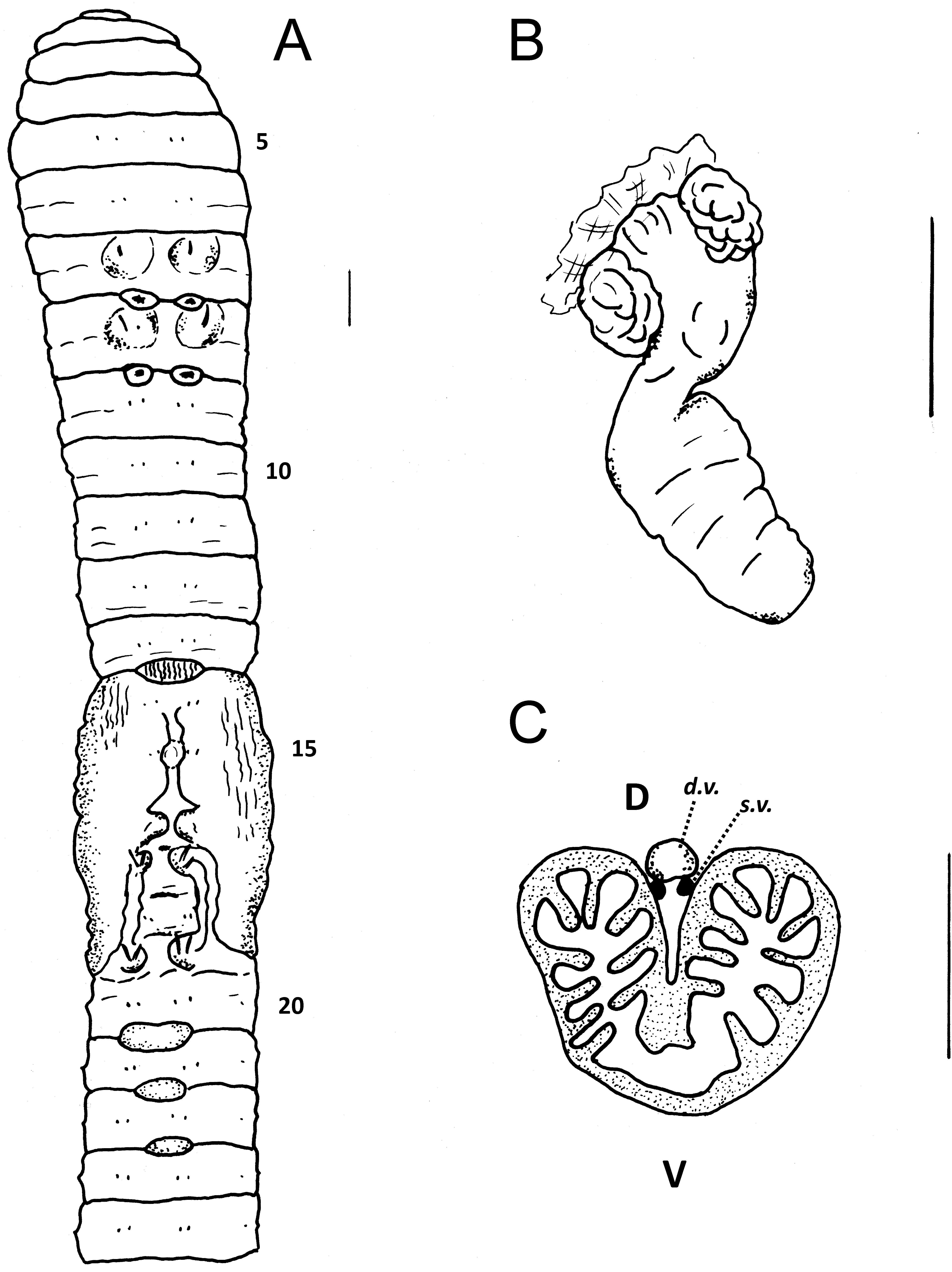
Description. External. Length 50 mm (holotype), 46 mm (paratype); width (postclitellar) 1.9 mm. Segments 141 (h), 127 (p). Pigment absent. Prostomium invaginated prolobous. Peristomium with several longitudinal grooves, reaching in paratype the second segment. Secondary annulations irregular, with one presetal and one postsetal in some segments. Setae eight per segment, closely paired in anterior region and quincuaxial in posterior region. Setal formula in anterior region (aa:ab:bc:cd:dd): 10: 2.7:1:9:2.9:23.7; 30: 2.8:1:3.3:1.7:16.2. In both individuals the first seta that shifts is seta c in 70; then seta d in 86, seta b in 87 or 89 and finally seta a in 91 or 98. Paratype with spermathecal setae on both sides of 7 and 8 (a or b) ( Fig. 3 View FIGURE 3 A) within ovoidal pouches of muscular tissue, those of 7 fixed to lateral walls by muscular stripes; in both segments only one functional seta and several small extra setae. Holotype with spermathecal seta on both sides of segment 7 and only on one side of 8 and 9.
Spermathecal setae slightly curved, ornamented in the last quarter ( Fig. 4 View FIGURE 4 B), characterized by alternated rounded crevices ( Fig. 4 View FIGURE 4 D); apex slightly concave and arrow-shaped; length of seta 0.7 mm. Paired penial setae (a and b) in 17 and 19, curved, length 0.68 mm, width 17 µm. Apex with two undulations and very thin ending ( Fig. 4 View FIGURE 4 A); two or three longitudinal rows of fine thorns just at beginning of first undulation, each row with 10–12 thorns; apex very delicate and easily broken, only observed in one of eight revised setae; small and irregular serrations present just before the beginning of apex undulations ( Fig. 4 View FIGURE 4 C). Setae a and b of 18 visible.
Clitellum light orange, saddle shaped, in 14–18; annular in 14, reaching a in 15 and 16 and limited by the genital zone in 17–18. Large dorsal pores present all along the body, first pore in 12/13. Spermathecal pores paired, large, in 7/8 and 8/9, centered in A; those of 8/9 slightly medial; in both individuals some filling observed. Female pores not recognized. Two pairs of prostatic pores in 17 and 19, over protuberances joined by parenthesis-shaped seminal grooves, which run outside B. Male pores in 18, in the deepest part of the genital region, slightly outside seta b. Genital marks of two kind, swellings and papillae ( Fig. 3 View FIGURE 3 A). Ovoidal swellings surrounding spermathecal setae. Unpaired, elliptical, mid-ventral papillae in 13/14 (with longitudinal ridges) and in 20/21, 21/22 and 22/23; the latter smooth and smaller, respectively reaching B, AB and A. Additional postsetal, paired marks in 16, with longitudinal ridges and extending from edge of clitellum to outside A.
Internal. Septa 5/6 very thin and membranous; 6/7–10/11 thin and slightly muscular; 11/12, 12/13 slightly muscular; septa 6/7–10/11 funnel shaped, imbricated and joined by 2–4 dorsal and lateral connective tissue fibers. One large gizzard in 5. Extramural calciferous glands present in 8–10, as dorso-lateral sacs that open widely into the esophagus; internally each sac with numerous, large lamellae with free margins ( Fig. 3 View FIGURE 3 C). Size of sacs: 10> 9 ≥ 8. Esophagus in 13 with scarce and small ventral and lateral ridges; in 12 and 11 with free margin lamellae projecting from the entire esophagus wall and occluding half of the lumen. Intestine begins in 13/14. Intestinal typhlosole starting very thin in 15, increasing abruptly in 19, reaching maximal size in 21; laminar, ending in 73 (holotype) or 81 (paratype). In 20–25 with lateral folds, slightly oblique in posterior direction. Smaller dorso-lateral typhlosoles in 19–25, at both sides of main typhlosole. Intestinal caeca absent.
Single dorsal vessel visible throughout. Supra-esophageal vessel visible in 8–12. Lateral hearts in 7, 8, 9 and 10; latero-esophageal hearts in 11 and 12. Ventral vessel present. A pair of extra parietal ventral vessels in 13, 14 and 15, running outside male gonoduct. Paired clusters of tufted micronephridia along a longitudinal row in segments 3–6, joined to both sides of pharynx; septal meronephridia difficult to see, in 11 one pair at each side, on septum 11/12. Parietal, closed meronephridia from 14 backwards, two (holotype) or three (paratype) on each side; in the holotype placed either in CD or outside D. The median ventral nephridium of last segments with a clear nephrostome.
Holandric. Testes and male funnels of 11 larger than those of 10; in both segments testes clearly recognized at both sides of mid-ventral line, joined to the anterior septum. Male funnels iridescent, not plicated but more or less solid, placed at both sides of the mid-ventral line of the posterior septum; in both segments dense coagulum strongly attached to funnels. Male gonoducts double, iridescent, running along body wall of 12–17 in AB, muscular in region 15–16, entering body wall in equator of 17, just below the penial setae pouches and in direction of midventral line. Two pairs of acinous seminal vesicles in 9 and 12, respectively fixed to septa 9/10 and 11/12; the anterior pair smaller and relatively flat; the posterior pair larger, voluminous and covering the esophagus. Two pairs of tubular prostates in 17 and 19, coiled, each gland limited to one segment or extending one segment backwards and fixed to the floor by connective tissue; the anterior pair slightly larger than the posterior one; muscular duct narrower and shorter (1/4) than the glandular part. Penial setae a and b of 17 and 19 within separate follicles that are joined to form an ovoid muscle pouch; these pouches fixed by muscular stripes to the lateral walls (one fiber) and to the floor (two fibers per follicle). Mid-ventral floor of 18 thickened. Undeveloped extra penial setae not present.
Ovaries one pair on the floor of 13, like large compacted bushes with numerous eggs dispersed all over, not in rows; female funnels one pair in 13, large and at both sides of the mid-ventral line. Two pairs of spermathecae in 8 and 9, 1.2 mm long, with two opposite sessile and ovoidal diverticles which project from the duct in the same segment of the ampulla ( Fig. 3 View FIGURE 3 B); each diverticle divided into two large wall chambers. Length of duct c. 1/2 of the pyriform ampulla; ampulla with several creases.
Etymology. The name makes reference to the type locality, that in nahuatl language means a river of stones.
Remarks. Ramiellona teapaensis sp. nov. belongs to the group of holandric species with last hearts in 12 and quincuaxial setae. It is most similar to R. tojolabala , differing mainly by the intestinal caeca (absent in R. teapaensis vs. present in R. tojolabala ), the shape of the spermathecae (with two opposite diverticles in the same segment of the ampulla vs. a single discoidal diverticle in the anterior segment of that of the ampulla) and the typhlosole (laminar vs. trifid).

No known copyright restrictions apply. See Agosti, D., Egloff, W., 2009. Taxonomic information exchange and copyright: the Plazi approach. BMC Research Notes 2009, 2:53 for further explanation.