
Microgomphus alani Kosterin, 2016
|
publication ID |
https://doi.org/ 10.11646/zootaxa.4701.3.4 |
|
publication LSID |
lsid:zoobank.org:pub:5B8B24FB-B6E5-4286-B9DF-6D1D60D05B67 |
|
persistent identifier |
https://treatment.plazi.org/id/FE5B8782-9940-835B-089E-E9FBFAEEFC35 |
|
treatment provided by |
Plazi |
|
scientific name |
Microgomphus alani Kosterin, 2016 |
| status |
|
Microgomphus alani Kosterin, 2016 View in CoL
Figs 1–7 View FIGURE 1 View FIGURE 2 View FIGURE 3 View FIGURE 4 View FIGURE 5 View FIGURE 6 View FIGURE 7
Heliogomphus selysi — Asahina (1986) View in CoL : 15 –16, figs 30–32, 37–38: (most probably a misidentified female from Thailand, Chieng Dao—description, drawings of the head, thorax, hind femora, vulvar lamina).
Microgomphus alani View in CoL , sp. n. — Kosterin (2016a): 341 View Cited Treatment –450, figs 1–4 (original description by two males from Koh Kong Province of Cambodia, collected in teneral state and hardened in captivity, differential diagnosis, habitat, distribution; illustrations of general habitus, wings, head, thorax, secondary genitalia including vesica spermalis and anal appendages).
Microgomphus jurzitzai View in CoL (misidentification)— Kosterin (2016b): 60 –61 (1 misidentified ♂ reported for Mondulkiri Province of Cambodia, the Monorom Waterfall area).
Heliogomphus View in CoL sp. (misidentification)— Kosterin (2016b): 73–75, fig. 58 (1 misidentified ♀ reported for Mondulkiri Province of Cambodia (the Buu Sraa Waterfal area); general habitus, head, thorax, fore wing base, genital lamina illustrated by photographs).
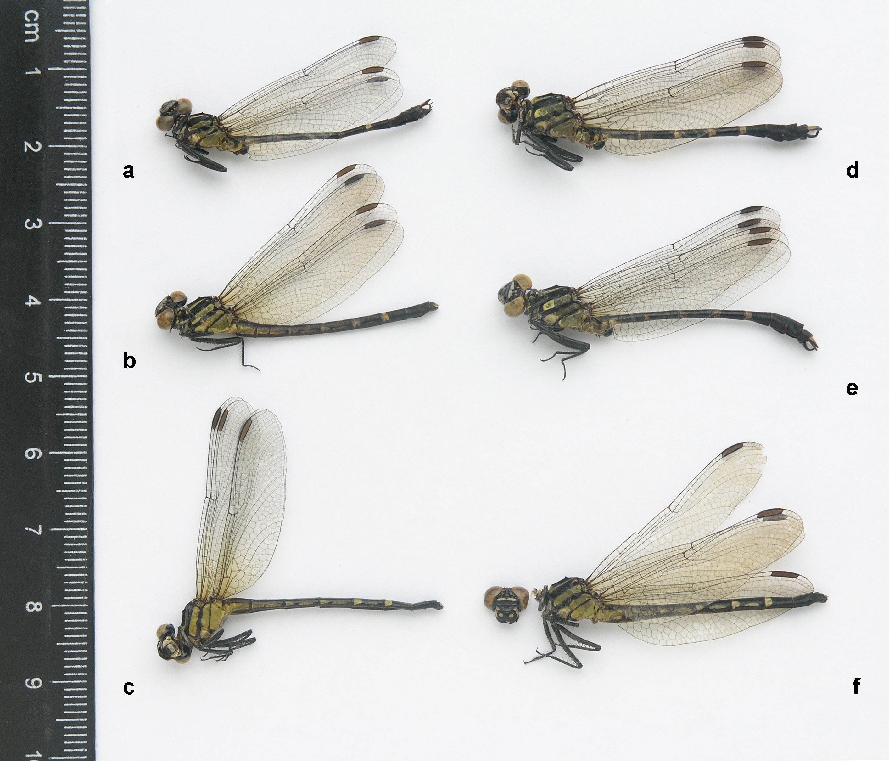
Specimens examined. Koh Kong Province: 1 ♂, the holotype (in alcohol)— Cambodia, Koh Kong Province, 17 km ENE of Koh Kong, ‘ Macromia Rivulet’, 11°40’17’’ N, 103°07’28’’ E [11.6714 N, 103.1244 E], 296 m a.s.l., 3 vi 2014 ( Kosterin 2016a: figs 1a,c, 2a–f,h, 3a–b,e–f, 4). 1 ♂, the paratype—Cambodia, Koh Kong Province, 6.5 km SW Thma Bang village, the ‘ Microgomphus River’, the slow reach upstream of the bridge, 11°38’42’’ N, 103°23’49’’ E [11.6450 N, 103.3969 E], 341 m a.s.l., 15 iv 2010 ( Kosterin 2016a: figs 1b; 2g; 3c–d). Phnom Kulen Mts, Siem Reap Province: 1 ♀ ( Figs 1c View FIGURE 1 , 2c View FIGURE 2 , 5b View FIGURE 5 , 6a,f View FIGURE 6 )— Cambodia, Siem Reap Province, Phnom Kulen Mts, the Siem Reap River just upstream of the bridge, 900 m W of Preah Ang Thom, 13.5662 N, 104.1019 E, 289 m a.s.l., 11 vi 2018. 1 ♂ ( Figs 1a View FIGURE 1 , 2a View FIGURE 2 , 3 View FIGURE 3 e–h, 4c)— Cambodia, Siem Reap Province, Phnom Kulen Mts, the Siem Reap River downstream of the waterfall, 600 m NNW of Preah Ang Thom, 13.5707 N, 104.1085 E, 256 m a.s.l., 17 vi 2018. 1 ♀ ( Figs 1b View FIGURE 1 , 2b View FIGURE 2 , 5c View FIGURE 5 , 6b,d,g View FIGURE 6 , 7c View FIGURE 7 )— Cambodia, Siem Reap Province, Phnom Kulen Mts, a brook, the left Siem Reap River tributary, 1.2–1.9 km SE of Preah Ang Thom, 13.551–554 N, 104.117–120 E, 301–330 m a.s.l., 30 vi 2018. Mondulkiri Province: 1 ♂ ( Fig. 7c View FIGURE 7 )— Cambodia, Mondulkiri Province, Monorom Waterfall area, the left grassy slope of the valley of the ‘Ochracea rivulet’, the left tributary of the main river, 3.5 km WSW of Sen Monorom, 12.4409 N, 107.1576 E, 654 m a.s.l., 13 vi 2014 ( Kosterin 2016b, misidentified as M. jurzitzai ; coordinates mentioned in that source for the relevant locality 4a were slightly incorrect). 1 ♀ ( Kosterin 2016b: fig. 58; Figs 2f View FIGURE 2 , 5a View FIGURE 5 , 6c,e View FIGURE 6 , 7c View FIGURE 7 )—Cam- bodia, Mondulkiri Province, just downstream of Buu Sraa Waterfall, the ‘ Loringae brook’ lowermost reaches, an open grassy area at the main river right bank, 12.5717 N, 107.4173 E, 432 m a.s.l., 3 viii 2016 ( Kosterin 2016b, misidentified as Heliogomphus sp.). 1 ♂ — Cambodia, Mondulkiri Province, 12.8 km NE of Sen Monorom, lower reaches of a brook—right tributary of Pulung (?) River, 12.5186–5189 N, 107.2931–2954 E, 529–544 m a.s.l., 25 vi 2018. 2 ♂♂ ( Figs 1 View FIGURE 1 d–e, 2d–e, 3a–d, 4a–b)— Cambodia, Mondulkiri Province, 16 km NNE of Sen Monorom, a small river, 12.584–587 N, 107.253–256 E, 400–420 m a.s.l., 26 vi 2018. 1 ♂ —the same place, 27 vi 2018.
All specimens are collected by the author. The holotype, one male collected on 26 vi 2018 and the female collected on 11 vi 2018 are kept in Naturalis Biodiversity Centre, Leiden, the Netherlands), the remaining specimens are in the author’s collection.
Variation in males. The holotype and paratype of M. alani , originating from the Cardamom Mts, are very small: HW 23 and 21.5 mm, Ab 25 and 23 mm, FW Ax 12–13 and 13–14, of which the second primary one is 5th and 4th/5 th, respectively ( Kosterin 2016a). The male from Phnom Kulen is only very slightly larger: HW 24 mm, Ab 26 mm, FW Ax 15, of which the second primary is 5 th /6th. However, the five males from Mondulkiri are distinctly larger: HW 26–27 (mean 26.6) mm, Ab 29.0–30.5 (mean 29.8) mm, FW Ax 13–15 (of which 5 th (7 cases) or 6 th (3 cases) are primary); they are thus 15–20% larger than the holotype .
A degree of inflation of the pale pattern correlates with size. The paratype, which is the smallest specimen, has no antealar spots on the mesepisterna ( Kosterin 2016a: fig.1b). The slightly larger holotype has their traces ( Kosterin 2016a: fig1a, 2a–b), the further, slightly larger male from Phnom Kulen, has well developed but small antealar spots ( Figs 1a View FIGURE 1 , 2a View FIGURE 2 , 3e,f View FIGURE 3 ), while these spots are relatively large in the males from Mondulkiri Province ( Figs 1 View FIGURE 1 d–e, 2d–e, 3a–b). In the type series males from the Cardamoms and the Phnom Kulen male all anteriolateral pale spots on S3–S6 are isolated from each other and dorsal pale streaks ( Fig. 2a View FIGURE 2 ) but in the Mondulkiri males those on S5–S6 (in one male only on S6) are fused to the anterior element of the dorsal streaks to form tripartite anterior spots ( Fig. 2e View FIGURE 2 ). The two males from the Cardamoms (the type series) ( Kosterin 2016a: fig. 1a), the male from Phnom Kulen ( Fig. 4c View FIGURE 4 ) and two males from Mondulkiri have no pale spots on S8 while three males from Mondulkiri Province have a very small, elongate and slanting, lateral spot at the S8 posterior margin, at the middle of its height in lateral view ( Fig. 4 View FIGURE 4 a–b).
Pterostigmata are dark brownish in all mature males, while pale brownish in the teneral males of the type series. Unlike these teneral males, the lower part of the pterothorax, the coxae, trochanters, femora and secondary genitalia in all mature males were covered with white pruinescence ( Fig. 1a,d View FIGURE 1 ). This was later washed out by acetone treatment ( Figs 2a View FIGURE 2 ,d–e, 3a,e) but is well preserved in a male of 13 vi 2014, which was not treated with acetone (not shown).
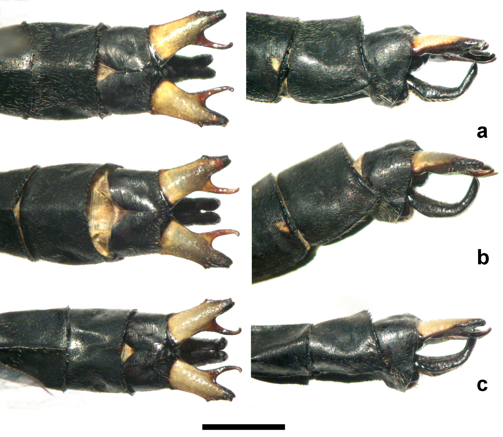
The anal appendages are deformed in the paratype and, as will be discussed below, not fully expanded in the holotype (although the original description was based on them, see Kosterin 2016a); hence the type series is not fully comparable with the presently available and here considered mature specimens. In these specimens, the cerci look identical in dorsal view ( Fig. 4 View FIGURE 4 a–c) and differ from those in the holotype ( Kosterin 2016a: fig. 2c) in having a divergent rather than parallel alignment, and having their inner branches slightly curved and protruding further behind than the main parts (‘outer branches’) of cerci, rather than strongly curved and not reaching the level of the main parts of cerci. The cercus main part has the same principal shape as in the holotype, with a bluntly bifid apex and a blunt outer tooth, but is longer. Interestingly, the epiproct in the holotype ( Kosterin 2016a: fig. c–d) and the male from Phnom Kulen ( Fig. 4c View FIGURE 4 ) is similar in both dorsal and lateral view, while in the Mondulkiri males it is more strongly curved up ( Fig. 4 View FIGURE 4 a–b). This difference is strong enough to suppose that these males are not conspecific with the males from the Cardamom and Phnom Kulen Mts., but the whole complex of evidence is not solid enough and made me to consider this as a case of geographical structural variation.
The posterior hamulus in lateral view is slightly curved apically in the holotype ( Kosterin 2016a: figs 2f, 3a), more strongly curved in the Phnom Kulen and Mondulkiri males ( Fig. 3d,h View FIGURE 3 ) and even more strongly hooked in the paratype ( Kosterin 2016a: figs 2g, 3c). The anterior hamulus is similarly simply curved in the type series and Phnom Kulen male ( Fig. 3h View FIGURE 3 ) but somewhat S-like curved and more robust in the Mondulkiri males ( Fig. 3h View FIGURE 3 ).
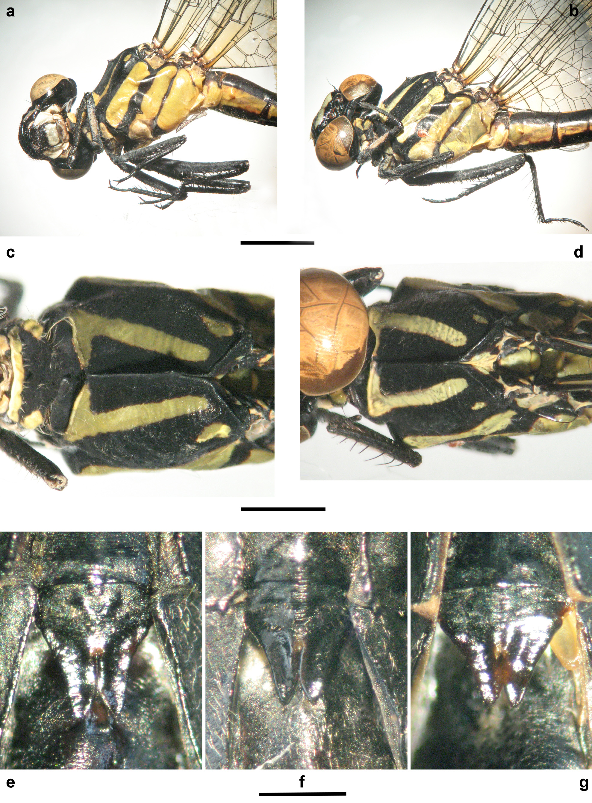
Female. Three females are available for study. The female collected in 2016 in Mondulkiri Province has already been briefly characterised, illustrated and compared to ‘ Heliogomphus svihleri ’ in Kosterin (2016b : fig. 56), who misidentified it as Heliogomphus sp. Like males from the same province, it is considerably larger than the females from Phnom Kulen Mts. as well as the males of the type series from the Cardamoms. Since the male from Phnom Kulen more closely resembles the holotype in size than those from Mondulkiri Province, one of the females from Phnom Kulen, that of 30 vi 2016 ( Figs 1b View FIGURE 1 , 2b View FIGURE 2 , 5c View FIGURE 5 , 6b,d,g View FIGURE 6 , 7c View FIGURE 7 ), was chosen for the description provided below, in spite of its aberrant asymmetrical occiput.
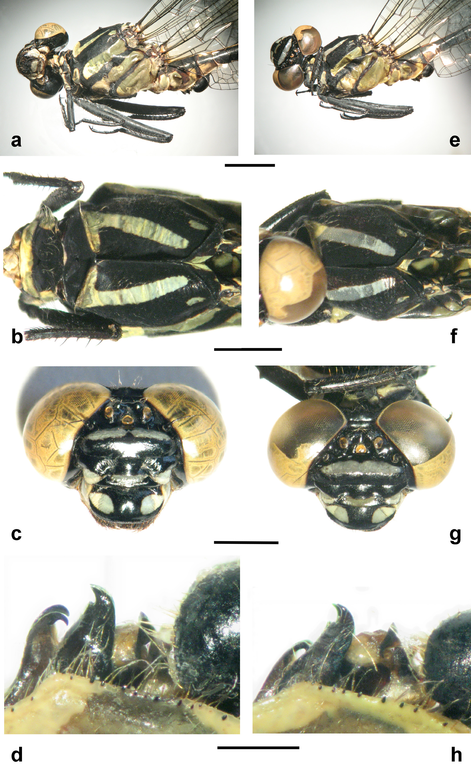
Head ( Figs 5c View FIGURE 5 ). Face black with pale yellow pattern. Labium pale but apical border of mentum and movable hooks black. Mandible bases pale. Labrum with a pair of large ovoid (tapering to external ends) yellow spots on either side. Anteclypeus with a broad rectangular central pale spot adjacent to its upper margin but leaving a black border along lower margin. Lower lateral parts of postclypeus broadly pale (these pale spots crossed by an additional seam separating lateroventral lobes of postclypeus). Frons dorsal surface with a broad pale stripe, broadest at middle. Vertex and occiput black. Vertex with a pair of bluntly conical tubercles behind ocelli, set twice as close to each other as to lateral ocelli and with a U-shaped depression between them. Occiput with a high blunt central hump (raising as high as eye tops), on the right side of which there is a narrow, pointed horn, the corresponding place to the left smooth, without a horn; occipital crest gently rises laterally towards eyes. Vertex and occiput with long sparse hairs; shorter hairs on sides of anteriolateral lobes of postclypeus, on labrum and mandibles.
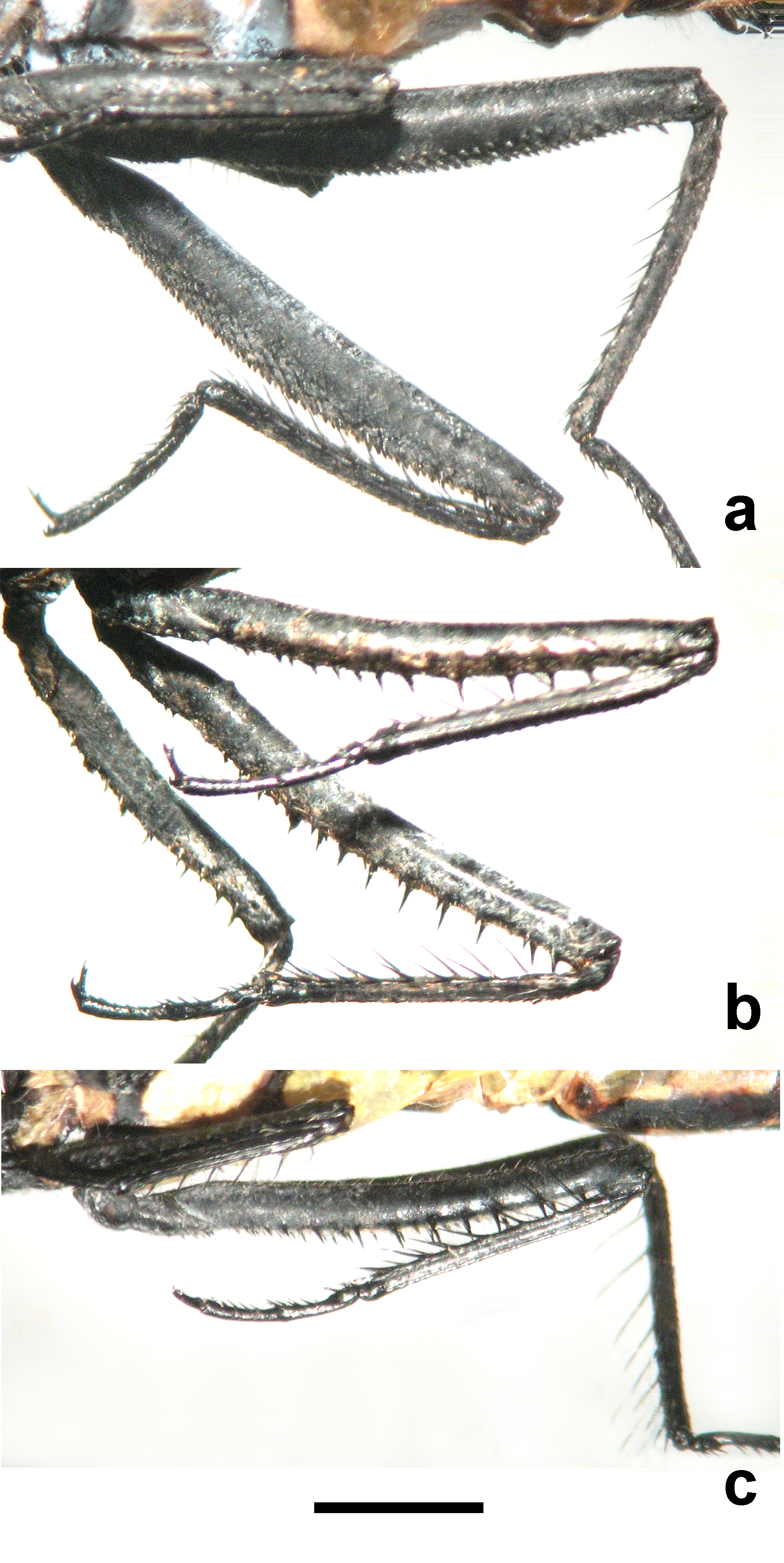
Thorax in life with a strong white pruinescence on coxae and lower parts of mesinfraepisternum and metinfraepisternum ( Fig. 1b View FIGURE 1 ), completely washed out post mortem by acetone treatment ( Figs 2b View FIGURE 2 , 6b View FIGURE 6 ). Prothorax black, anterior lobe and large spots at sides of median lobe yellow. Pterothorax ( Figs 1b View FIGURE 1 , 2b View FIGURE 2 , 6b, d View FIGURE 6 ) black and pale yellow. Its dorsum ( Fig. 6d View FIGURE 6 ) black, dorsal yellow stripes broad, posteriorly rounded, not reaching alar sinus, anteriorly fused to broad stripes of mesothoracic collar at their lower ends forming two inverted 7s; collar stripes narrowly interrupted with black at medial carina. Antealar spots well expressed but rather small, elongate. A broad yellow stripe occupies ca 2/3 of mesepimeron width; it is shifted towards its anterior margin so that the anterior of the two dark remaining borders is only half as wide as the posterior, wavy one. Mesepisternum also with a broad yellow stripe occupying ca 2/3 of its width, which is strongly shifted to its posterior margin, with only a narrow black border there ( Fig. 6b View FIGURE 6 ). Metepimeron yellow but its anterior border and anteroventral corner black; posterior margin with a short and narrow black border in its upper part. Mesinfraepisternum black with a long vertical yellow spot in its lower part; metinfraepisternum pale with anterior and posterior black borders ( Fig. 6b View FIGURE 6 ). Generally, sides of pterothorax yellow with two parallel black stripes along mesopleural and metapleural sutures, the former thrice as broad, with wavy anterior margin producing a projection narrowly embracing the spiracle with black ( Figs 1b View FIGURE 1 , 2b View FIGURE 2 , 6b View FIGURE 6 ). Subalar ridges black but that of metepisternum mostly yellow. Ventral side yellow. Coxae black anteriorly, yellow posteriorly. Legs black but outer side of profemur with a broad long yellowish spot occupying ca 2/3 of its length. Metafemur reaches ca 0.4 of S2 length, its flexor surface with two rows of robust and sparse spines, becoming shorter but not denser towards its base; between these rows there is a third median row of small, very short and more densely set spinulets. Mesofemur with smaller but similarly sparse long spines, without distinct intermittent row of fine spinulets, profemur with three rows of very small, short spinulets. Trochanters and femora were covered with a slight white pruinescence ( Fig. 2b View FIGURE 2 ), later washed out by acetone.
Wings ( Fig. 2b View FIGURE 2 ) hyaline with a very slight brownish infumation, basally with a slight yellowish tint gradually disappearing towards triangles. Venation black but outer surface inside nodes whitish. FW Ax (1 st and 5 th primary) 14 (left)–15 (right), HW Ax 10; FW Px 9 (right)–10 (left); HW Px 8 (right)–9(left). Crossveins between Arc and R1–R4 junction: 6 (left)–7 (right) above Rs and 4 (left)–5 (right) below Rs on FW, 3 (left)–4 (right) above Rs and 3 below Rs on HW. One cubito-anal crossvein in all wings. Tornus smoothly curving. Pterostigmata brown, almost transparent, in FW covering 4–4.5 cells below, in HW 3.5–4 cells, bordering longitudinal veins inflated, black.
Abdomen black with yellow maculation ( Figs 1b View FIGURE 1 , 2b View FIGURE 2 ). S1–S6 with yellow dorsal stripes throughout their length, with uneven margins, becoming narrower on subsequent segments so that those on S1–S3 rather broad, those on S4–S5 narrow with a broader base and tapering to apex, that on S6 linear, hardly noticeable, with a small roundish spot at anterior segment margin. Laterally, S1 with yellow ventrolateral parts occupying ca 2/3 of segment height; dorsal black colour produces a narrow protrusion into this yellow part at about middle of its upper margin. S2 with broad, yellow lateral stripes with even margins throughout its length. S3–S6 with anteriolateral spots expanding along segment anterior margins (especially on S3), and ending before medial suture, that on S3 here truncated, other rounded. In line with these spots and behind them (and medial suture), there are isolated lateral stripes twice as short as respective segments; that on S3 with a truncated anterior end, other ends rounded. On S3, the anteriolateral spot and further stripe are separated by just a black streak along midsegmental suture, on S4–S6 by a broader gap. S7 with large anteriolateral spots occupying ¼ of segment length and a shorter dorsal spot almost fused to them, separated only with vague and irregular blackish streaks. Rest of abdomen black. Cerci narrowly pointed, yellowish, paraprocts (short structures as seen between cerci) also yellowish. Sternites with a slight white pruinescence ( Fig. 1b View FIGURE 1 ), washed out by acetone.
Vulvar lamina outline approaches an equilateral triangle but with incised apex as split into two lobes for 60% of its length, anterior margin somewhat convex and lateral margins slightly concave; inner margins of lobes convex so that lobes almost touch each other for 2/3 of their length.
Measurements (mm). Hindwing 23; abdomen without appendages 25; total length (with head and appendages) 36.
Variation in females. The second female from Phnom Kulen, of 11 vi 2018, has 0.5 mm shorter hind wing but 2 mm longer abdomen ( Figs 1c View FIGURE 1 , 2c View FIGURE 2 ) (HW 25.5, Ab 28.5, total length 37; FW Ax 14). The latter may be related to its somewhat immature state, while the female described above was obviously fully mature as collected during oviposition ( Fig. 1c View FIGURE 1 ). The second Phnom Kulen female has vestigial yellow maculation also on S8: a small triangular anteriodorsal spot, lateral streaks at anterior segment margin accompanied by a pair of tiny elongate spots just above them at some distance from segment margin. In this female the paraprocts are also dark greyish.
The only female from Mondulkiri Province (of 3 viii 2016) ( Fig. 2f View FIGURE 2 ) is ca 10% larger than the one used for description (HW 27.5, Ab 29.5; total length 41; FW Ax 14/15). Its pale stripe across the frons is interrupted at the middle ( Fig. 5a View FIGURE 5 ); cerci and paraprocts whitish. Although treated with acetone, this specimen has a strong while pruinescence of the coxae and prothorax lower surface.
Both these females have a symmetrical occiput with two horns at either side of the central hump ( Fig. 5 View FIGURE 5 a–b).
Main differences from males. Besides the reproductive organs, the females of M. alani differ from males in having a central hump and a pair of horns on the occiput (which is simple and straight in the male; Fig. 3c,g View FIGURE 3 ), a pair of strong blunt tubercles behind the ocelli (vs gentle swellings set more apart from each other in males; Fig. 3c,g View FIGURE 3 ). The metafemur bears two rows of strong and sparse spines in females (with an intermittent row or fine spinulets) but only fine and dense spinulets in males, as discussed below. In maculation, the main difference of females from males is the presence of long lateral (subventral) pale stripes behind the short anteriolateral spots on S3 – S6. Profemur has a large yellowish area in females but is black in males.
Differential diagnosis for females. The prominent central hump on the occipital plate is unique among the known females of Microgomphus spp. The female head sculpture of Microgomphus telyphonus Lieftinck, 1929 (presently considered a subspecies of M. chelifer Selys, 1858 ) was described (and illustrated) as follows: “The free margin of the occipital plate, in frontal view, with a low but sharply crested elevation, directed upwards under right angle; this margin slightly concave in the middle, with a row of 5 – 6 robust, irregular black spines on either side; anterior surface with a low, central tubercle, just behind the two lateral ocelli” ( Lieftinck 1929: 126 – 127). Fraser (1934) stated that the female occiput in Microgomphus troquatus (Selys, 1854) is “with a low, slightly concave crest, which is bordered with about ten minute spines” (that is like in M. chelifer telyphonus ). He described that of M. lilliputians Fraser, 1923 as “simple, sinuous, a little notched at its middle”, that or M. (?) minusculus (Selys, 1878) as “simple, straight”, but provided no information on the female head sculpture of Microgomphus souteri Fraser, 1924 , M. (?) verticalis (Selys, 1873) and M. loogali Fraser, 1923 , that could imply absence of notable structures. The female of Microgomphus wijaya Lieftinck, 1940 is said to have the “occiput as in the opposite sex”, in which “occipital plate straight posteriorly, armed with a row of microscopical, erect, finely pointed denticles along margin” ( Lieftinck 1940: 98, 100). The female of M. phewataali has “occipital margin with two small close-set tubercles less prominent than in male, upper frons ... peaked in middle with shallow cleft” (Coniff & Singh Limbu 2018: 282). The head of the female of M. farrelli is not described except for the statement of “absence of occipital horns (presence in M. svihleri )...”, the illustration shows a simple straight occiput margin ( Makbun & Fleck 2018: 445, fig. 10).


No known copyright restrictions apply. See Agosti, D., Egloff, W., 2009. Taxonomic information exchange and copyright: the Plazi approach. BMC Research Notes 2009, 2:53 for further explanation.
|
Kingdom |
|
|
Phylum |
|
|
Class |
|
|
Order |
|
|
Family |
|
|
Genus |
Microgomphus alani Kosterin, 2016
| Kosterin, Oleg E. 2019 |
Microgomphus jurzitzai
| Kosterin, O. E. 2016: 60 |
Heliogomphus selysi —
| Asahina, S. 1986: 15 |