
Acantholaimus marliae, Manoel, Alex, Silva, Maria Cristina Da & Esteves, André M., 2017
|
publication ID |
https://doi.org/ 10.11646/zootaxa.4258.3.3 |
|
publication LSID |
lsid:zoobank.org:pub:AD166CBA-0191-4C8E-ABDC-31B60C6D5758 |
|
DOI |
https://doi.org/10.5281/zenodo.6010381 |
|
persistent identifier |
https://treatment.plazi.org/id/543F0D7C-FFDE-E269-FF75-8F38FD47B7A0 |
|
treatment provided by |
Plazi |
|
scientific name |
Acantholaimus marliae |
| status |
sp. nov. |
Acantholaimus marliae sp. n.
( Table 1; Figs 2–7 View FIGURE 2 View FIGURE 4 View FIGURE 5 View FIGURE 6 View FIGURE 7 )
Material studied. Type specimens: The holotype and one paratype (female) are deposited in the National Museum of Rio de Janeiro ( MNRJ), Brazil . The juvenile paratypes and paratype females are deposited in the Meiofauna Laboratory , Zoology Department, Universidade Federal de Pernambuco (LMZOO-UFPE), Brazil . Holotype male (MNRJ 350); allotype female (MNRJ 351); two paratype females (186–187 NM LMZOO-UFPE) and three juveniles (188–190 NM LMZOO-UFPE).
Type locality. Material collected in June 2009, from the Potiguar Basin (03°00’00”S, 038°45’00”W). Sediment: fine to coarse bioclastic. Sampling: Van Veen grab.
Etymology. The species name is given in tribute to Marli Maria da Silva, mother of the first author.
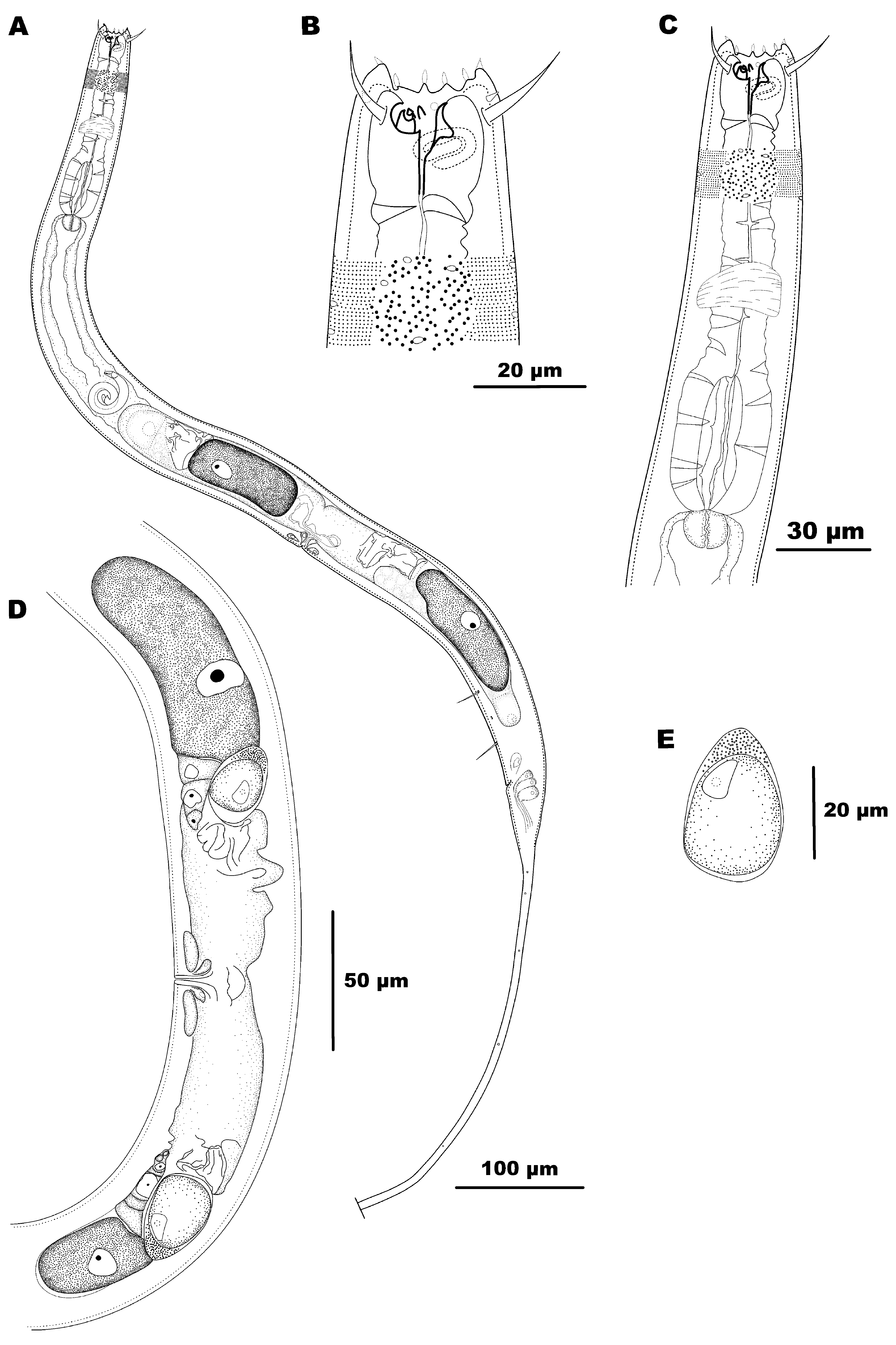
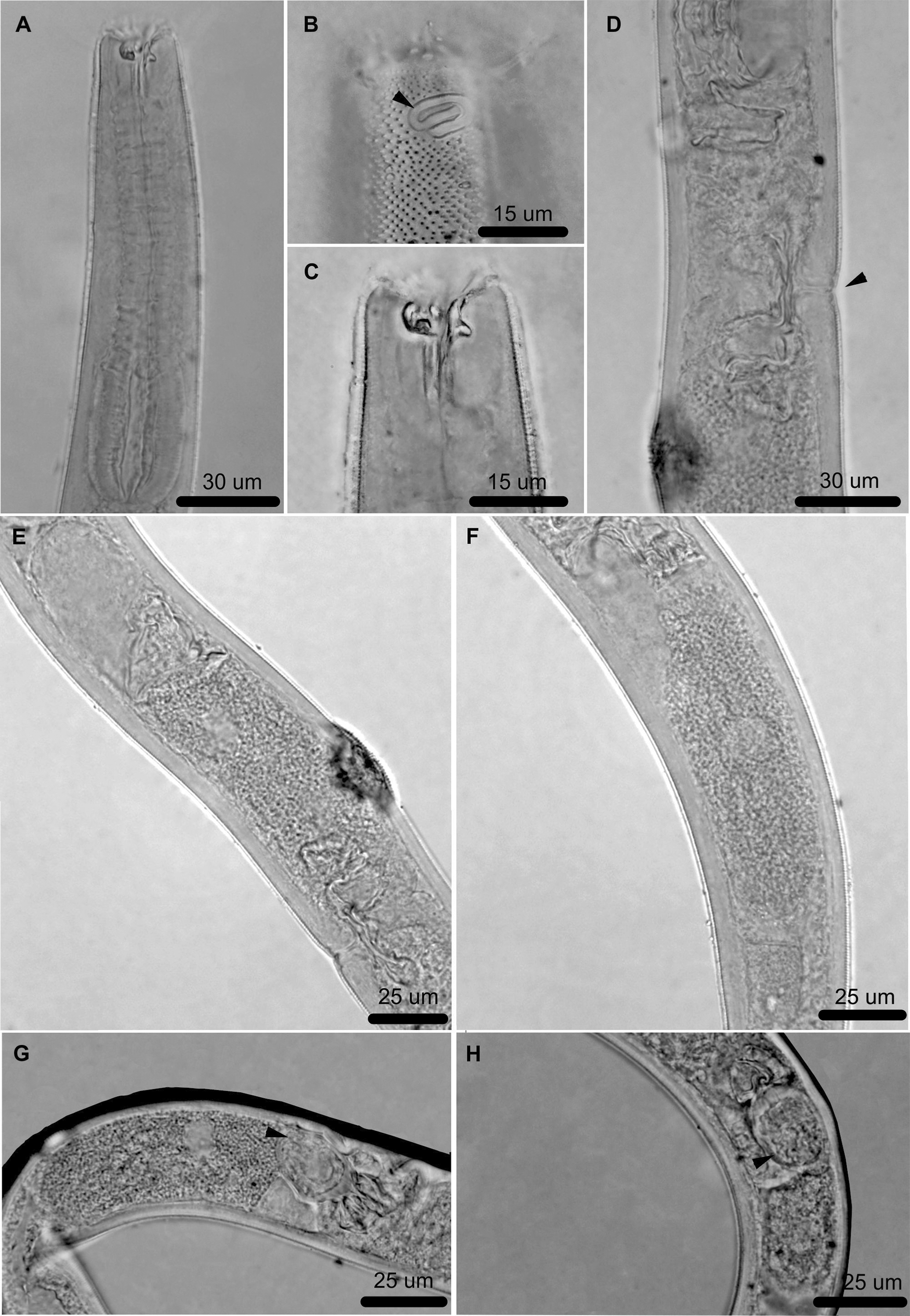
Holotype male. Body cylindrical and attenuated at extremities, 684 µm long excluding tail ( Figs 2 View FIGURE 2 A, 3A). Cuticle densely punctuated with dots arranged in transverse rows ( Figs 2 View FIGURE 2 B, 3B). Lateral differentiation beginning at posterior border of fovea amphidialis, extending over pharynx until rear bulb portion, absent on rest of body. Lateral differentiation with larger dots, dispersed and more widely spaced than median ones, which are more densely arranged (Fig 3C). Cuticular pores oval and randomly scattered, extending along entire body length. Anterior sensilla difficult to see (first and second rings) and arranged according to following pattern 6+6+4: six inner labial papilliform sensilla, six outer labial papilliform sensill and four cephalic setiform sensilla (25 µm long), corresponding to 89% of head diameter ( Fig 2 View FIGURE 2 B). Ventrally spiral fovea amphidialis (about 1.5 turns) located below cephalic setae and with transversely wider spiral shape, 7 µm in height and 13 µm wide, comprising 48% of corresponding body diameter and located 10 µm behind anterior end ( Figs 2 View FIGURE 2 B, 3D). Two pairs of cervical setae 11- 15 µm, located posterior to each fovea amphidialis. Somatic setae (arranged in four sublateral longitudinal rows) present from posterior end of fovea amphidialis along entire body except for filiform part of tail. Buccal cavity relatively long. Cheilostom possesses 12 rugae. Five solid teeth, one large dorsal tooth measuring 6 µm and four small subventral teeth ( Figs 2 View FIGURE 2 C, 3E). Most protuberant of subventral teeth (3 µm) shaped as "bottle opener". Remaining three teeth tiny and difficult to see. Pharyngostom about 9 µm long. Pharynx (130 µm long) muscular, cylindrical, expanded at level of pharyngostom, and forming well-developed basal bulb (39 µm diameter) at its proximal end, occupying 77% of corresponding body area ( Fig 2 View FIGURE 2 D). Cardia embedded in intestine. Nerve ring situated at 41% of the pharyngeal region length ( Fig 2 View FIGURE 2 D). Ventral gland and secretory-excretory pore not observed. Reproductive system with single anterior outstretched testis on right-hand side of intestine (155 µm), occupying about 25% of body length (excluding tail). Spicules curved, expanded and flattened in proximal portion ( Figs 2 View FIGURE 2 E, 3F). Gubernaculum in form of tapered rod, its proximal portion slightly curved; bifurcated at its distal end ( Figs 2 View FIGURE 2 E, 3G). Apophysis absent. Precloacal supplements absent. Three caudal glands. Tail conical-cylindrical with long filiform portion, with two rows of circular pores; distal portion of the tail is broken off.
Allotype female. Largely similar to male, except somatic setae that are generally absent (fewer somatic setae than in holotype, seen only in female paratype 2, arranged similarly to those in male). Body measuring 804 µm in length to anal region, and maximum diameter 54 µm ( Figs 4 View FIGURE 4 A, 5A). Cuticular pores over entire body, with similar size, shape and arrangement to those in male. Outer labial and cephalic sensilla equivalent to 20% and 74.5% respectively of corresponding body diameter ( Fig 4 View FIGURE 4 B). Fovea amphidialis with same measurements as in male, and occupying 46% of corresponding body width ( Figs 4 View FIGURE 4 B, 5B). Buccal cavity similar to that of male ( Figs 4 View FIGURE 4 B, 5C). Pharyngostom about 9 µm long. Pharynx similar to that of male ( Fig 4 View FIGURE 4 C). Nerve ring at 47% of the pharyngeal region length. Basal bulb occupying 75% of corresponding body area. Ventral gland and secretory-excretory pore not observed. Cardia embedded in intestine. One nematode (unidentifiable) observed in gut contents. Vulva located 516 µm from anterior end, at 64% of body length excluding tail ( Fig 5 View FIGURE 5 D). Two opposite, reflexed and antidromous ovaries (anterior ovary lying to right of intestine, posterior ovary to left of intestine) ( Figs 5 View FIGURE 5 E, 5F). Anterior and posterior ovary measuring respectively 220 and 320 µm. In reproductive ducts of allotype, a large spermatozoa (24 x 21 µm) was found. This structure is most clearly visible in reproductive ducts of paratype 1 ( Figs 4 View FIGURE 4 D, 5G, 5H), where two spermatozoids were found, measuring about 30 x 20 µm ( Fig 4 View FIGURE 4 E). Three caudal glands. Tail conicalcylindrical with long filiform portion, with two rows of circular pores; distal portion of the tail is broken off.
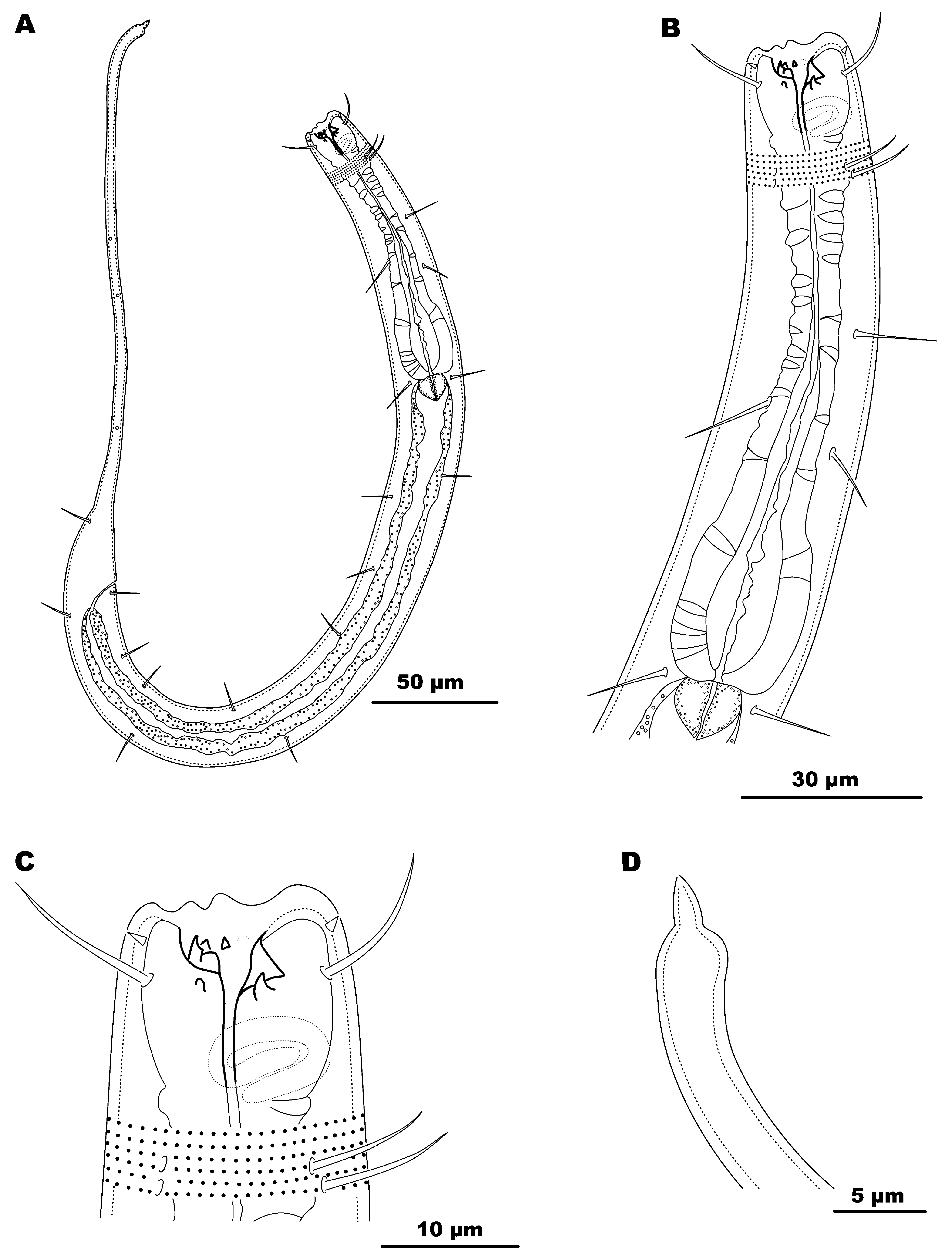
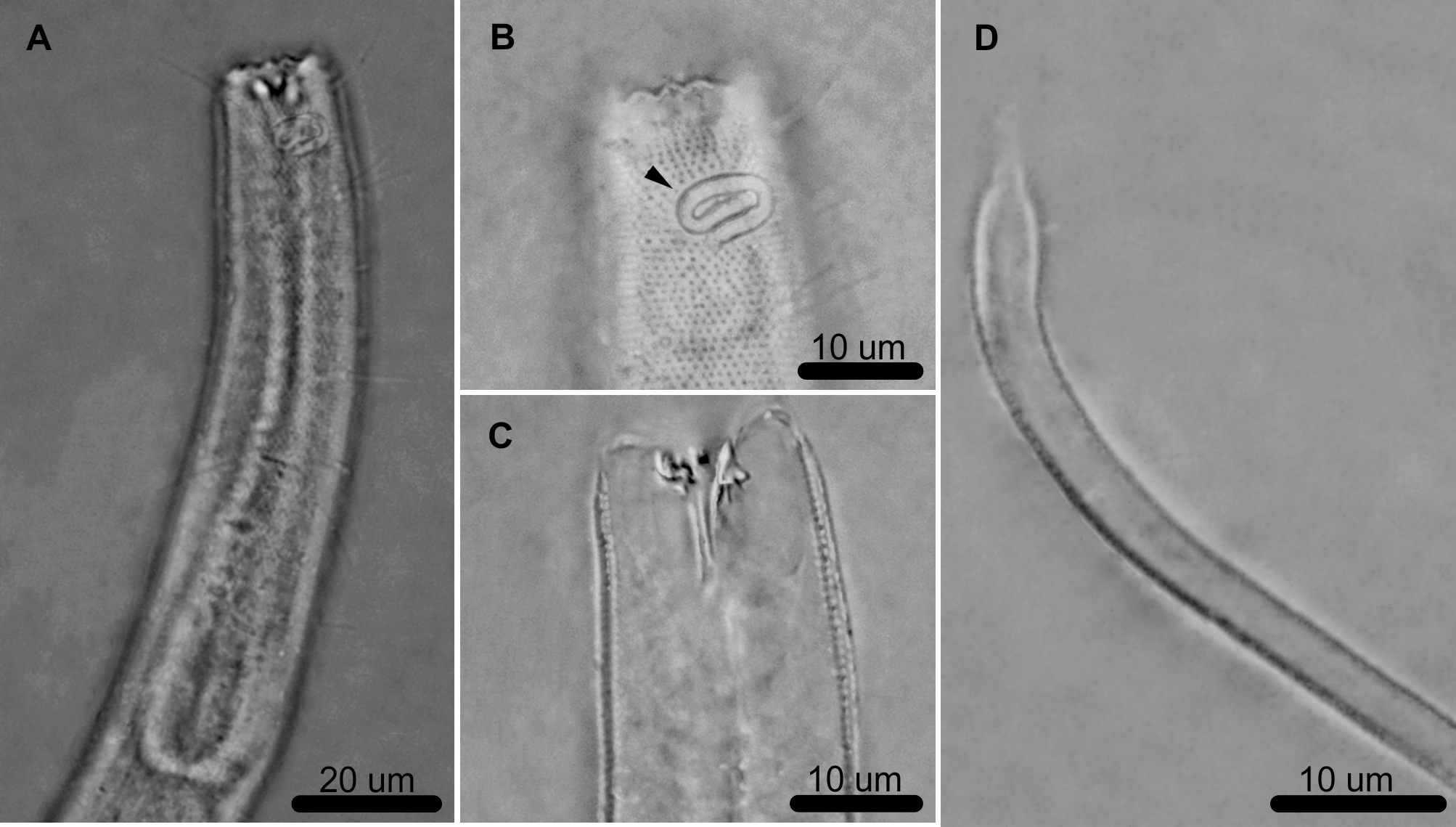
Paratype juvenile. The juveniles analyzed may be in the early stage of development, because the genital primordium is not visible. Juvenile sharing features of both adults. Body cylindrical and attenuated at extremities (618 µm long) ( Figs 6 View FIGURE 6 A, 7A). Cuticle densely punctuated with dots arranged in transverse rows, but without lateral differentiation ( Fig 6 View FIGURE 6 B). Cuticular pores not observed (except on filiform part of tail). First and second sensilla circles, as well as nerve ring and secretory-excretory pore not visible. Fovea amphidialis occupying 49% of corresponding body width ( Figs 6 View FIGURE 6 C, 7B). Somatic setae arranged similarly to male, though fewer in number. Buccal cavity similar to that seen in adults ( Figs 6 View FIGURE 6 C, 7C). Pharyngostom approximately 7 µm long. Pharynx similar to that in adults (93 µm), however, with narrowing at 39% of length. Basal bulb occupying 76% of corresponding body area. Tail conical-cylindrical with filiform end portion ( Figs 6 View FIGURE 6 D, 7D), with two rows of circular pores. Spinneret short.
Diagnosis. Acantholaimus marliae sp. n. is characterized by possessing numerous somatic setae arranged in four sublateral longitudinal rows, buccal cavity with five solid teeth (four subventral and one dorsal), the largest tooth dorsally located and the most protuberant tooth between the subventral shaped as a "bottle opener". It differs from all other species of the genus in having a spiral fovea amphidialis, wider than long, here considered as a differential characteristic and unique for the genus.
Differential diagnosis. The new species shares with Acantholaimus arthrochaeta Miljutina & Miljutin, 2012 the following features: the arrangement of the somatic setae along the body (4 sublateral longitudinal rows); dorsal tooth well developed and of similar length (5–6 µm in A. arthrochaeta and 6–7 µm in A. marliae sp. n.). The largest of the four subventral teeth is also similar in length (5 µm in A. arthrochaeta and 3–5 µm in A. marliae sp. n.). The length of the spicules (31–41 µm in A. arthrochaeta and 32 µm in A. marliae sp. n.) and anal body diameter (20–38 µm in A. arthrochaeta and 21–34.5 µm in A. marliae sp. n.) are also similar. However, A. arthrochaeta has distinct
characteristics such as a longer body without the tail (839–1055 µm vs 582–804 µm in A. marliae sp. n.), and lateral differentiation extending along the entire body (except filiform part of tail); while in A. marliae sp. n. the lateral differentiation begins at the posterior border of the fovea amphidialis, extends over the pharynx until basal bulb level and is absent on the rest of the body. In A. arthrochaeta the pharynx forms a poorly developed bulb, whereas A. marliae sp. n. has a well-developed basal bulb; in the former, the anterior sensilla are jointed (inner labial sensilla bipartite, outer labial sensilla tripartite), whereas in A. marliae sp. n. these structures are not jointed; and in A. arthrochaeta the somatic setae are sometimes clavate, whereas the new species has only setae of edged form. Furthermore, the testis occupies about 40–50% of the pre-anal body length, whereas in A. marliae sp. n. it occupies about 25%. The spicules described for A. arthrochaeta are more curved, longer when measured along an arc (41–56 µm vs 36 µm in A. marliae sp. n.).
Acantholaimus maks Gerlach, Schrage & Riemann, 1979 shares with the new species features such as: the number of teeth in the buccal cavity (5), dorsal tooth well developed and of similar length (6 µm in A. maks and 6– 7 µm in A. marliae sp. n.), diameter of the fovea amphidialis (11–14 µm and 11–13 µm A. marliae sp. n.), as well as the percentage that this structure occupies in the corresponding area of the body (37–50% and 37–49% in A. marliae sp. n.); and anal body diameter (27–45 µm and 24–35 µm in A. marliae sp. n.). Yet, A. maks differs from A. marliae sp. n. in the following features: longer body without tail (1180–1307 µm vs 582–804 µm in A. marliae sp. n.); lateral differentiation absent; longer spicules along the chord (45–60 µm vs 32 µm in A. marliae sp. n.); and the testis occupying 40% of the pre-anal body length, whereas the corresponding structure occupies about 25% in A. marliae sp. n.
The species of Acantholaimus first described from shallow water, A. polydentatus Gerlach, 1951 shares with the new species: the length of the cephalic setae (20–22 µm in A. polydentatus and 21–25 µm in A. marliae sp. n.); maximum diameter of the body (22–48 µm and 24–54 µm in A. marliae sp. n.); index b’ (4–5 and 3–5 in A. marliae sp. n.); ratio %v’ (70% and 63–73% in A. marliae sp. n.); and numerous somatic setae occurring over the entire the body except the filiform portion of the tail. However, these species differ in the shape and proportion occupied by the fovea amphidialis (50–70% in A. polydentatus vs 37–49% in A. marliae sp. n.) and the distance of this structure from the anterior end (5 µm in A. polydentatus vs 10–16 µm A. marliae sp. n.). Moreover, in A. polydentatus the spicules are smaller (24 µm along the chord in A. polydentatus vs 31.5 µm along the chord in A. marliae sp. n.) and the gubernaculum is absent.

| MNRJ |
Museu Nacional/Universidade Federal de Rio de Janeiro |
No known copyright restrictions apply. See Agosti, D., Egloff, W., 2009. Taxonomic information exchange and copyright: the Plazi approach. BMC Research Notes 2009, 2:53 for further explanation.
|
Kingdom |
|
|
Phylum |
|
|
Class |
|
|
Order |
|
|
Family |
|
|
Genus |
Acantholaimus marliae
| Manoel, Alex, Silva, Maria Cristina Da & Esteves, André M. 2017 |
Acantholaimus maks
| Gerlach, Schrage & Riemann 1979 |
A. polydentatus
| Gerlach 1951 |