
Agdistis marionae, M., 2012
|
publication ID |
https://doi.org/ 10.21248/contrib.entomol.62.2.447-457 |
|
DOI |
https://doi.org/10.5281/zenodo.4812924 |
|
persistent identifier |
https://treatment.plazi.org/id/CC2E8782-FFA8-3008-78C5-FF0D17866052 |
|
treatment provided by |
Carolina |
|
scientific name |
Agdistis marionae |
| status |
sp. nov. |
Agdistis marionae View in CoL sp. n.
Holotype, : „ St. Helena Island, Manati Bay , Great Shaking Rocks, reared from Frankenia portulacifolia , caterpillars/pupa collected on 30. Dec. 2010, hatched Jan. 2011, leg. Annalea M. Beard “. GU 6451 Ar. Coll. NHMW (in Coll. Arenberger).
Paratypes: 1 : with the same data as holotype. GU 6468 Ar. 1 : South Atlantic Ocean , St. Helena Island, Great Shaking Rocks, Manati Bay, 31.12.2011, leg. Miss Annalea Beard. GU 6475 Ar. Barcoding CCDB-11447. Sample MNVD-11447-A08; : Barcoding CCDB- 11447. Sample MNVD-11447-A07. 1 : St. Helena Island, Man and Horse, Joan Hill, reard from Frankenia portulacifolia , January 2011, leg. Annalea M. Beard. GU 6469 , 6512 Ar. 2 : South Atlantic Ocean , St. Helena Island, Joan Hill , Man and Horse, 28.12.2011, leg. Miss Annalea Beard. Barcoding CCDB-11447. Sample MNVD-11447-A011. 2 : ditto. GU 6476 Ar. Barcoding CCDB-11447. Sample MNVD-1147-A10. In coll. NHMW (in Coll. Arenberger) and in Coll. NHML .
Etymology:
The new species is dedicated to Mrs. Marion Rose Beard, mother of the discoverer of this beautiful Pterophoridae .
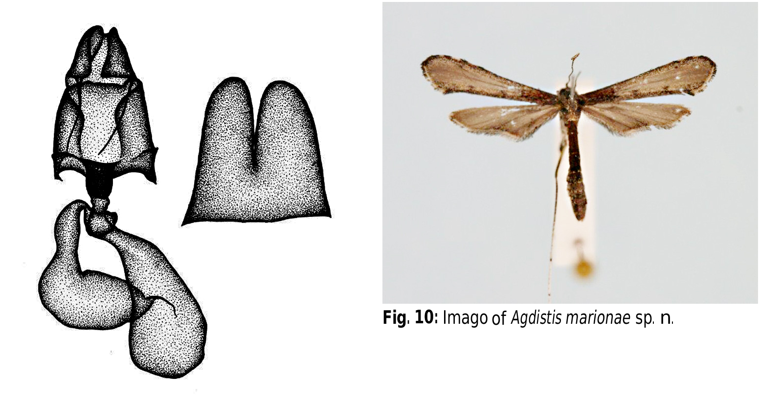
Diagnosis ( Fig. 10 View Fig ):
Wingspan: 14-15 mm. Forewings grey brown, from the basis to the area without markings strongly darkened, at the end of the dark area with 2 dots lying one on top of the other. Costal- and inner margin a little lighter, but strewn with many dark brown scales. Costal margin with 3 dark dots, the space between whitish. Beneath the first costal dot is another dot. Lower fold margin with two strong patches. Outer margin with light brown basal line, top of the fringe dark brown. Hindwings single-coloured brown. Head, thorax and abdomen dark brown. Thorax tergites with trapeziform white markings. Antennae brown – whitish ringed. Tarsi of hind legs light brown with dark brown ends.
Genitalia, ( Fig. 6 View Fig ): Valves asymmetrical. Both with a pointed process at a quarter of the inner margin. Left valve at half of its lenght weakly shovel-shaped widened. Right valve small, stripshaped, then nodularly widened and emarginate at the end. Costal arms similar on both sides, with a thin stem and globular end. Tegumen clasp-shaped. Uncus elongate, end with two tips. Caudal margin of sternite 7 roundishly cut, edges of the cutting with short, outwards pointed tip, each. First quarter of aedeagus extremely thin, further course slightly arcuate.
Genitalia, (Fig. 8): Antrum strongly sclerotized, cup-shaped, opened wide in caudal direction, ostium convex. From the corners of the antrum, on both sides arises one long, arcuate seta directed to the end of the body. Corpus bursae sack-like. Apophyses anteriores very short, tooth-shaped. Apophyses posteriores bristle-shaped, about twice as long as papillae anales. Sternite 8 deeply cut.
Biological notes:
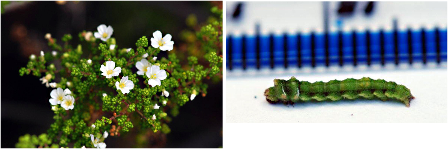
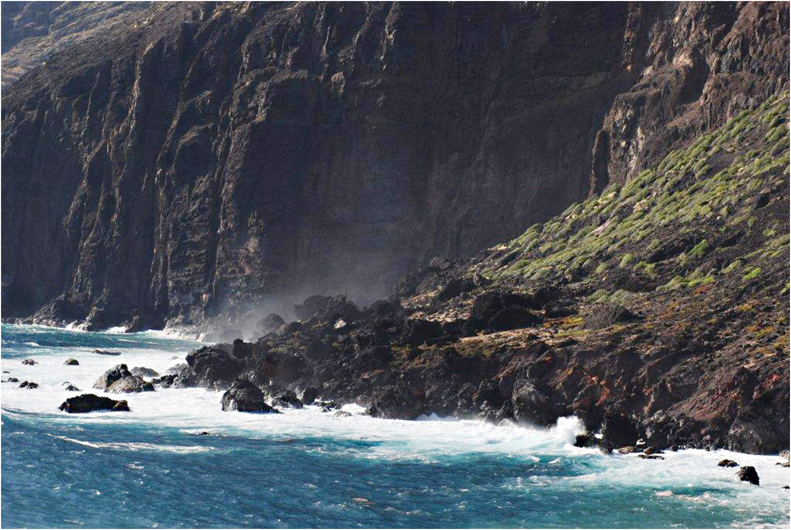
The unconcealed larvae and pupae of Agdistis marionae sp. n. were found on plants of the endemic species Fankenia portulacifolia (Roxb.) Sprengel ( Fig. 12 View Fig ) growing on dry cliffs at the shore of St. Helena (Great Shaking Rocks ( Fig. 11 View Fig ) and Joan Hill, Man and Horse) from December to April. No larvae were found in May. The larvae feed on leaves, rarely on flowers, and prefer large plants of more than 40 cm height. Duration of the pupal stage is 10 to 12 days under laboratory conditions.
Distribution: Endemic to St. Helena.
Remarks:
The new species was compared with all hitherto known Agdistis species from tropical regions. Agdistis marionae sp. n. and the other Agdistis species do not agree. The new species is on no account identical with A. santahelenae Wollaston, 1879 , which is endemic to St. Helena, too. In A. marionae sp. n. it is conspicuous, that there are only two patches at the lower fold margin. Characters, which are only found in A. marionae sp. n. Conspicous are the two dots lying one on top of the other at the end of the darkened basal field. In the male genitalia the valves are slimmer than in santahelenae and the end of the right valve is emarginate. In the female genitalia the antrum is widened in caudal direction, the ostium is convex and on both sides arises one sclerotized seta each. In santahelenae the setae are missing and the antrum is cylindrically formed.
Descriptions of larva and pupa of Agdistis marionae sp. n.
Materials and Methods:
The descriptions of larva and pupa of Agdistis marionae sp. n. are based on 2 mature and one submature larvae preserved in 70 % ethanol, one ethanol preserved pupa, one desiccated pupa and several pupal exuvia. Morphological observations and illustrations were made with the aid of both compound and stereomicroscopes, and a camera lucida. The terminology of sclerites and the nomenclature of setae and other sensilla follow Hasenfuss & Kristensen, 2003. The description refers to the left side of the body.
For comparisons, the as yet unpublished findings on the larva and pupa of Agdistis tamaricis (Zeller, 1847) from Tour de Valat (Camargue/South France), reared from eggs on Tamarix spec. were noted in brackets, when the characters were different from those of Agdistis marionae .
Mature larva ( Figs 1-3 View Fig View Fig View Fig , 13):
General. Unconcealed living larval type, length ca. 12 mm, head width 0.8 mm, antenna and mouthparts directed ventrally. The dorsal part of the head and nearly the whole surface of the body (including the unsclerotized coxae of the thoracic legs) exhibit densely arranged granules ( Fig. 2C, D View Fig ). The larva has only the regular general pattern of setae and other sensilla, no irregular setae are present. Most dorsal and lateral setae are more or less stout, club-like or flattened, and pressed to the body surface ( Fig. 2C, D View Fig ).
The dorsal part of the prothorax (T1) is hood-like extending over the head. Prominent fingerlike dorsal projections are on the thoracic segments T1 and T2. Segment T1 exhibits two projections, the anterior one bears seta XD1 and the posterior the seta D2; a small wartlike elevation bears XD2 (T1- Fig. 2A View Fig ). The large finger-like projection of T2 bears the setae D1 and D2 (T2- Fig. 2A View Fig ); the corresponding projection on T3 is smaller (T3- Fig. 2A View Fig ). In the submature instar, the dorsal projections of T1 and T2 are small, not larger than on T3. Small wart-like elevations are located, on the abdominal segments A1-A9, at the sites of D1, D2, and less distinct at the site of SD1 ( Fig. 2A View Fig ). On A9, the warts of D1 and D2 are fused and form together with the corresponding warts of the opposite side an unpaired flat unity ( Fig. 2B View Fig ). [The dorsal projections of A. tamaricis are different in the following characters: the large fingerlike projection on T2 bears three setae (D1, D2 and SD2); the corresponding projection on T3 and the prothoracic projections XD1 and D2 are small, wart-like; a small unpaired wart-like elevation without setae is located dorsally at the anterior margin of T2; on A2, A5 and A8, the separated warts of D1 and D2 are elongated to small finger-like projections; the unpaired wart of A9 is large and finger-like].
Color pattern: Head capsule dark brown, frontal side less dark. Body light yellowish green with a white line along the lateral sides (stigmatale) which is more or less interrupted in the posterior region of the segments. White larger vertically elongated spots are laterally on the prominent dorsal projections of segment T2 and T3. In darker specimens a line of blackish spots are dorsally applied to the white line/spots, especially pronounced in the interruptions of the stigmatale. The cuticular granules appear as white or light colored small dots on a darker background. The white especially densely arranged ones are composing the white spots.
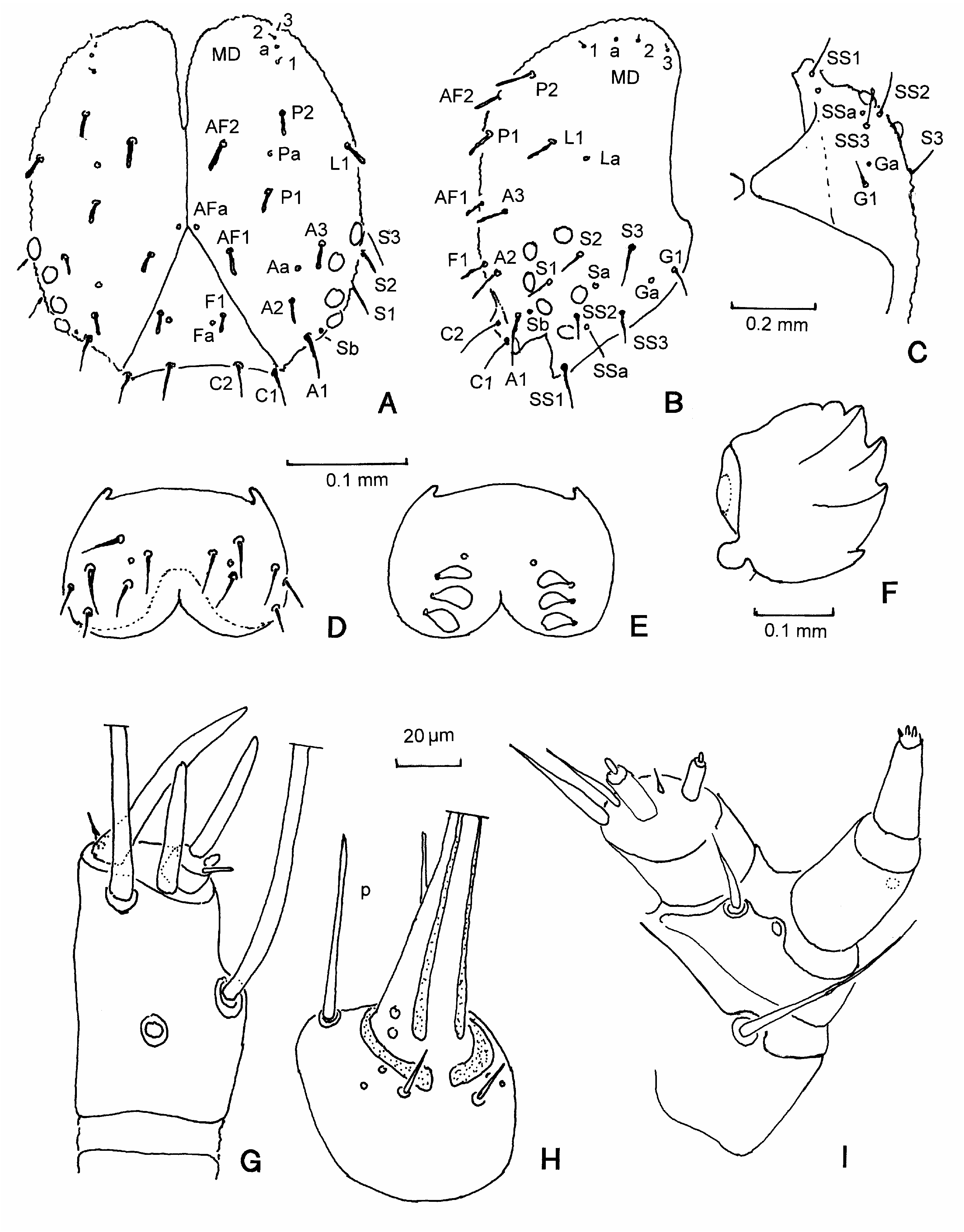
Head ( Fig. 1 View Fig )
Head capsule ( Fig. 1 View Fig A-C) with deep epicranial notch, the ecdysial line laterad of AF-setae is missing, the capsule splits upon ecdysis to pupa closely along the inverted Y-sulcus, puncture Pb absent. 6 stemmata present. Seta G1 long (in A. tamaricis microscopical). – Antenna ( Fig. 1G View Fig ): with the complete general pattern of sensillae. - Labrum ( Fig. 1D View Fig ): lateral margin well sclerotized, not widened antero-laterally, only one puncture (in A. tamaricis , the lateral sides are membranous and antero-laterally widened, 2 punctures). Epipharynx ( Fig. 1E View Fig ): the 3 scale-like sensilla arranged in a straight line. Mandible ( Fig. 1F View Fig ): only with marginal teeth, on the lateral side one puncture and one seta present (in A. tamaricis , on the median side with a conspicuous dentate ridge). - Maxilla ( Fig. 1I View Fig ): palpus maxillaris 4-segmented, its median lobe without the row of 3 ventral sensilla. - Labium ( Fig. 1H View Fig ): prementum ventrally with a small seta and 2 punctures, the dorsally located palpus labial reduced to a single long seta (p), 2 punctures on the base of the spinneret.
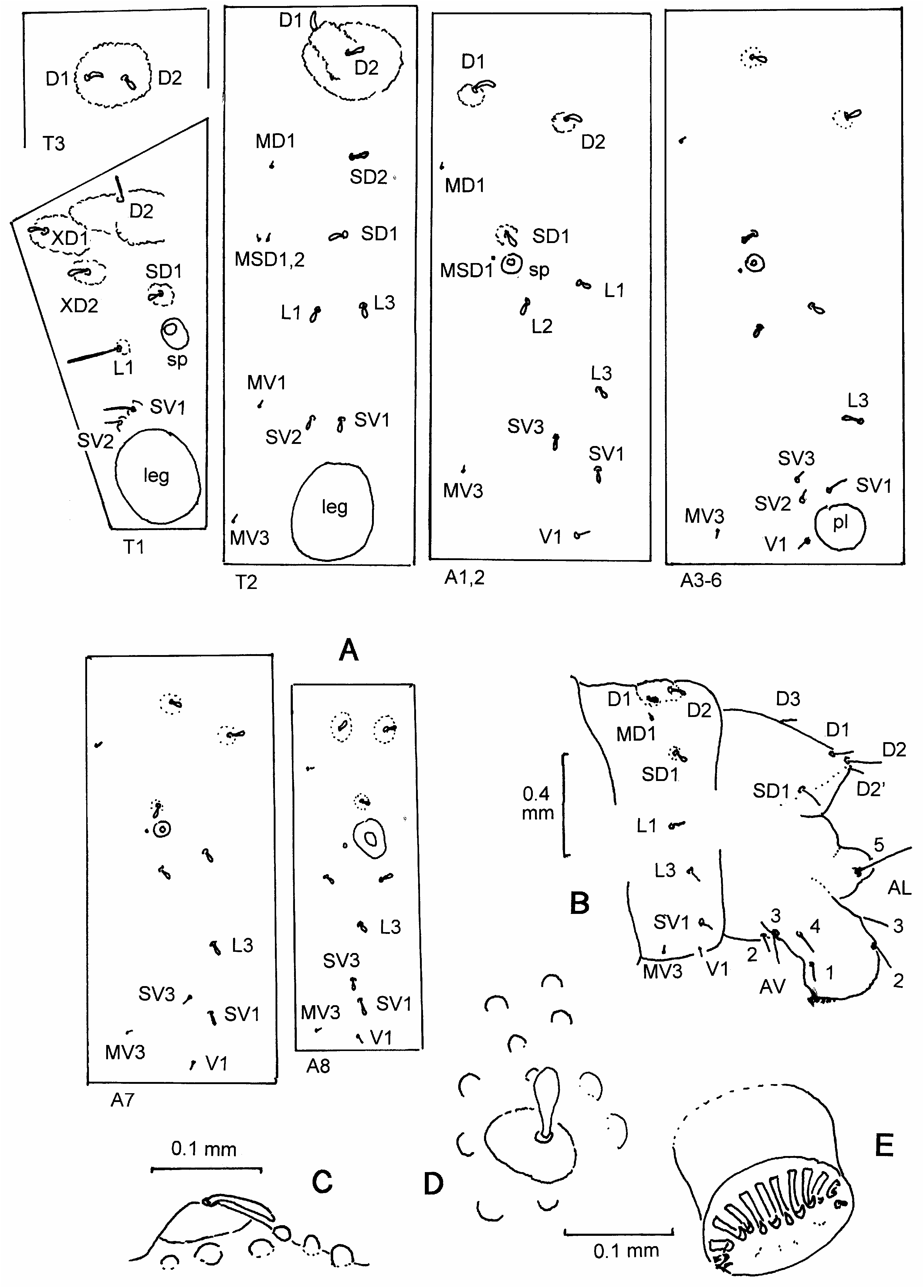
Thorax and abdomen ( Fig. 2 View Fig )
Tergal shields on T1 and A10 absent. The spiracles are slightly elevated on a conical peritrema (sp- Fig. 2A View Fig ). Pinacula of setae inconspicuous.
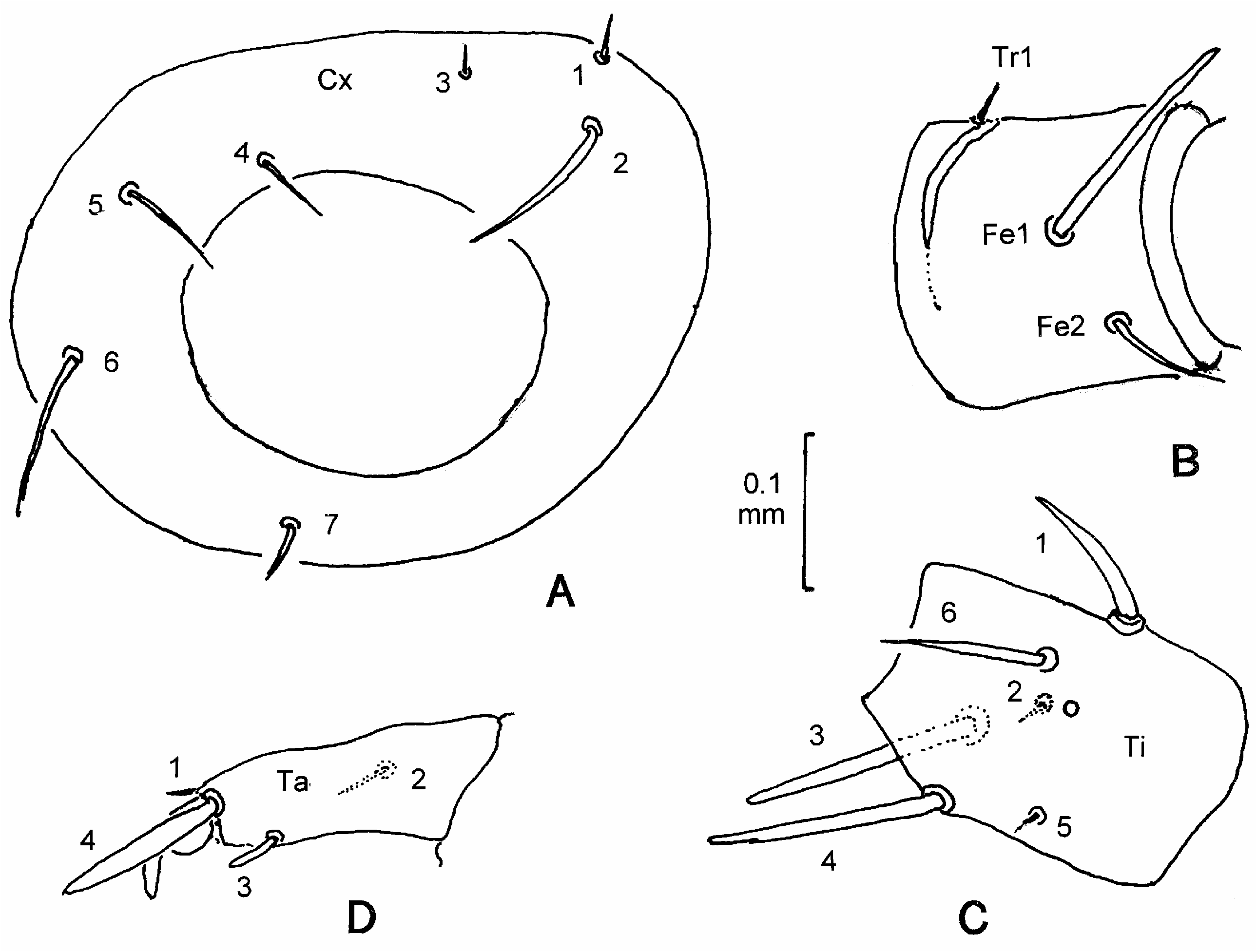
Segments of the thoracic legs: Coxa unsclerotized, seta Cx8 absent ( Fig. 3A View Fig ). The sole seta T1 and no punctures present on the trochanter ( Fig. 3B View Fig ), the trochanter is as usual only partly separated from the femur by a membranous strip. In contrast to coxa and trochanter, all setae of the general pattern are present on femur, tibia and tarsus ( Fig. 2 View Fig B-D).
Prolegs with crochets arranged in a median uniordinal penellipse, gradually smaller towards both ends ( Fig. 2E View Fig ). The number of crochets are 13, 14/15, 17/18, 20/21 and ca. 23 on segments A3, A4, A5, A6 and A10, respectively.
Chaetotaxy of the thoracic (T1, T2, T3) and abdominal (A1-A10) segments ( Fig. 2A, B View Fig ): No microscopical setae were found on T1. All other microscopical setae are present with the exception of MV2 on T2 and T3 (all present in A. tamaricis ). MSD1 is without a discernible shaft (well visible in A. tamaricis ); MD1 is on A 8 in the unusual position just below D1 ( Fig. 2B View Fig ). From the normally 6 prothoracic tactile setae and 3 punctures dorsal from the spiracle, only 4 setae were detected, their homologies are somewhat uncertain (T1 - Fig. 2A View Fig ). The prespiracular L-group on T1 is unisetose. V1 is absent on the thoracic segments. Yano, 1963 and Wasserthal,1970 figured V 1 in their setal maps as present on the thorax in Pterophoridae . Contrary to this, Hasenfuss (unpubl. observations) stated the absence of V1 on segment T1, T2 and T3 not only in Agdistis marionae sp. n. but also in Agdistis tamaricis , Agdistis bennetii (Curtis, 1833) , Pterophorus pentadactylus (Linnaeus, 1758) , Emmelina monodactyla (Linnaeus, 1758) , and an Oxyptilus spec. Segments T2 and T3 are identical except for the fact, that the common wart of D1 and D2 is much smaller on T3 than on T2 ( Fig. 2A View Fig ).
On A1-A8, the setae L1 and L2 are widely separated, L3 being unisetose (two L3-setae in A. tamaricis on A1-A6, the anterior one in some distance before the other). On A9, only L1 and L3 are present, L2 is missing. The SV-group on T2/T3 is bisetose. SV3 is present on the abdominal segments A1-A8 and SV2 only on A3-A6. (SV3 is absent on A 8 in A. tamaricis ). An additional dorsal seta (D2) is present on A10, its pair is located between and somewhat behind the pair of D2 ( Fig. 2B View Fig ). On the ventral part of A10 no punctures were found, and setae AL1 and AL4 are missing. ( A. tamaricis differs in having in the AL-group, the full set of 5 setae and one puncture).
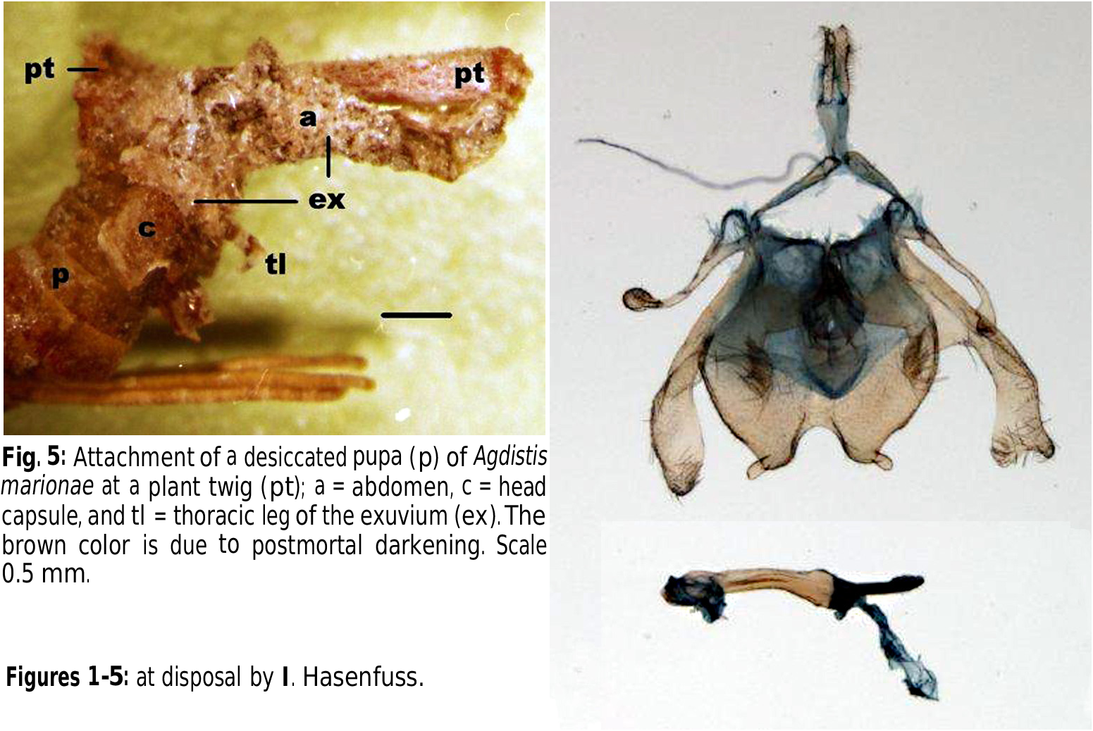
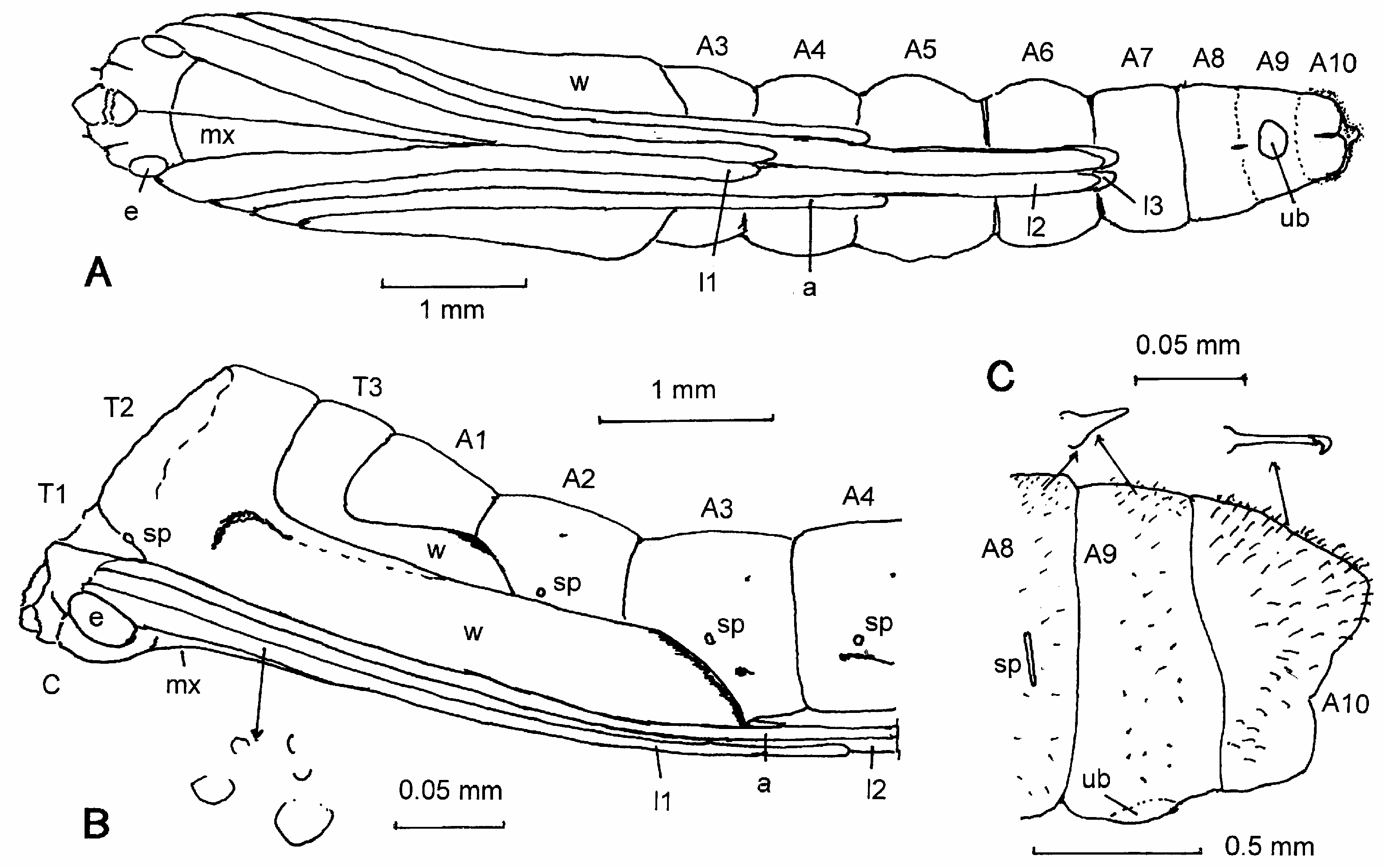
Pupa ( Figs 4 View Fig , 5 View Fig , 14 View Fig ):
Length ca. 9 mm. The surface of the head, thorax and appendages is uneven, partly sculptured with granules ( Fig. 4B View Fig below); the surface of A1-A9 is armed with small spinules. A9 bears ventrally an unpaired rounded bulge or prominence (ub - Fig. 4 A, C View Fig ). Segment A10 is short, stout, and not elongated; the spinules are modified to small hooked spines (not setae!) especially on the dorsal and (less numerous) on the lateral surface ( Fig. 4C View Fig ); the ventral surface and the region around the anus are without such spines. [In other Pterophoridae , the segment is elongated, gradually tapering to the end and form ventrally together with A9 an oblique plane. The lateral and posterior margin of the plane bears hooked spines; an additional group of such spines is located mid-ventrally on A9 at the margin of a rounded prominence (Hasenfuss unpublished observations, see Mosher, 1916, Yano, 1963). The prominence is supposedly the homologue of the similar smoth strucure up in Fig. 4A, C View Fig of the present species. Apparently, the marginal hooked spines of A10 are likewise homologous with the dorsal and lateral spines of Agdistis marionae sp. n.]. The spiracles on T2 and A2-A7 are functional, on A1 covered by the wings, and on A8 collapsed ( Fig. 4B, C View Fig ). Nearly all larval setae are maintained in the pupa (as in other Pterophoridae , see Yano, 1963), even the form is the same as in the larva. – The pupa is lightly colored with few dark spots ( Fig. 4B View Fig ): a curved line at the base and the margin of the fore wings, a line on the corner of the hind wings, a line or spot below the spiracle on A3-A6 and a small subdorsal spot in A3-A6.
The pupa is attached to a twig with the dorsal side (!) of its last abdominal segment ( Fig. 5 View Fig ). The attachment is due to the dorsal hooked spines which are hooking to a web made by the larva before pupation. Since, the larva and hence its exuvium always cling to a twig with its prolegs on the ventral side, the attachment of the pupa is inverse. During ecdysis, both the larval had capsule and the thoracic cuticle remain on the ventral side of the end of the pupal abdomen. More posteriorly, the exuvium is twisted so that the crochets of the prolegs remain hooked to the web at the twig. It may be that during this processes the pupa hangs at the exuvium by the cuticle of the proctodaeum and hooks itself into the web by wriggling and rotating the abdomen thus causing the 180° turn of the pupa around its length axis and twisting the exuvium. All three, the anterior part of the exuvium, the end of the pupa, and the twig are glued together by a hardened secretion. [The pupa of A. tamaricis is attached to the side of a twig by hooking the marginal hooked spines of the ventral plane of A9 and A10 to a small web (Hasenfuss, unpublished observation)].


No known copyright restrictions apply. See Agosti, D., Egloff, W., 2009. Taxonomic information exchange and copyright: the Plazi approach. BMC Research Notes 2009, 2:53 for further explanation.