
Agyneta ledfordi, Dupérré, Nadine, 2013
|
publication ID |
https://doi.org/ 10.11646/zootaxa.3674.1.1 |
|
publication LSID |
lsid:zoobank.org:pub:981F80ED-96D7-40C7-8A3C-677954416A2E |
|
DOI |
https://doi.org/10.5281/zenodo.6162414 |
|
persistent identifier |
https://treatment.plazi.org/id/038D6700-FFC3-5697-118C-02B7AB16B6A4 |
|
treatment provided by |
Plazi |
|
scientific name |
Agyneta ledfordi |
| status |
sp. nov. |
Agyneta ledfordi View in CoL new species
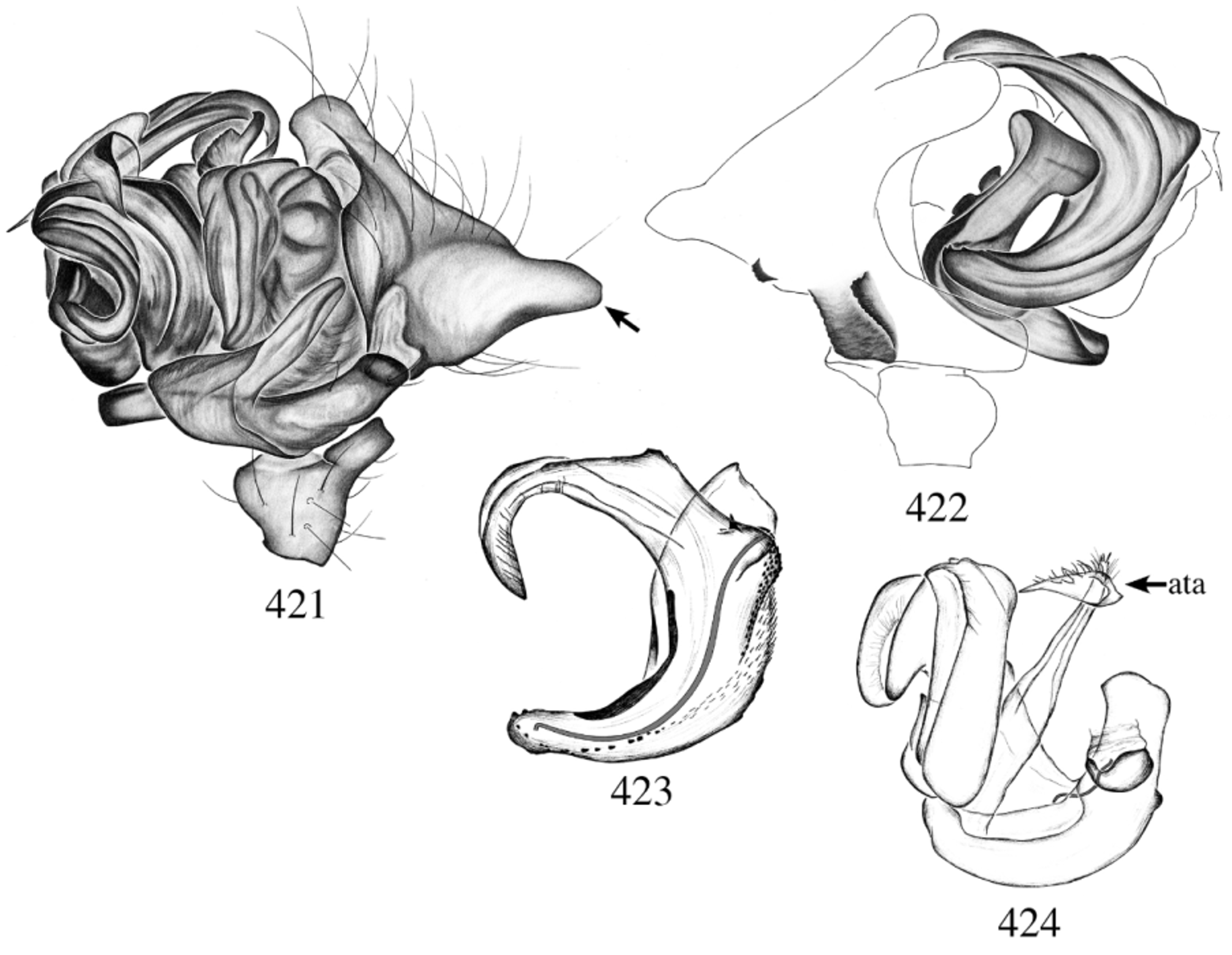
Figs 421–424 View FIGURES 421 – 424 , map 27
Type material: Male holotype from Florida, Dade county, Everglades National Park, Long Pine Key, 08.vi- 26.viii.1986, pinelands, malaise and FIT, S. J. Peck ( AMNH). EXAMINED.
Etymology: The specific name is a patronym in honor Dr. Joel Ledford, great arachnologist and friend.
Diagnosis: Males can be distinguished from all other species by the very large conical extension of the cymbium ( Fig. 421 View FIGURES 421 – 424 arrow), from A. hedini by the squared, rugose ventral cymbial tubercle ( Fig. 422 View FIGURES 421 – 424 ), pointed in the latter ( Fig. 403 View FIGURES 402 – 411 ).
Description: Male: Total length 1.56; carapace length 0.76, width 0.61.
CEPHALOTHORAX: (Specimens faded) carapace light yellow with V-shap gray mark on pars cephalica. Sternum light yellow. Clypeus height 2. Chelicerae light yellow, excavated; ~ 20 seta-tipped tubercles; promargin three teeth, retromargin two denticles. Endites with seta-tipped tubercles. Cheliceral stridulatory organ with ~26 striae, well spaced throughout. ABDOMEN: Uniformly off-white. LEGS: Light yellow; leg I total length: 3.52; leg III total length: 1.88; Tm I: 0.24, Tm IV: absent. GENITALIA: Palpal tibia with large, smooth retrolateral tibial apophysis; dorsal apophysis absent; two retrolateral trichobothria and a dorsal one ( Fig. 421 View FIGURES 421 – 424 ). Cymbium with large conical extension; glabrous depression present ( Fig. 421 View FIGURES 421 – 424 ); dorsal cymbial tubercle small and rugose; ventral tubercle, large and rugose; prolateral notch very deep ( Fig. 422 View FIGURES 421 – 424 ). Paracymbium apical pocket small, anterior pocket long and curved, posterior pocket absent ( Fig. 421 View FIGURES 421 – 424 ). Embolus tip curved; row of small spines basally; rugose ventrally; Fickert’s gland absent; ventral lamella absent; thumb reaching beyond embolus proper ( Fig. 423 View FIGURES 421 – 424 ). Embolus proper set apically, small prong near base of embolus proper ( Fig. 423 View FIGURES 421 – 424 ). Anterior terminal apophysis large with long protrusions; posterior terminal apophysis and lamella characteristica difficult to separate, fused, large and C-shaped ( Fig. 424 View FIGURES 421 – 424 ).
Female: Unknown.
Other material examined: Six males paratypes collected with the holotype. Distribution: Southeastern USA, Florida.
The llanoensis View in CoL group includes 4 species, A. llanoensis ( Gertsch & Davis 1936) View in CoL , A. bronx View in CoL n. sp., A. paquini View in CoL n. sp. and A. serrata ( Emerton 1909) View in CoL .
This group show some interesting habitat adaptation, as three species are often found in caves. A. llanoensis View in CoL is found in caves all over Texas, A. serrata View in CoL is mostly found on the surface but also been found in some caves in Texas, and A. bronx View in CoL was found in a cave in Missouri.
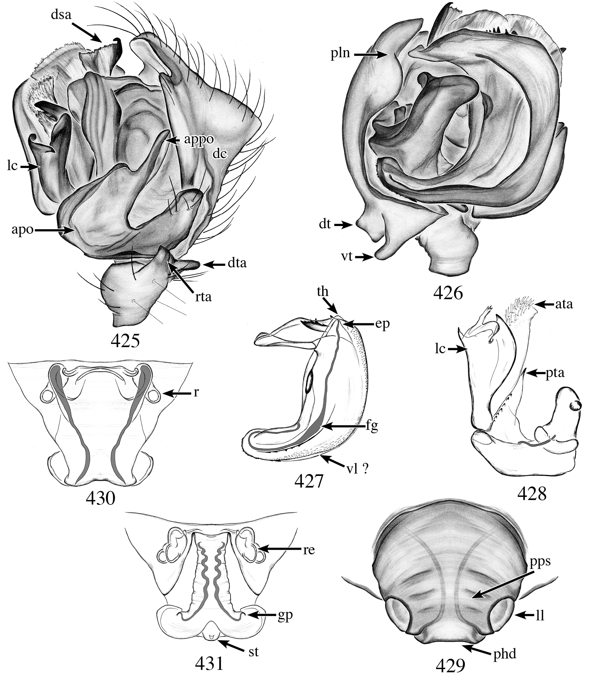
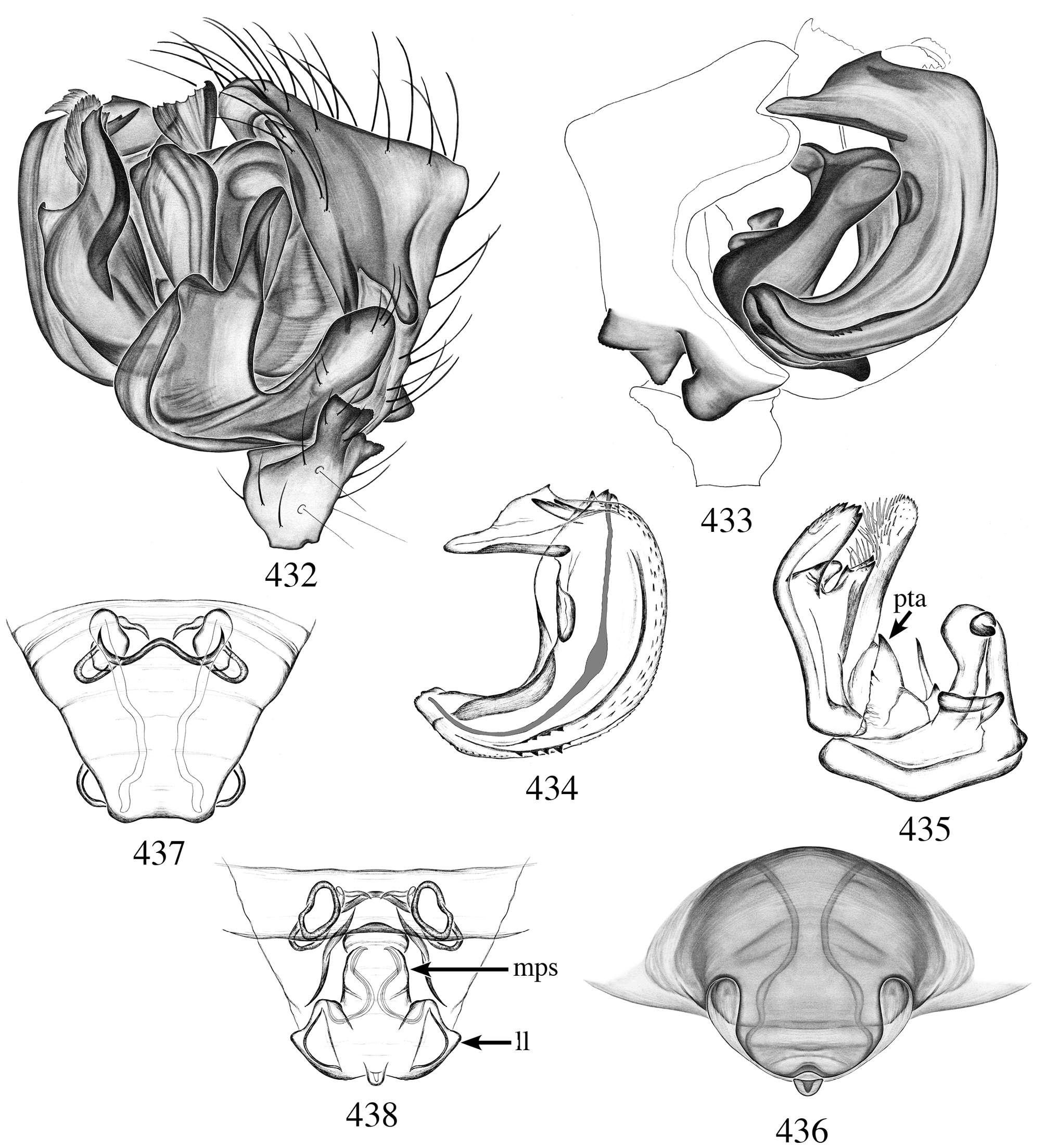
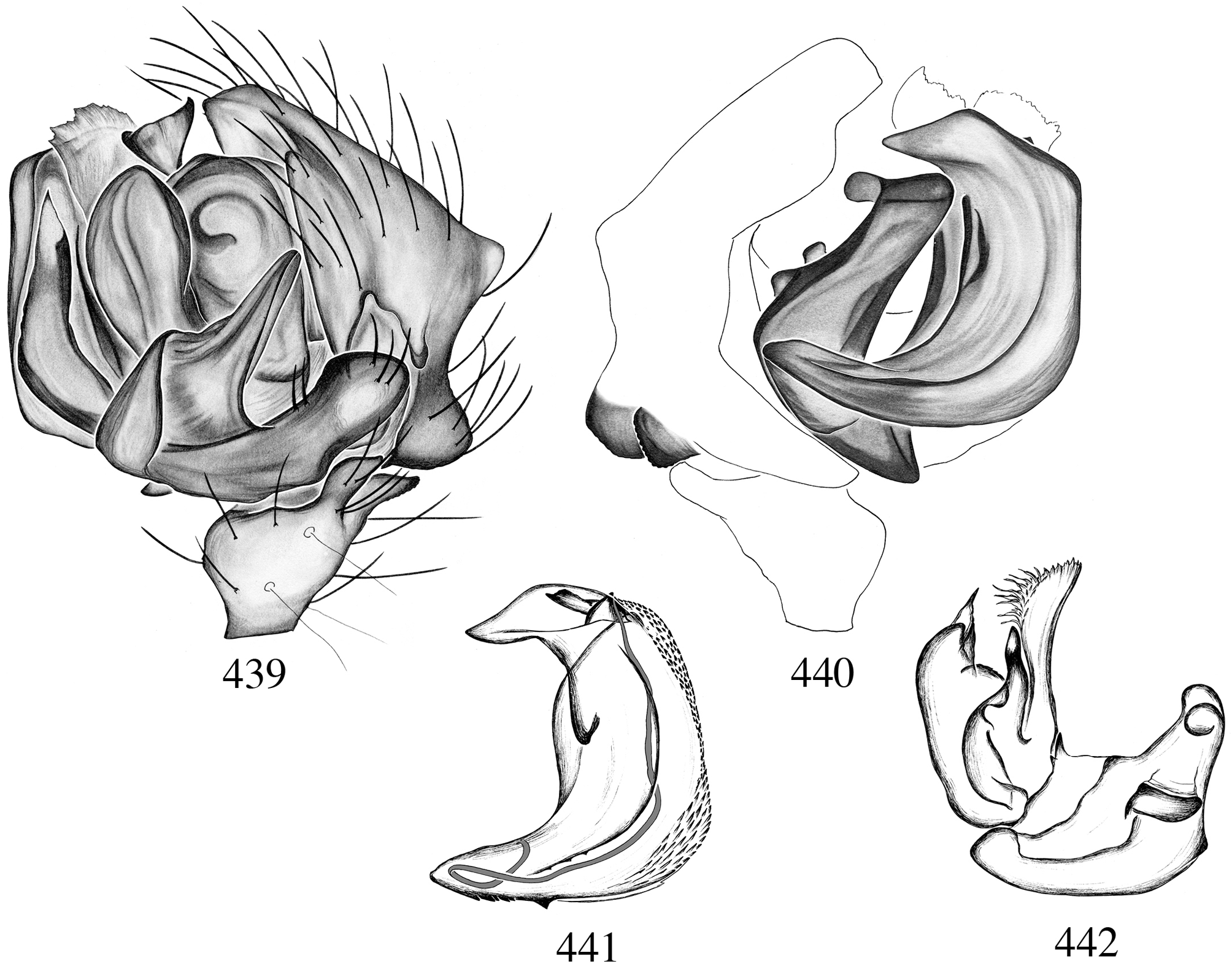
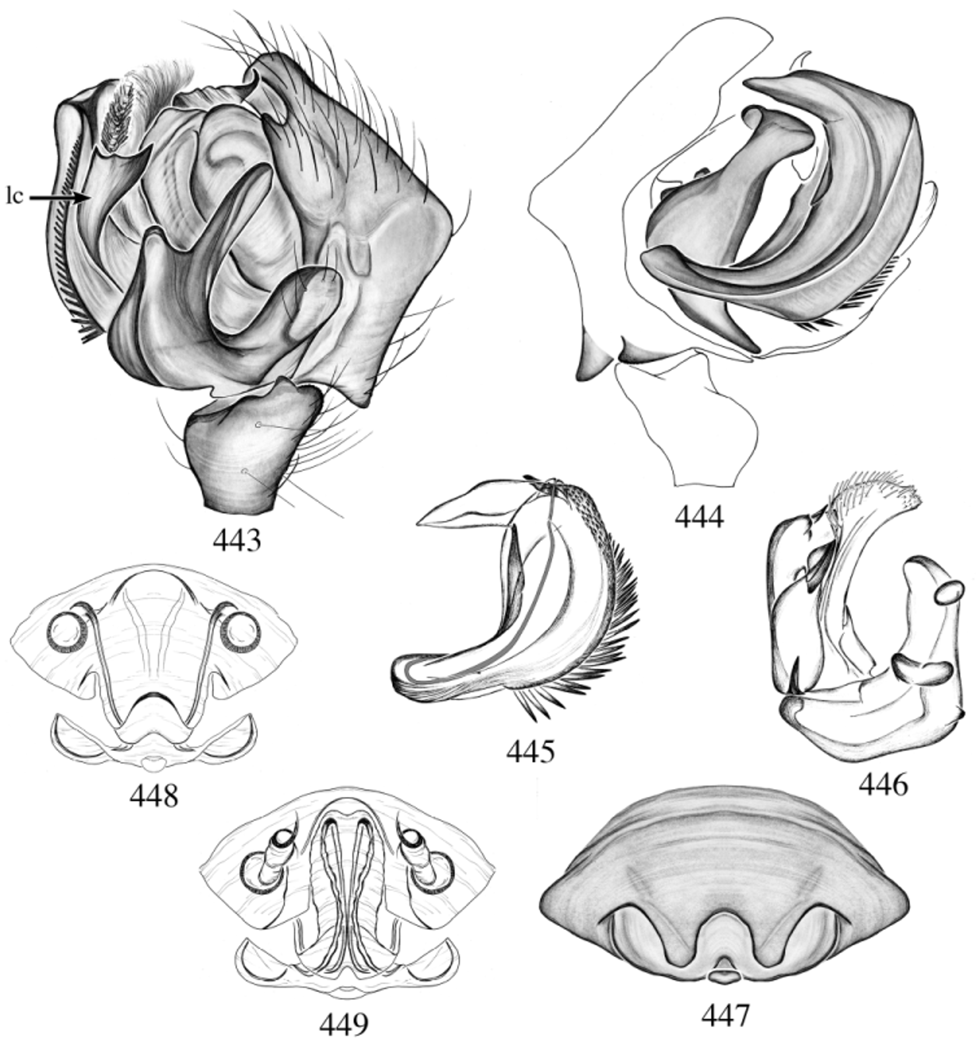
Members of this group are share a unique character, the presence of a large prong close to the embolus proper, in between the latter and the thumb ( Figs 427 View FIGURES 425 – 431 , 434 View FIGURES 432 – 438 , 441 View FIGURES 439 – 442 , 445 View FIGURES 443 – 449 ). Furthermore, the group is characterized by the combination of these characters; male chelicerae not excavated, seta-tipped tubercles absent; males and females without abdominal pattern. Palpal tibia with two retrolateral trichobothria and a dorsal one; paracymbium with long curved anterior pocket ( Figs 425 View FIGURES 425 – 431 , 432 View FIGURES 432 – 438 , 439 View FIGURES 439 – 442 , 443 View FIGURES 443 – 449 ). Cymbium with dorsal and ventral tubercles present ( Figs 426 View FIGURES 425 – 431 , 433 View FIGURES 432 – 438 , 440 View FIGURES 439 – 442 , 444 View FIGURES 443 – 449 ). Ventral lamella highly reduced or absent; thumb reaching the embolus proper; embolus proper set apically on a horizontal ridge reaching the tip of the embolus ( Figs 427 View FIGURES 425 – 431 , 434 View FIGURES 432 – 438 , 441 View FIGURES 439 – 442 , 445 View FIGURES 443 – 449 ); radical division with well developed, wide anterior terminal apophysis with long curved protrusions ( Figs 428 View FIGURES 425 – 431 , 435 View FIGURES 432 – 438 , 442 View FIGURES 439 – 442 , 446 View FIGURES 443 – 449 ). Females are characterized by their extremely wide median part of scape and short lateral lobes ( Figs 429 View FIGURES 425 – 431 , 436 View FIGURES 432 – 438 , 447 View FIGURES 443 – 449 ).

| AMNH |
American Museum of Natural History |
No known copyright restrictions apply. See Agosti, D., Egloff, W., 2009. Taxonomic information exchange and copyright: the Plazi approach. BMC Research Notes 2009, 2:53 for further explanation.
|
Kingdom |
|
|
Phylum |
|
|
Class |
|
|
Order |
|
|
Family |
|
|
Genus |
Agyneta ledfordi
| Dupérré, Nadine 2013 |
A. llanoensis (
| Gertsch & Davis 1936 |
A. serrata (
| Emerton 1909 |