
Anisogomphus yanagisawai, Sasamoto, Akihiko, 2015
|
publication ID |
https://doi.org/ 10.11646/zootaxa.3904.3.8 |
|
publication LSID |
lsid:zoobank.org:pub:DA54DD29-A766-4129-8AB2-89B210B83AE9 |
|
DOI |
https://doi.org/10.5281/zenodo.6098338 |
|
persistent identifier |
https://treatment.plazi.org/id/F45987E7-FFEF-FFC1-FF6F-FA469AC3B39C |
|
treatment provided by |
Plazi |
|
scientific name |
Anisogomphus yanagisawai |
| status |
sp. nov. |
Anisogomphus yanagisawai View in CoL sp. nov.
Figures 1–12 View FIGURES 1 – 2 View FIGURES 3 – 10 View FIGURES 11 – 12
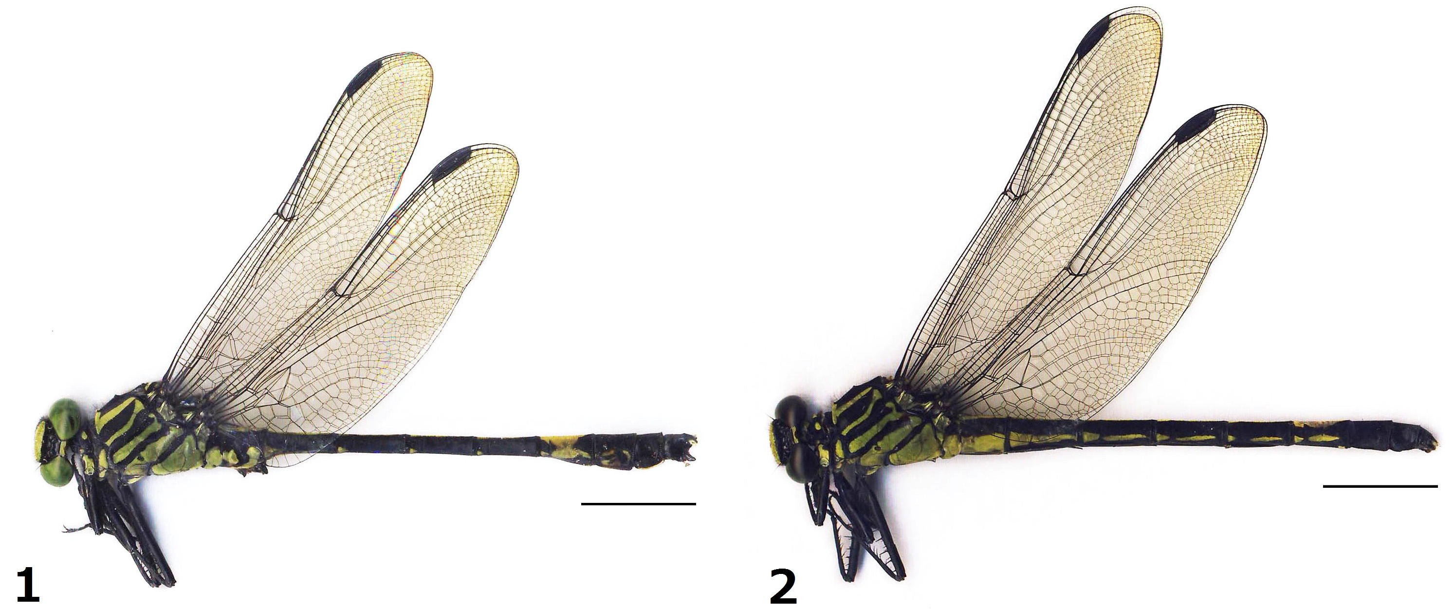
Material examined. Holotype: ♂ ( Fig. 1 View FIGURES 1 – 2 ) (NSMT-I-Od-15803), Doi Inthanon (18º54' N, 98º52' E, ca. 1,400 m a.s.l.), Ban Luang, Chiang Mai Prov., Thailand, Takashi Yanagisawa leg., 6. VI. 2013. Paratype: 1 ♀, 2. VI. 2012; 4 ♀, 3. VI. 2012; 3 ♀, 4. VI. 2012; 2 ♀ 2. VI. 2013; 1 ♂, 6. VI. 2013, 1 ♂ 1 ♀ ( Fig. 2 View FIGURES 1 – 2 ) (NSMT-I-Od-15804), 8. VI. 2013. All collecting localities are the same as that of holotype. The holotype and one female paratype will be deposited in the National Museum of Nature and Science, Tokyo. The other specimens are preserved in Mr. Yanagisawa’s and author’s private collections.
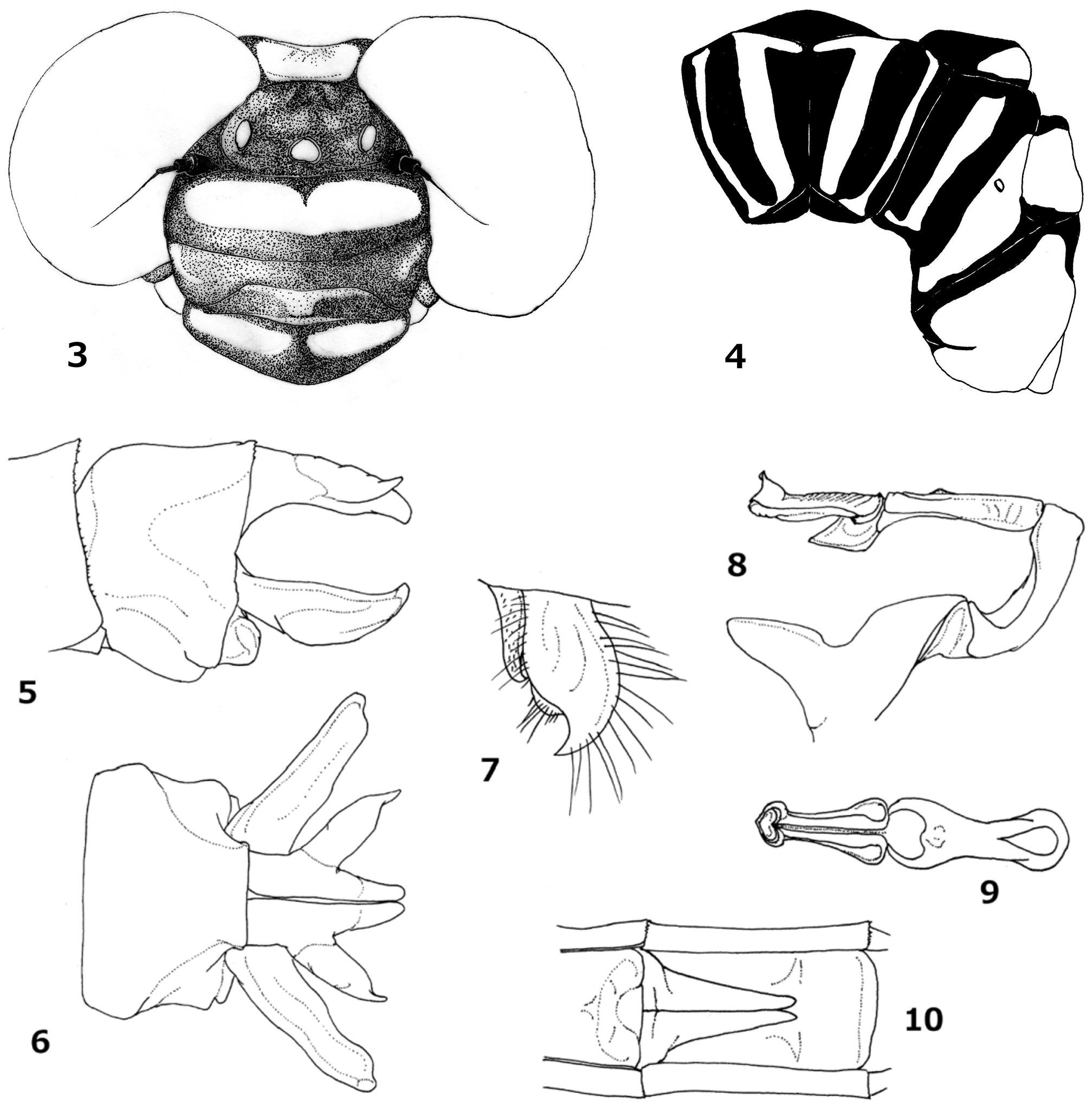
Holotype Male: Head ( Fig. 3 View FIGURES 3 – 10 ) black with yellow markings with brownish setae. Labium black in median lobe and pale yellow in lateral lobes; mandible black; labrum with a pair of transverse ellipsoid yellow spots; genae yellow except for black on upper margin; anteclypeus with yellow in upper margin and median part; postclypeus with a pair of small triangular spots laterally; frons medially with a slight depression and with a broad yellow band on dorso-anterior surface; eyes moss-green in life; vertex and antennae black; occiput slightly depressed in middle, yellow with posterolateral edge black.
Prothorax black with yellow on anterior margin of anterior lobe, a dorsal spot and a pair of lateral ones on median lobe. Synthorax ( Fig. 4 View FIGURES 3 – 10 ) bicolored, black and yellow, as follows: mesepisternum with an anterior yellow stripe, running obliquely, attenuated ventrad and nearly touching antealar ridge, confluent below with collar stripe; antehumeral yellow stripe slender and weakly undulating, slightly expanded on ventral extreme to form fan shape, and barely touching or very close to anterior stripe; laterally synthorax yellow with broad black stripes on humeral, 1st and 2nd sutures. Stripes on humeral and 1st suture connected at ventral and dorsal margins of mesepimeron; metastigma black; mesokatepisternum and metakatepisternum both yellow with black on anterior half; poststernum black. Legs black, outer surface of coxae yellow, inner surface of fore-femora pale yellow; femora with numerous small spines and two rows of 3 or 4 long spines distally; tibiae with 5–8 rows of medium length spines.
Wings hyaline with black veins; primary (thick) antenodal veins first and fifth; an incomplete basal antenodal vein, i.e. only crossing subcostal space, present in all wings. Triangle in forewing entire; in left hind wing once crossed, but not crossed in right wing; anal triangle 3-celled. Pterostigma brown, about 4 mm in length, overlying about 4 cells in both wings. Nodal index 11:16::15:11/11:11::11:11.
Abdomen ( Fig. 1 View FIGURES 1 – 2 ) basal two segments relatively thick, then constricted and slender on S3–6, expanded from S7 and broadest at distal margin of S8, tapering in S9–10. Coloration black with yellow markings as follows: S1 with yellow on ventral half. S2 with an irregular round spot anteroventrally including auricle, and with an elongate ellipsoidal spot posteroventrally. S3–6 each with small notched markings on anteroventral corner. S1–6 with longitudinal stripe running continuously on dorsum, a part of which on S1 and S2 is irregularly broad and constricted, the posterior part uniformly very thin. S7 with a large rhomboid marking on dorsum and irregular semi-circular markings ventrolaterally, both obscurely merging laterally. S8 with vestigial poorly demarcated spots ventrally. S9 and S10 wholly black.
Anal appendages ( Figs. 5 & 6 View FIGURES 3 – 10 ) black with whitish yellow outer branch of cerci. Cerci and epiproct are nearly the same length, a little shorter than S10, in lateral view. Cercus with a prominent outer branch in dorsal view; cercus in dorsal view of an elongated triangular shape, main branch straight, ending obtusely, turned slightly downward in lateral view, a pair of the cerci disposed closely to each other; outer branch thick, arising from the middle of cercus, directing transversely and obliquely posterolaterad and suddenly constricted to a sharpened apex in dorsal view, this turned upward in lateral view. Epiproct bifid and strongly divergent in ventral view; very gently undulate along inner margin and of an almost uniform width before distal portion constricted to a blunt apex ( Fig. 6 View FIGURES 3 – 10 ), in lateral view gently hooked upward ( Fig. 5 View FIGURES 3 – 10 ).
Accessory genitalia ( Fig. 7 View FIGURES 3 – 10 ) as follows; anterior hamulus broad basally, tapering, sharpened apically and hooked posteriad; posterior hamulus robust with large hook at apex turning anteroventrally.
Penile organ ( Figs. 8 & 9 View FIGURES 3 – 10 ); vesicle triangularly shaped, excavated ventrally; 2nd segment gently curved; 3rd slender with swollen distal and proximal portions; 4th (distal) tubular shaped with an opening in distal portion, and with a curtain-like lobule in baso-ventral part.
Variations in paratype males. There are minor variations among two paratype males, compared with the holotype. Antenodal veins 16–20 in forewing, 11–13 in hind wing; postnodal veins 12–14 in both forewing and hind wing. Markings on S7 somewhat variable, sometimes dorsal and lateral markings partly fused.
Paratype female: General maculation pattern ( Fig. 2 View FIGURES 1 – 2 ) similar to that of male; however, there are several differences as follows: yellow maculation on head sometimes fading, especially that of postclypeus. Occiput depressed medially, more excavated than in male but lacking additional processes. Hind femora and tibia with a pair of rows of 4–6 extra-long spines. Antenodal veins 15–19 in forewing, 11–14 in hind wing; postnodal veins 11–16 in forewing, 12–15 in hind wing. Abdomen almost uniformly cylindrical, without conspicuous expansion in distal segments, S1–7 with clearer and broader ventrolateral markings, almost continuous throughout, only interrupted at anterior 1/3 and posterior margin on each of S3–7. S8 wholly black. Cerci about same length as S10, basally thick, then distal third constricted and ended in filament, bright yellow and slightly blackish laterally.
Valvula vulvae ( Fig. 10 View FIGURES 3 – 10 ) slender and triangular, branched in distal part and each tip slightly turning laterad, reaching beyond middle of 9th sternite.
Measurements (mm). Holotype male: total length (TL) 56.5; abdomen (including anal appendages) (Abd) 42.3; hind wing (HW) 35. Paratype males: TL 53.5–57.5; Abd 38.5–39.5; Hw 32.5–33.0. Paratype females: TL 48.5–54; Abd 35.5–40; Hw 32–33.5.
Etymology. The species name, a noun in the genitive case, is dedicated to Mr. Takashi Yanagisawa, who discovered the holotype male and paratype specimens.
Diagnosis. The characteristic morphology of the cerci ( Figs. 5, 6 View FIGURES 3 – 10 ) is the most obvious difference from the other species of this genus. The cerci are straight and disposed closely to each other along their inner margin; the outer branch is thick and pointed apically, arising from the middle of the cercus and projected in a horizontal plane posterio-obliquely.
Among the other species, of which the male is known, none has such characteristics. The anal appendages of Anisogomphus forresti ( Morton, 1928) from Yunnan have a similar morphology, but the outer branch is small and arises more distally on the cercus. In A. jinggangshanus Liu, 1991 , from Jiangxi, P. R. China, A. wuzhishanus Chao, 1982 , from Hainan and A. vulvaris Yousuf & Yunus, 1977 , from Pakistan, only the female has been described ( Liu, 1991; Zhao [= Chao], 1990; Yousuf & Yunus, 1977). The female of A. yanagisawai sp. nov. is differentiated from these species by the thoracic maculation and morphology of the valvula vulvae. In addition, Wilson (2005) recorded a female specimen of an unnamed Anisogomphus from Guangxi, China, which has similarities in thoracic maculation and shape of valvula vulvae to those in A. yanagisawai sp. nov., but differences in the yellow stripe on labrum, i.e. continuous in the former but separated in the latter. The true identity of the former will be revealed by the discovery of its male.
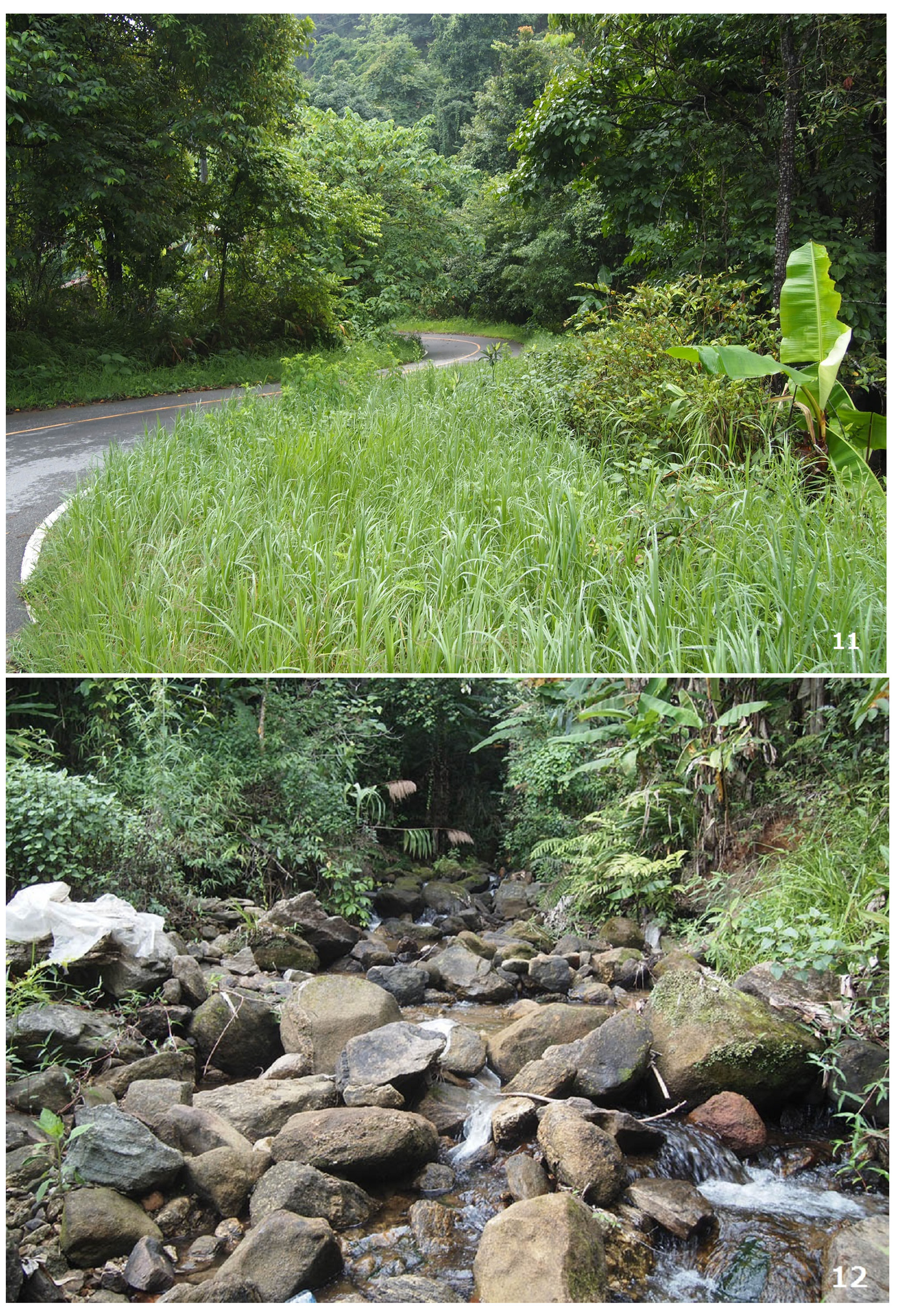
Habitat. According to Mr Yanagisawa, these specimens were collected on paved roads ( Fig. 11 View FIGURES 11 – 12 ) beside a running stream ( Fig. 12 View FIGURES 11 – 12 ). The males were found to settle on leaves of roadside trees. By contrast, females were seen to fly swiftly over the road. One oviposited in a small stagnant part of the stream ( Fig. 12 View FIGURES 11 – 12 ), tapping the tip of her abdomen on the water repeatedly, flying in a circle. In the end, she flew off and did not perch nearby. They appeared from 09:00 to 14:00 hours only in sunny conditions. Unfortunately, mating behaviour was not observed. At the same time and location, Anotogaster gregoryi Fraser, 1924 , and Macromia moorei Selys, 1874 , were also observed.
Distribution. Chiang Mai Province, northern Thailand.
Comments. The species of this genus are mostly characterized by the morphology of the male cercus. This new species also shows distinct features of the cercus, as noted in the diagnosis. Among congeners, the closely disposed straight cerci are also found in A. forresti and Himalayan A. occipitalis (Selys, 1854) , so this may indicate that they share a phylogenetic affinity, though the structure of the branch of the cercus is unique in each species. This new species is the second member of the genus Anisogomphus from Thailand, following A. pinratani , and both are the southernmost known members of this genus. This being the case, Doi Inthanon can be regarded as a region of special interest as the southernmost outpost of an East Asian fauna.
Curiously, earlier some researchers had obtained only female specimens, and also in this time, females were observed much more frequently than males. The activity patterns of the male may be investigated by future field work.
No known copyright restrictions apply. See Agosti, D., Egloff, W., 2009. Taxonomic information exchange and copyright: the Plazi approach. BMC Research Notes 2009, 2:53 for further explanation.