
Antoonops sarae, Fannes, Wouter, 2013
|
publication ID |
https://doi.org/ 10.11646/zootaxa.3709.6.2 |
|
publication LSID |
lsid:zoobank.org:pub:98FE3306-8862-4C89-B735-37515E84BE9E |
|
DOI |
https://doi.org/10.5281/zenodo.6145396 |
|
persistent identifier |
https://treatment.plazi.org/id/E57CAF63-FF91-FFEF-FF75-FE68A76EFE64 |
|
treatment provided by |
Plazi |
|
scientific name |
Antoonops sarae |
| status |
sp. nov. |
Antoonops sarae View in CoL n. sp.
( Figs. 10–16 View FIGURE 10 View FIGURE 11 View FIGURE 12 View FIGURE 13 View FIGURE 14 View FIGURE 15 View FIGURE 16 , 17 View FIGURE 17. A D)
Type material. CAMEROON: Adamaoua: holotype female, Tchabal Mbabo, 7°25’N, 12°49’E, 1500 m, April 11, 1983, hand catch, litter in gallery forest near mountain stream, R. Bosmans (MRAC 241.395, PBI_OON 32985). Paratype: 1 female, same locality, 1200 m, April 8, 1983, hand catch, litter in gallery forest, R. Bosmans (MRAC 241.396, PBI_OON 32986).
Other material examined. CAMEROON: Adamaoua: 2 females, same data as paratype, used in part for SEM (MRAC 241.397, PBI_OON 32982).
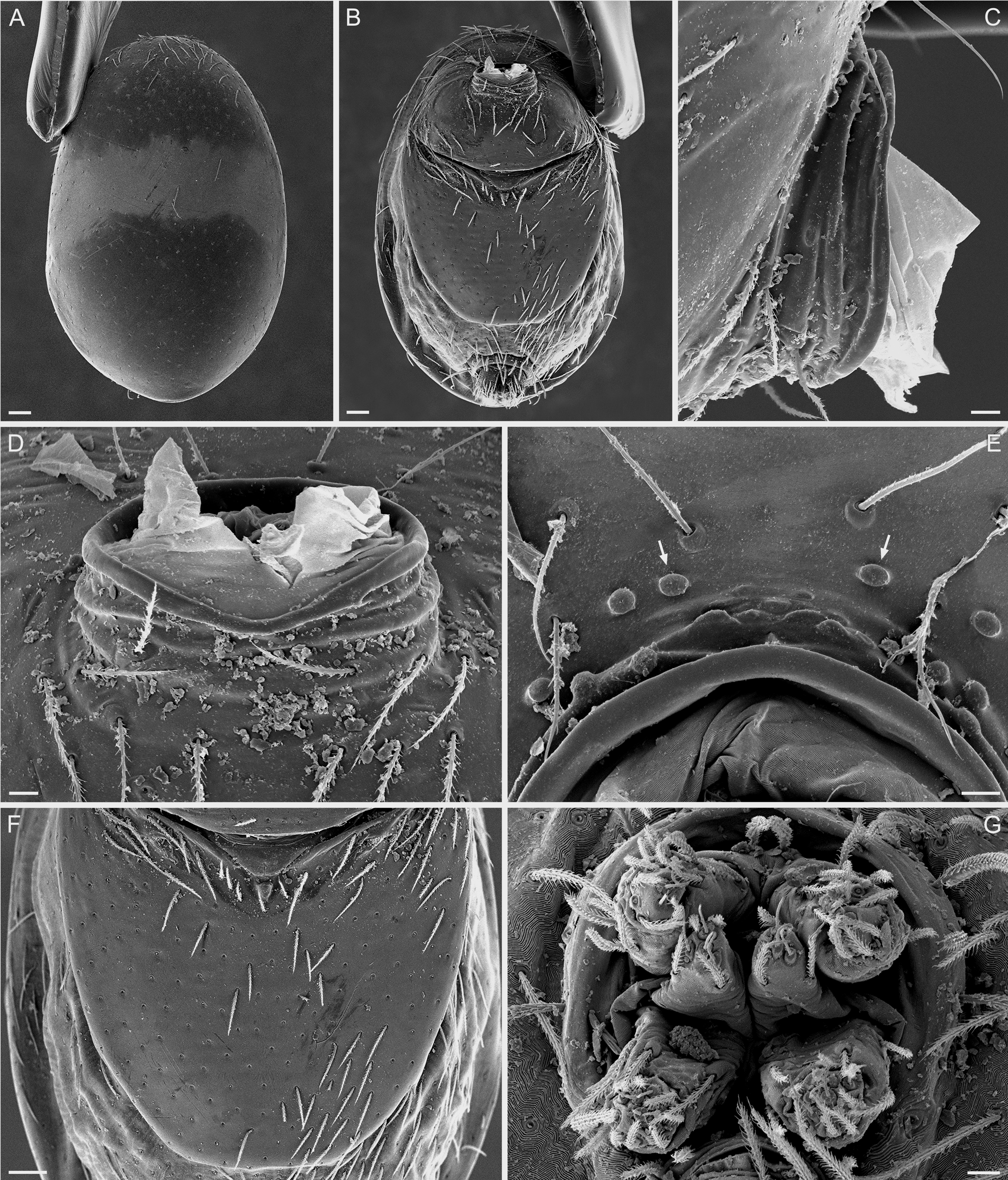
Etymology. Named after my sister-in-law, Sara Verpooten, in recognition of her support. Diagnosis. Females can be recognized by the long postepigastric scutum that leaves less than 1/4 of the
abdomen length uncovered ( Fig. 10 View FIGURE 10 E). They can also be recognized by their genitalia ( Figs. 10 View FIGURE 10 F, 15, 16). Description. Male. Unknown.
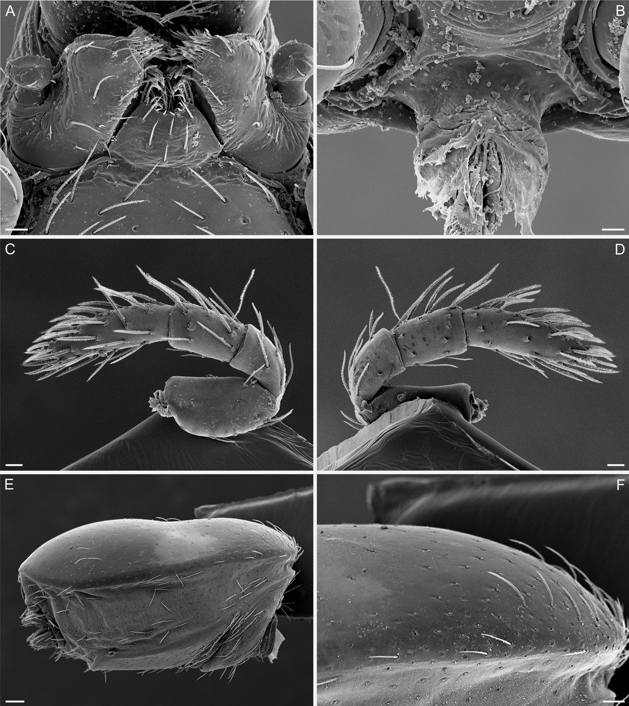
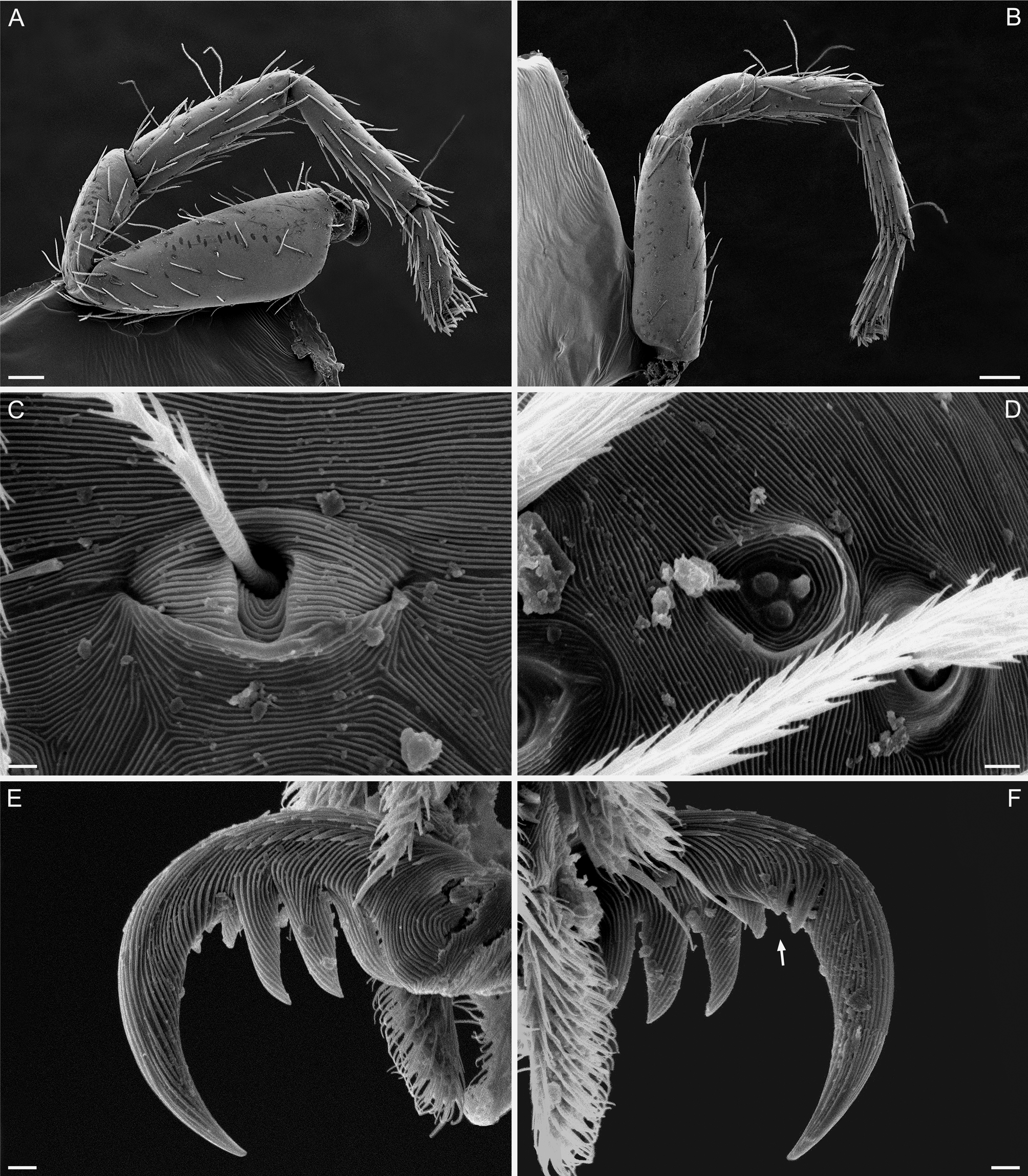
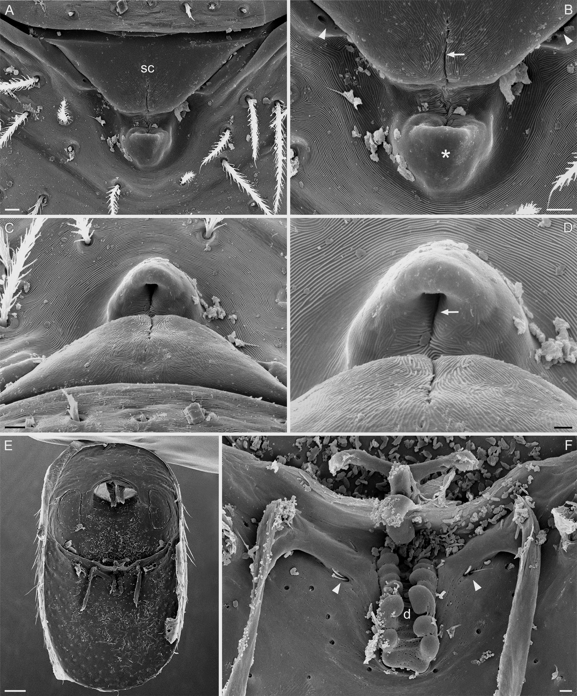
Female ( Figs. 10–16 View FIGURE 10 View FIGURE 11 View FIGURE 12 View FIGURE 13 View FIGURE 14 View FIGURE 15 View FIGURE 16 , 17 View FIGURE 17. A D). TL 1.44. Habitus as in Fig. 10 View FIGURE 10 A. Cephalothorax: Carapace brown, without any pattern, ovoid in dorsal view, completely covered by microsculpture except anterior part of dorsal surface, thorax without depressions or radiating rows of pits, posterolateral edge without pits, posterior margin not bulging below posterior rim, posterolateral surface without spikes; lateral margin straight, rebordered ( Figs. 10 View FIGURE 10 B, 11A, B). Clypeus sinuous in front view, high, ALE separated from edge of carapace by their radius or more, median projection absent ( Fig. 11 View FIGURE 11 D). Six eyes, small; ALE-ALE: separated by more than their diameter; ALE-PLE: separated by less than PLE radius; PME-PME: almost touching; PLE-PME: separated by PME diameter; posterior eye row recurved in dorsal view ( Fig. 11 View FIGURE 11 D, E). Sternum longer than wide, yellow-brown, fused to carapace, surface smooth, median concavity absent, with radial furrows between coxae I-II, II-III, III-IV, without posterior hump, setae abundant ( Figs. 10 View FIGURE 10 C, 11C, F). Chelicerae, endites and labium yellow-brown. Anterior face of paturon unmodified, with sparse setae ( Fig. 11 View FIGURE 11 B); fang shape normal, without prominent basal process, tip unmodified. Endites same as sternum in sclerotization, unmodified ( Fig. 12 View FIGURE 12 A). Labium same as sternum in sclerotization, shape as in Fig. 12 View FIGURE 12 A. Female palp brown, without spines; femur approximately twice as long as patella; patella about as long as tibia, without prolateral row of ridges, without leaf-like setae; tibia with three dorsal trichobothria; tarsus not expanded, twice as long as tibia ( Fig. 12 View FIGURE 12 C, D). Pedicel: With one dorsal and one ventral sclerite; dorsal sclerite flat, posteriorly drawn out into a point, not fused to prosoma, without special modifications; ventral sclerite Ushaped, covering ventral and lateral sides of pedicel, anteriorly fused to prosoma, without special modifications ( Fig. 12 View FIGURE 12 B). Abdomen: Ovoid, without long posterior extension, with dorsal constriction ( Figs. 10 View FIGURE 10 D, 12E, 13A). Book lung covers large, ovoid, without setae, anterolateral edge unmodified ( Fig. 10 View FIGURE 10 F). Posterior spiracles not connected by groove. Pedicel tube short, without triangular extensions, without fringe of setae ( Fig. 13 View FIGURE 13 C, D); scutopedicel region without ridges or denticles, with small oval glands ( Fig. 13 View FIGURE 13 E), plumose hairs absent, matted setae on anterior ventral abdomen in pedicel area absent. DS strongly sclerotized, covering full length of abdomen, no soft tissue visible from above, brown, without white transverse band, not fused to ES, anterior half without projecting denticles, surface smooth ( Figs. 10 View FIGURE 10 D, 12E, F, 13A). ES strongly sclerotized, light brown, surrounding pedicel, small lateral sclerites absent. PES strongly sclerotized, light brown, long, leaving less than 1/4 of abdomen length uncovered, not fused to ES ( Figs. 10 View FIGURE 10 E, 13B, F). Lateral apodemes visible through integument ( Fig. 10 View FIGURE 10 F). Spinneret scutum present, incomplete ring, with fringe of needle-like setae. Anal scutum present. Dense patch of setae anterior to spinnerets absent. Interscutal membrane with setae. Colulus small, sclerotized, sporting two setae. ALS bisegmented, with one major ampullate gland spigot and three piriform gland spigots; PMS unisegmented, with four spigots; PLS bisegmented, with six spigots ( Fig. 13 View FIGURE 13 G). Legs: Base color white-yellow, femora I-IV with distal darkening, tibiae I-IV and metatarsi III-IV with basal darkening; without spines; femur IV not thickened, same size as femora I-III, patella plus tibia I shorter than carapace; tibia I without dorsal row of ridges, tibia III with group of specialized setae on ventral apex. Tarsal claws examined with SEM; superior claws I-II with 3-4 large, proximally situated teeth on lateral surfaces, about 10 small, distally situated teeth on median surfaces; superior claws III-IV with 2-4 large, proximally situated teeth on lateral surfaces, about 3 small, distally situated teeth on median surfaces ( Fig. 14 View FIGURE 14 E, F); inferior claw absent. Trichobothria examined with SEM; each leg with four dorsal trichobothria: one on proximal tibia, two on distal tibia, one on distal metatarsus ( Fig. 14 View FIGURE 14 A, B); bothrium with ridges, aperture internal texture grate-like ( Fig. 14 View FIGURE 14 C). Tarsal organ exposed, legs I-II with three receptors ( Fig. View FIGURE 14
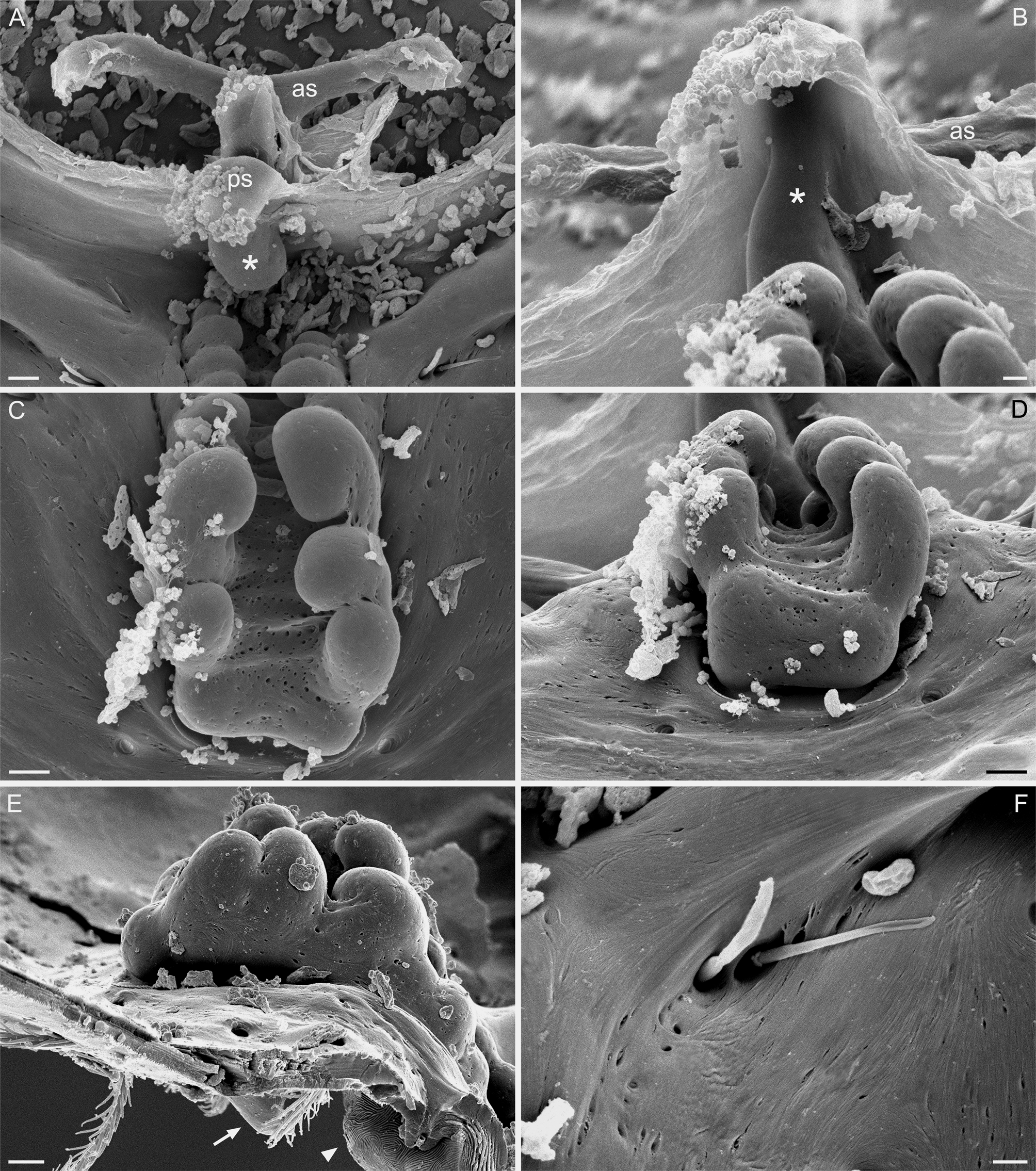
14D), legs III-IV with two receptors. Genitalia: External genitalia as in Fig. 15 View FIGURE 15 A–D, with a prominent scape-like structure; in posterior part of scape a short longitudinal groove (arrow in Fig. 15 View FIGURE 15 B). Posterior of the scape a small cone-like structure ( Fig. 15 View FIGURE 15 B); in anterior surface of cone a narrow groove ( Fig. 15 View FIGURE 15 D; this groove may be a continuation of the groove in the scape). Internal genitalia consisting of apodemes, two uterine sclerites, and a genital duct ( Fig. 15 View FIGURE 15 F). Anterior uterine sclerite T-shaped ( Fig. 16 View FIGURE 16 A). Posterior uterine sclerite plate-like. Proximal part of genital duct relatively broad, with two rows of finger-like protrusions ( Fig. 16 View FIGURE 16 C–E); distal part of duct narrow, ascending along posterior uterine sclerite, lacking protrusions ( Fig. 16 View FIGURE 16 A, B). On either side of duct a pair of small tubular structures, presumably glands ( Figs. 15 View FIGURE 15 F, 16F); these glands open to the outside via small pores situated lateral of the scape (arrowheads in Fig. 15 View FIGURE 15 B).
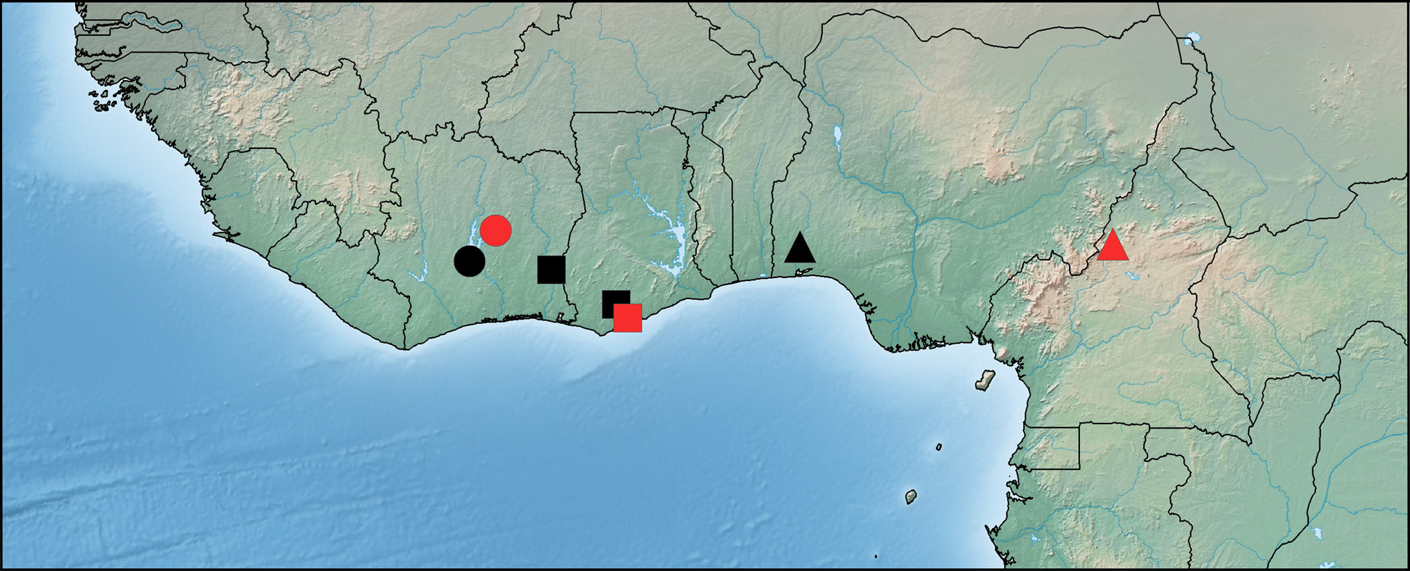
Distribution. Known only from the type locality ( Fig. 1 View FIGURE 1 ).


No known copyright restrictions apply. See Agosti, D., Egloff, W., 2009. Taxonomic information exchange and copyright: the Plazi approach. BMC Research Notes 2009, 2:53 for further explanation.