
Aphelonema brevata Caldwell, 1945
|
publication ID |
https://doi.org/ 10.5852/ejt.2020.717.1097 |
|
publication LSID |
lsid:zoobank.org:pub:A03063E4-23C7-4084-BDB6-7495687FFDC5 |
|
DOI |
https://doi.org/10.5281/zenodo.4330349 |
|
persistent identifier |
https://treatment.plazi.org/id/4C61685F-FF9D-5F28-A429-904745A1FBB9 |
|
treatment provided by |
Valdenar |
|
scientific name |
Aphelonema brevata Caldwell, 1945 |
| status |
|
Aphelonema brevata Caldwell, 1945 View in CoL
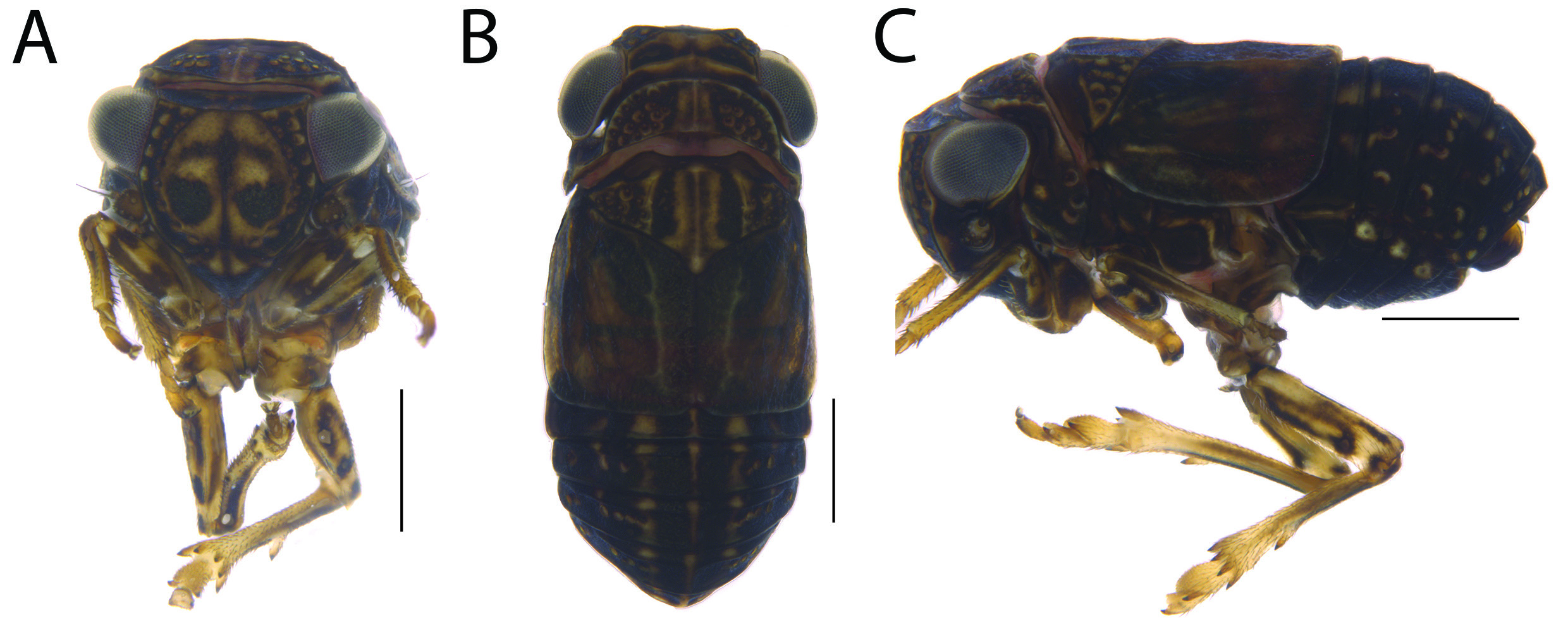
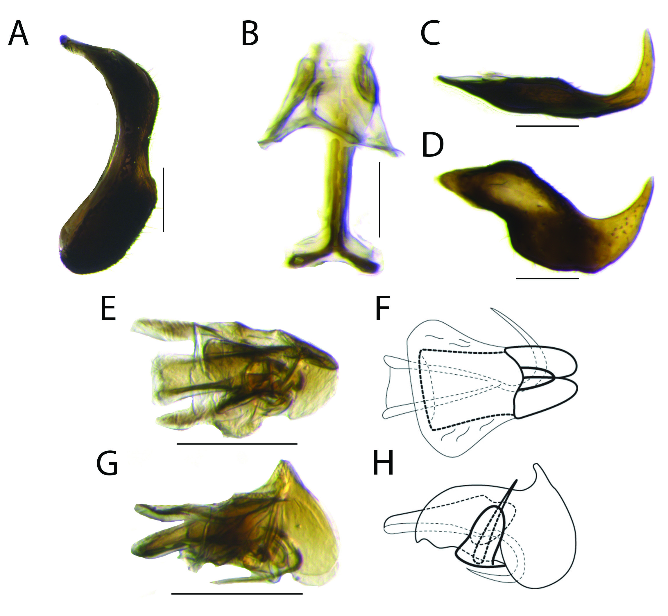
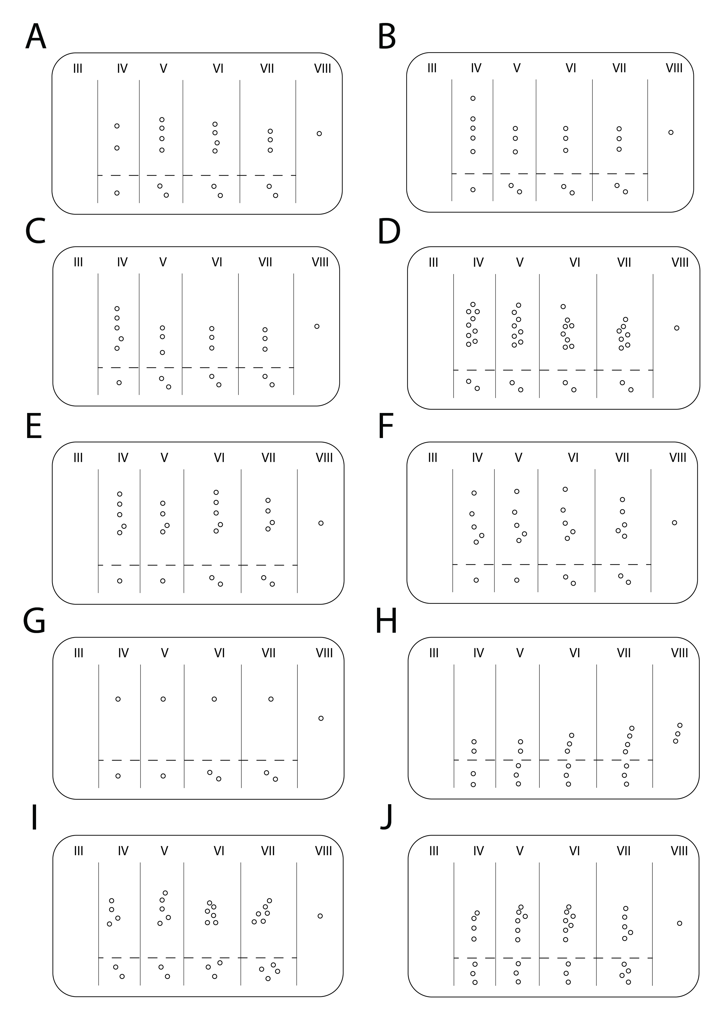
Figs 1–2 View Fig View Fig , 26A View Fig
Aphelonema brevata Caldwell, 1945: 94 View in CoL , pl. I: fig. 6.
Aphelonema (Protrocha) brevata View in CoL – Emeljanov 1996: 835 (proposed new subgenus for Aphelonema View in CoL ). Protrocha brevata – Gnezdilov 2013: 212.
Diagnosis
Body mainly light brown with some regions dark brown ( Fig. 1 View Fig ); central plate dark brown with pair of swirl-shaped light brown maculae ( Fig. 1A View Fig ); sides of frons with two rows of sensory pits on each side ( Fig. 1C View Fig ); clypeus not swollen ( Fig. 1A, C View Fig ), with median carina ( Fig. 1A View Fig ); lateral lobe of pronotum with approximately three sensory pits arranged in group ( Fig. 1C View Fig ); abdominal tergites ( Fig. 1C View Fig , 26A View Fig ) with row of sensory pits followed by single isolated ventral sensory pit (tergite IV) or by isolated pair of diagonally aligned ventral sensory pits (tergites V to VII).
Material examined
Holotype
MEXICO • ♂; Toluca Rd. ; 24 Nov. 1938; J.S. Caldwell leg.; NMNH USNMENT 01513543 (based on photographs).
Other material
MEXICO • 1 ♂; “MICH” [Michoacán], E Morelia, route 15, Km 18; 19.68392º N, 101.00981º W; 2050 m a.s.l.; Oct. 2005; R. Rakitov leg.; sweep; DNA voucher ENT4920; INHS GoogleMaps .
Description
BODY LENGTH. Male = 2.4 mm.
COLORATION. Body mainly light brown with some regions dark brown ( Fig. 1 View Fig A–C). Vertex ( Fig. 1B View Fig ) with pair of dark brown maculae. Central plate ( Fig. 1A View Fig ) dark brown with pair of swirl-shaped light brown maculae; median carina dark brown. Gena and lateral lobe of pronotum ( Fig. 1C View Fig ) dark brown. Clypeus dark brown with pair of light brown triangular maculae ( Fig. 1A View Fig ). Pronotum ( Fig. 1B View Fig ) dark brown with three median light brown stripes connected to stripes on mesonotum. Mesonotum ( Fig. 1B View Fig ) with additional pair of lateral light brown stripes. Forewing ( Fig. 1B View Fig ) brown. Legs ( Fig. 1A, C View Fig ) light brown with several dark brown maculae. Abdomen ( Fig. 1 View Fig B–C) dark brown with three median light brown interrupted stripes in dorsal view.
HEAD AND THORAX. Vertex ( Fig. 1B View Fig ) hexagonal, shorter than half its width, almost as long as pronotum; posterior margin slightly elevated. Frons ( Fig. 1A View Fig ) with median carina and pair of sublateral carinae; sublateral carinae convergent and almost fused to each other ventrally ( Fig. 1A View Fig ); central plate ( Fig. 1A View Fig ) as long as wide at widest portion, not visible in dorsal view ( Fig. 1B View Fig ), not extending anteriorly beyond sublateral carinae in lateral view ( Fig. 1C View Fig ); sides of frons partially visible in frontal view ( Fig. 1A View Fig ) and almost fused above clypeus, with two rows of sensory pits on each side in lateral view ( Fig. 1C View Fig ): anterior row with eight sensory pits, ventral pair slightly displaced; posterior row with four sensory pits, most ventral one slightly isolated. Clypeus ( Fig. 1C View Fig ) not swollen, with median carina. Ocelli absent. Eye oblong. Antenna short, with several small circular structures visible on pedicel. Pronotum ( Fig. 1B View Fig ) semicircular, shorter than half its width, with median carina; median portion of disc without sensory pits; lateral portion of disc with 14 to 15 sensory pits; lateral lobe of pronotum ( Fig. 1C View Fig ) with three sensory pits arranged in group. Mesonotum ( Fig. 1B View Fig ) with median carina and pair of lateral carinae; region between lateral carinae depressed and without sensory pits; region laterad of lateral carinae with nine to 11 sensory pits. Brachypterous, with reduced venation. Legs simple, with carinae, setose; tibia III with single median spine.
ABDOMEN. Terga with longitudinal carina. Tergite III ( Figs 1C View Fig , 26A View Fig ) without sensory pits. Tergite IV ( Figs 1C View Fig , 26A View Fig ) with row of two sensory pits followed by single isolated ventral one. Tergites V and VI ( Figs 1C View Fig , 26A View Fig ) with row of four sensory pits followed by isolated ventral pair aligned diagonally. Tergite VII ( Figs 1C View Fig , 26A View Fig ) with row of three sensory pits followed by isolated ventral pair aligned diagonally. Tergite VIII with one sensory pit ( Fig. 26A View Fig ).
MALE TERMINALIA. Pygofer ( Fig. 2A View Fig ) with anterior margin deeply concave; posterior margin with dorsal third rounded, middle third concave, ventral third wide and rounded. Connective ( Fig. 2B View Fig ) inverted Y-shaped, with support bridge with dorsal flap. Style ( Fig. 2 View Fig C–D) hook-like; anterior portion pointed; dorsal margin with slight protuberance on median third ( Fig. 2D View Fig ); caudal portion strongly curved anterodorsally in lateral view ( Fig. 2D View Fig ) and mesad in dorsal view ( Fig. 2C View Fig ); ventral margin ( Fig. 2D View Fig ) truncate between anterior and middle portion, irregularly rounded posteriorly; middle portion longer than high, setose; apex serrated ( Fig. 2D View Fig ). Phallobase ( Fig. 2 View Fig E–H) sclerotized, symmetrical, with two defined lobes; apex with pair of lobes fused in dorsal view ( Fig. 2 View Fig E–F) and rounded in lateral view ( Fig. 2 View Fig G–H); sides expanded and rounded at half-length of aedeagus in dorsal view ( Fig. 2 View Fig E–F); with dorsal process near to apex in lateral view ( Fig. 2 View Fig G–H), surrounding almost all aedeagus length. Aedeagus ( Fig. 2 View Fig E–F) apex narrow and open dorsally, with pair of aedeagal hooks, one curved to side of aedeagus and visible in dorsal view ( Fig. 2 View Fig E–F), another visible in lateral view ( Fig. 2 View Fig G–H) and curved ventrally to other side of aedeagus. Suspensorium V-shaped.
Remarks
This species was originally placed in Aphelonema but later transferred to Protrocha by Emeljanov (1996). However, the species treated herein shares more characteristics of Aphelonema , according to the diagnostic features given by Emeljanov (1996, see Discussion), such as (1) lateral lobe of pronotum with no fewer than two, but usually with three or more sensory pits ( Fig. 1C View Fig ); (2) sides of frons in upper half with two parallel rows of sensory pits ( Fig. 1C View Fig ); and (3) abdomen with sensory pits aligned in one row and with an isolated pair of ventral sensory pits ( Fig. 26A View Fig ). Based on this combination of characters we propose that this species returns to its original combination. Its original description is short, includes only a superficial illustration of the male terminalia, and does not include information about abdominal sensory pits or female terminalia. The single male specimen at hand was identified based on the original description, illustrations of male terminalia made by Caldwell (1945), and photographs of the head, thorax and male terminalia of the holotype. However, the abdomen of the holotype was lost so redescription of the distribution of sensory pits on this structure was based on the specimen at hand. Unfortunately, the anal tube of the studied specimen was damaged during dissection.
No known copyright restrictions apply. See Agosti, D., Egloff, W., 2009. Taxonomic information exchange and copyright: the Plazi approach. BMC Research Notes 2009, 2:53 for further explanation.
|
Kingdom |
|
|
Phylum |
|
|
Class |
|
|
Order |
|
|
Family |
|
|
Genus |
Aphelonema brevata Caldwell, 1945
| de Freitas, Abner S., Dietrich, Christopher H. & Takiya, Daniela M. 2020 |
Aphelonema (Protrocha) brevata
| Gnezdilov V. M. 2013: 212 |
| Emeljanov A. F. 1996: 835 |
Aphelonema brevata
| Caldwell J. S. 1945: 94 |