
Apobaetis pucupi, Cruz, Paulo Vilela & De-Souza, Marcia Regina, 2014
|
publication ID |
https://doi.org/ 10.11646/zootaxa.3866.4.9 |
|
publication LSID |
lsid:zoobank.org:pub:D1DB8398-EC11-41FE-96FF-AEC5F475E696 |
|
DOI |
https://doi.org/10.5281/zenodo.6144409 |
|
persistent identifier |
https://treatment.plazi.org/id/524487D3-FFBE-1271-ABA7-9258CB7AF986 |
|
treatment provided by |
Plazi |
|
scientific name |
Apobaetis pucupi |
| status |
sp. nov. |
Apobaetis pucupi sp. nov.
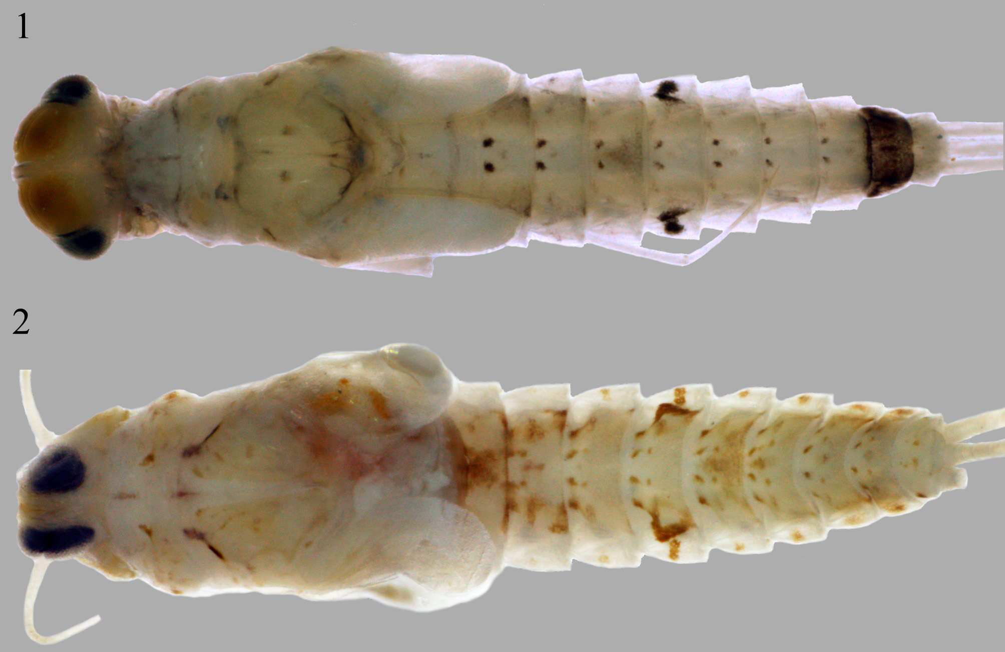
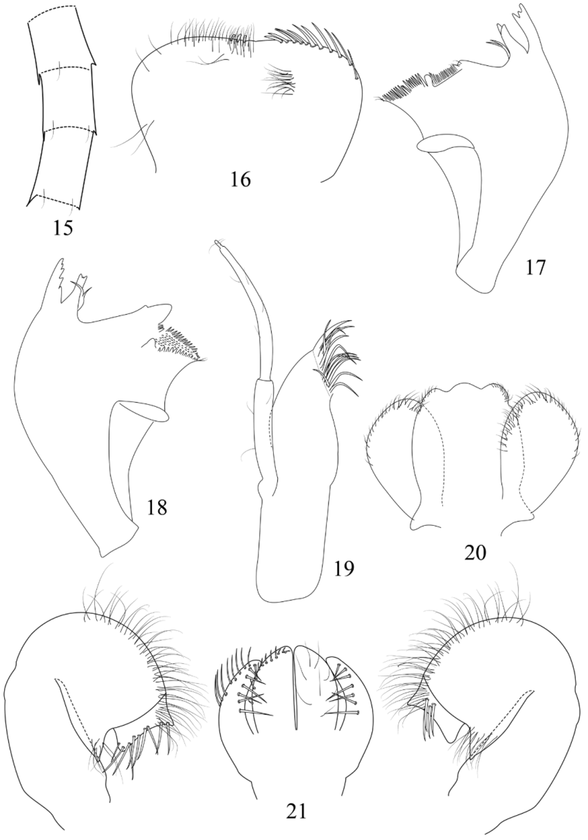
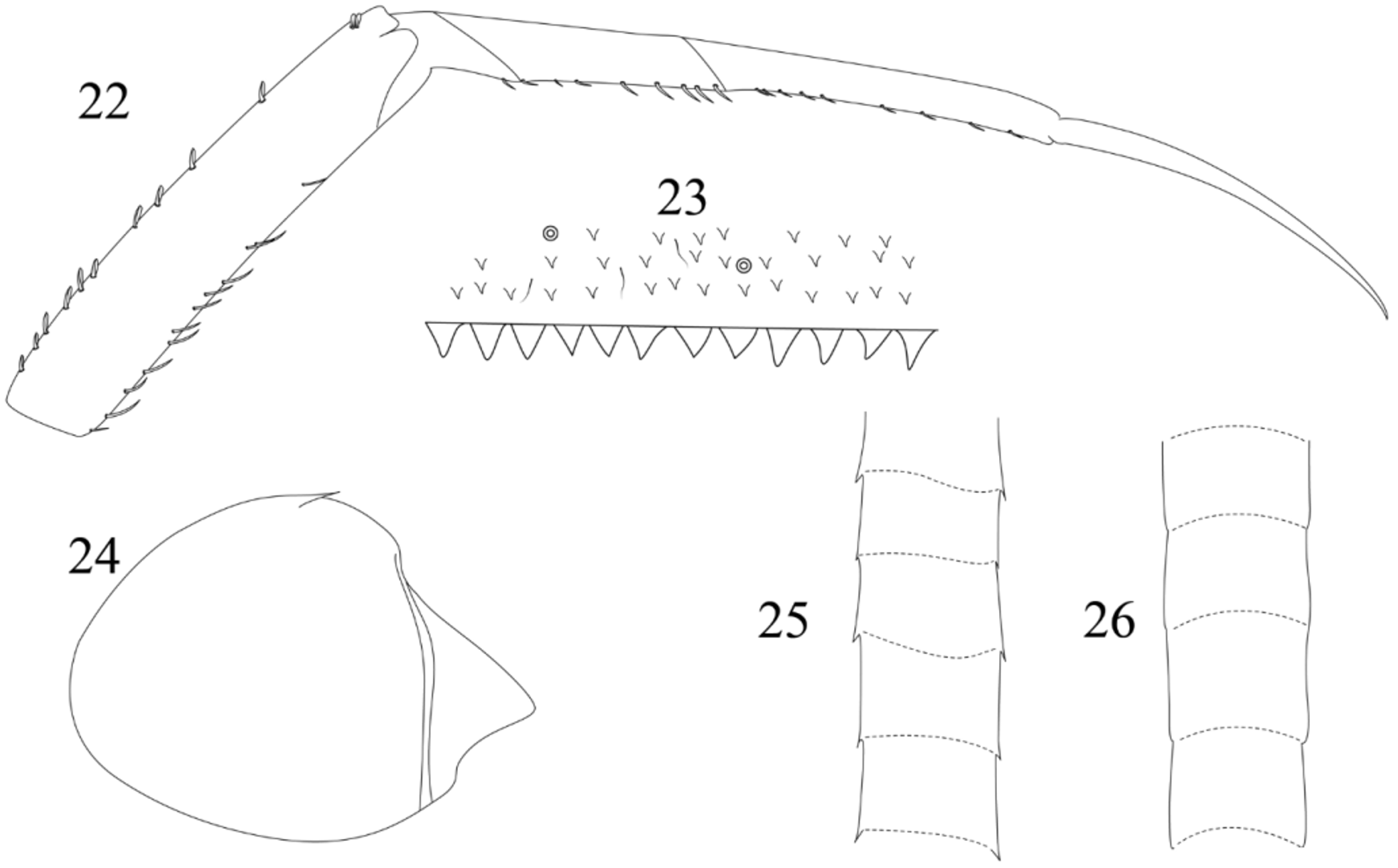
( Figures 2 View FIGURES 1 – 2 , 15–26 View FIGURES 15 – 21 View FIGURES 22 – 26 )
Diagnosis. Mature nymph. 1) distal margin of labrum medially with spatulate setae; 2) maxillary palp 1.50× length of galea-lacinia ( Fig. 19 View FIGURES 15 – 21 ); 3) segment II of maxillary palp with an apical constriction ( Fig. 19 View FIGURES 15 – 21 ); 4) apical margin of lingua with three lobules ( Fig. 20 View FIGURES 15 – 21 ); 5) segment II of labial palp with pointed and apically directed distomedial projection ( Fig. 21 View FIGURES 15 – 21 ); 6) segment III rectangular, length 0.40× width ( Fig. 21 View FIGURES 15 – 21 ); 7) tarsal claw 1.54× length of tarsus ( Fig. 21 View FIGURES 15 – 21 ); 8) posterior margin of abdominal terga with pointed spines ( Fig. 22 View FIGURES 22 – 26 ); 9) paraproct with one marginal spines, posterolateral extension without spines ( Fig. 24 View FIGURES 22 – 26 ).
Description. Mature female nymph. Length of body: 3.00 mm; cerci, terminal filament and antenna broken. Body coloration ( Fig. 2 View FIGURES 1 – 2 ). Head. Colorations: light yellow. Antenna light yellow. Turbinate portion of compound eyes light yellow. Thorax. Light yellow. Foreleg. Femur, tibia and tarsus light yellow. Femur with one light brown mark at middle on anterior surface and one light brown mark on apex dorsal margin. Tibia with one light brown mark on base ventral margin. Abdomen. Terga light yellow, segments II, III and VI brown medially, segment V brown near lateral margin, all terga with four spots on anteromedial margin and one mark laterally ( Fig. 2 View FIGURES 1 – 2 ).
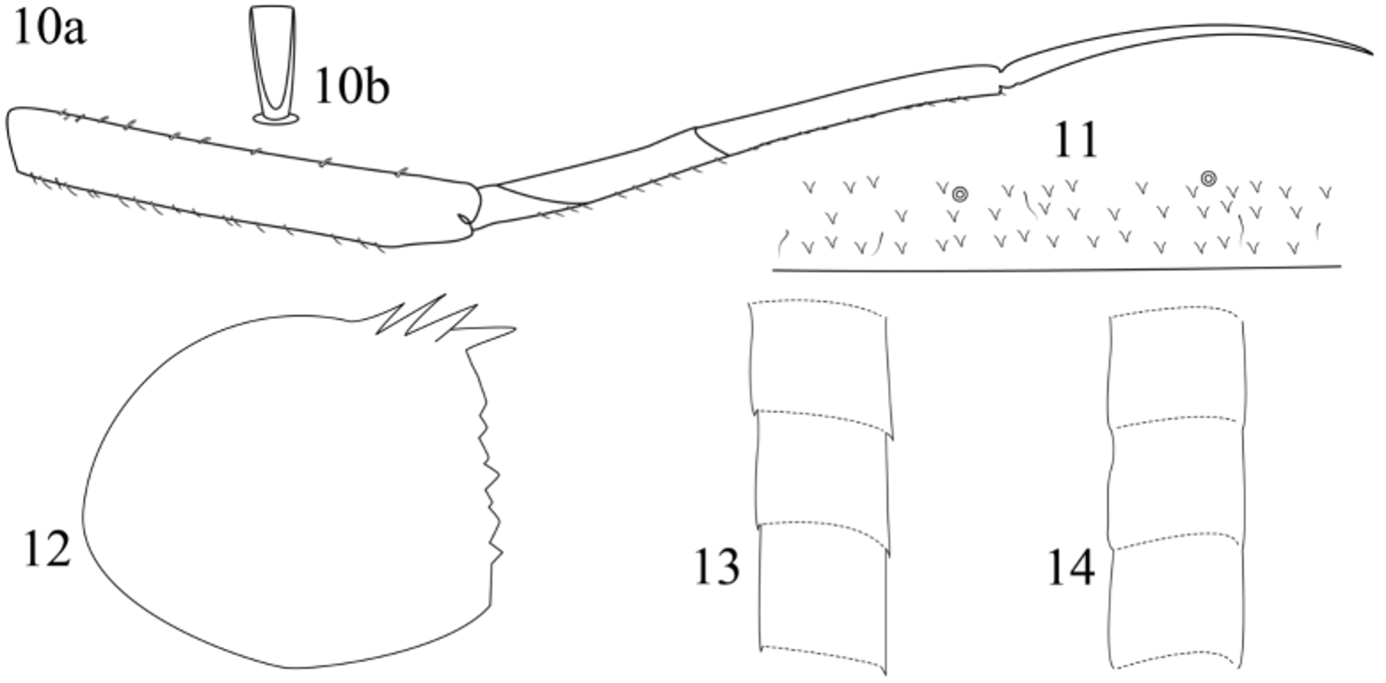
Body morphology. Head. Antenna with minute spines and fine, simple setae on apex of each segment ( Fig. 15 View FIGURES 15 – 21 ). Frons with two keels. Labrum ( Fig. 16 View FIGURES 15 – 21 ). Rectangular, broader than long; length about 0.69× maximum width; distal margin with shallow medial emargination; ventral surface with robust spine-like setae on anterolateral and distal margin; dorsal surface with three to four short and spatulate setae medially near distal margin; dorsal surface near distal margin covered by long and thin setae. Right mandible ( Fig. 17 View FIGURES 15 – 21 ). Incisors deeply cleft in two sets; outer and inner set of incisors respectively with 3 and 2 denticles; prostheca slender, bifurcated at middle; margin between prostheca and mola concave; tuft of spine-like setae at base of mola present; denticles of mola not constricted; apex of mola with two simple setae; lateral margin convex. Left mandible ( Fig. 18 View FIGURES 15 – 21 ). Incisors deeply cleft in two sets; outer and inner set of incisors respectively with 5 and 3 denticles; prostheca robust, bifid at middle, inner lobe slender, outer lobe robust; margin between prostheca and mola concave; tuft of spine-like setae at base of mola absent; subtriangular process wide; denticles of mola not constricted; lateral margin convex. Hypopharynx ( Fig. 20 View FIGURES 15 – 21 ). Lingua subquadrangular with three lobules, without apical tuft of setae and slightly longer than superlingua; superlingua not expanded; short, fine, simple setae scattered over distal margin of lingua and superlingua. Maxilla ( Fig. 19 View FIGURES 15 – 21 ). Maxillary palp long, 1.50× length of galea-lacinia; segment II 1.40× length of segment I, apex with constriction; maxillary palp with fine and simple setae scattered over surface. Labium ( Fig. 21 View FIGURES 15 – 21 ). Glossa basally broad, narrowing apically and longer than paraglossa; inner margin bare; apex with three short spine-like setae; outer margin with eight spine-like setae; ventral surface covered with thin and long setae. Paraglossa curved inward; apex bare; outer margin with one row of eight robust spine-like setae; dorsal surface with one longitudinal row of seven robust spine-like setae near inner margin; ventral surface with one longitudinal row of seven robust spine-like setae at middle. Labial palp with segment I 0.87× length of segments II and III combined; segment I covered with micropores; segment II with pointed and apically directed distomedial projection, outer margin and distomedial projection covered with fine, long and simple setae; inner margin bare; segment III rectangular, length 0.40× width, covered with fine, long and simple setae on outer margin, ventral surface with four robust spine-like setae near outer margin, distal margin with one row of ten robust spine-like setae. Thorax. Foreleg ( Fig. 22 View FIGURES 22 – 26 ). Ratio 1.8:1:1.1:1.3. Forefemur. Length about 4.90× maximum width; dorsally with row of 12 short concave and apically straight setae; apex with two concave and apically rounded setae (similar to Fig. 11 View FIGURES 10 – 14 a, but with apex rounded instead); ventrally with row of elongated spine-like setae. Tibia. Dorsally bare; ventrally with one row of nine short spine-like setae. Tibio-patelar suture present. Tarsus. Dorsally bare; ventrally with one row of ten short spine-like setae. Tarsal claws 1.54× longer than tarsus, row of denticles absent. Abdomen. Terga surface covered by scale-like triangular spines, micropores and short, fine and simple setae; posterior margin with regular spines ( Fig. 23 View FIGURES 22 – 26 ). Sterna white. Gills lost. Paraproct with one marginal spine, posterolateral extension without spines ( Fig. 23 View FIGURES 22 – 26 ). Cerci with small lateral spines on all segments ( Fig. 25 View FIGURES 22 – 26 ), terminal filament without spines ( Fig. 26 View FIGURES 22 – 26 ).
Etymology. The specific name is derived from the Tupi-Guarani roots Pucu —long and Pi —claw. Tupiguarani is a language spoken by an indigenous tribe that inhabits the Brazilian litoral.
Material examined. Holotype: nymph on slide, BRAZIL, state of Minas Gerais, Lima Duarte, road to Ibitipoca after bridge, 26.x.2011, S21º 47’55.0’’ / W043º49’27.7’’, Cruz P.V. and De-Souza M.R. (colls.).
No known copyright restrictions apply. See Agosti, D., Egloff, W., 2009. Taxonomic information exchange and copyright: the Plazi approach. BMC Research Notes 2009, 2:53 for further explanation.