
Asterocheres astroidicola, Conradi & Bandera & López-González, 2006
|
publication ID |
https://doi.org/ 10.1080/00222930600774210 |
|
persistent identifier |
https://treatment.plazi.org/id/305E8783-FFA1-DC2F-FE5A-44A682FAFB92 |
|
treatment provided by |
Carolina |
|
scientific name |
Asterocheres astroidicola |
| status |
sp. nov. |
Asterocheres astroidicola n. sp.
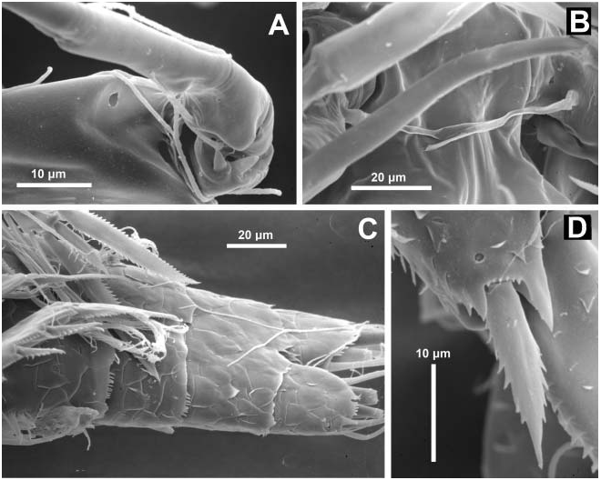
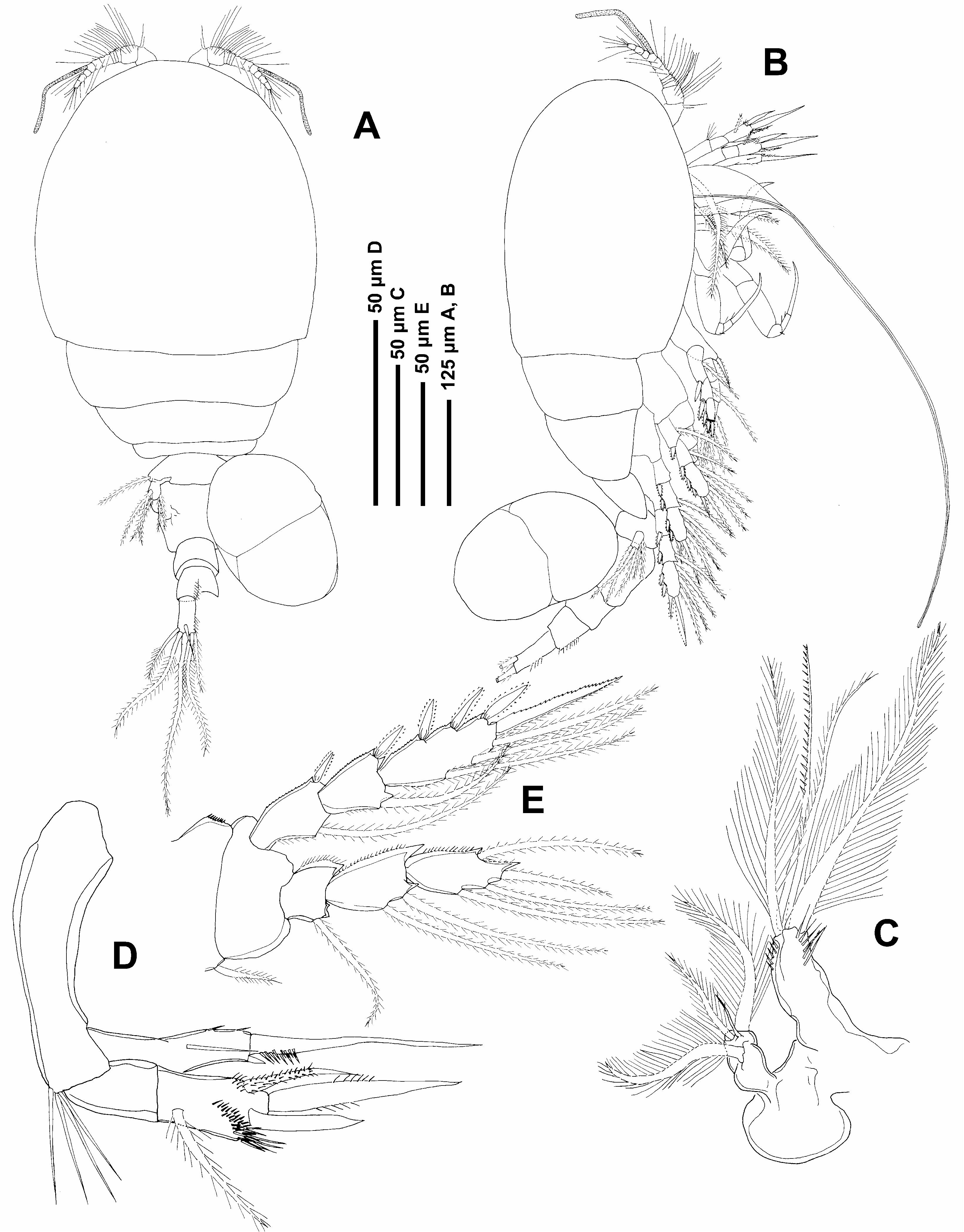
( Figures 6–10 View Figure 6 View Figure 7 View Figure 8 View Figure 9 View Figure 10 )
Material examined
MNCN 20.04/7578 holotype, one adult female associated with the scleractinian Astroides calycularis, Tarifa Island, 36 u 019N, 5 u 379W, 10–20 m depth, July 1999; MNCN 20.04/ 7579 allotype, one adult male, with the same sampling data as the holotype; BEIM ( COP 501 View Materials ), three adult females, with the same sampling data as the type material .
Description
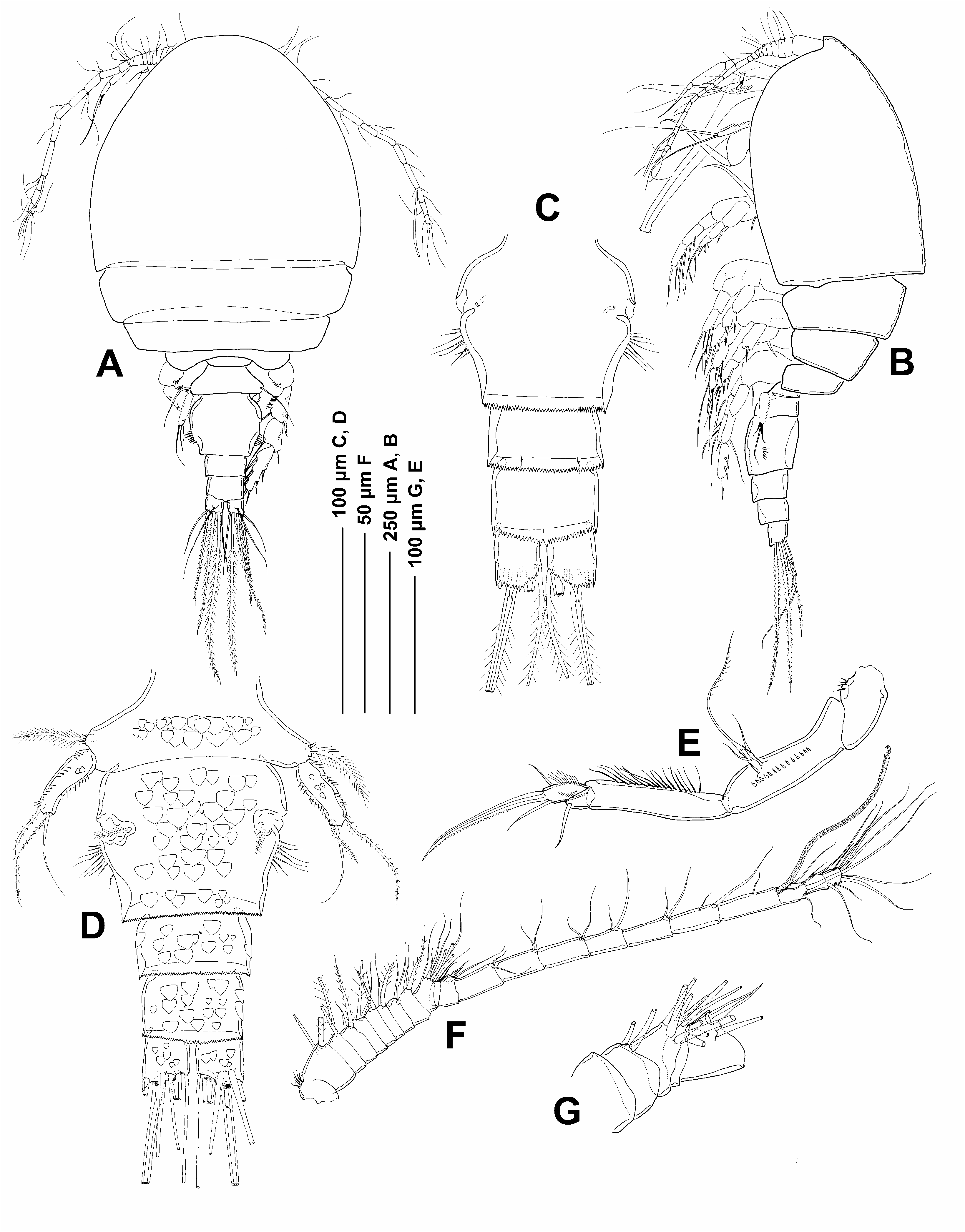
Female. Body cyclopiform, slender with cephalothorax oval and cylindrical urosome ( Figure 6A, B View Figure 6 ). Body length 750 Mm (650–790 Mm) and width 420 Mm (390–450 Mm), based on four specimens. Ratio of length to width of prosome 1.19:1. Ratio of length of prosome to that of urosome 2.1:1. Prosome comprising cephalothorax fully incorporating first pedigerous somite and three free pedigerous somites. Urosome four-segmented comprising leg 5-bearing somite, genital double somite and two free abdominal subquadrate somites. Somite bearing leg 5 ( Figure 6D View Figure 6 ) wider than long, with some spinules on its lateral surface. Dorsal surface of free abdominal somites and posterior part of double somite ornamented with large, flattened epicuticular scales, arranged in irregular, overlapping rows ( Figures 6D View Figure 6 , 10C View Figure 10 ). Posterior margins of all somites ornamented with hyaline frills with more or less serrated margins. Genital double somite about 1.25 times wider than long, bearing genital apertures, paired gonopores located laterally. Lateral margin of double somite ornamented with fringe of long spinules located about midway along double somite, posterior to gonopores level ( Figure 6C View Figure 6 ). Each genital area armed with one plumose seta. Integumental pores and sensilla present on urosomal somites ( Figure 6C View Figure 6 ). Caudal rami slightly longer than wide, ornamented dorsally with epicuticular scales; armed with six setae.
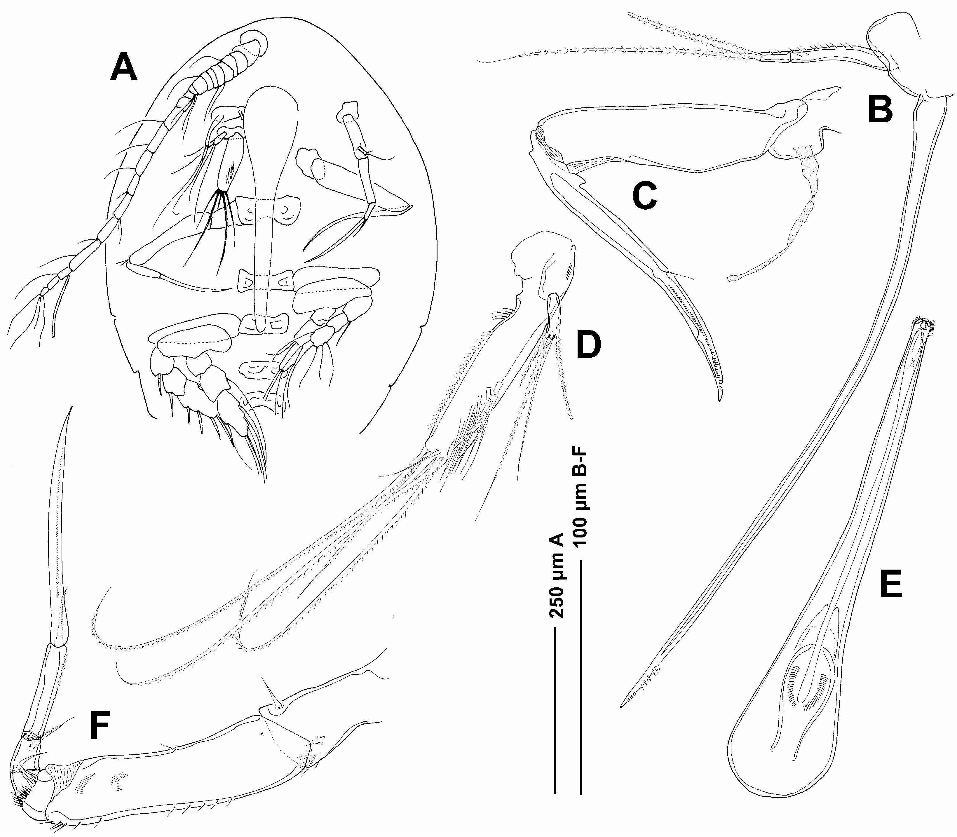
Antennule 21-segmented ( Figures 6F, 6G View Figure 6 ); segmental fusion pattern as follows: 1(I), 2(II), 3(III), 4(IV), 5(V), 6(VI), 7(VII), 8(VIII), 9(IX–XII), 10(XIII), 11(XIV), 12(XV), 13(XVI), 14(XVII), 15(XVIII), 16(XIX), 17(XX), 18(XXI), 19(XXII–XXIII), 20(XXIV– XXV), 21(XXVI–XXVIII). Segments 1–8 each with two setae; segment 9 with eight setae; segment 10–17 each with two setae; segment 18 with two setae plus an aesthetasc; segment 19 with two setae; segment 20 with three setae; segment 21 with seven setae. Segment 10 (XIII) ( Figure 6G View Figure 6 ) reduced, partly overlapped by distal expansion of compound segment 9 (IX–XII). Antenna ( Figure 6E View Figure 6 ) biramous, 380 Mm long with a small unarmed coxa ornamented with tuft of spinules and a large unarmed basis with fine spinule row. Exopod small, one-segmented, bearing two lateral and one apical setae. Endopod three-segmented; first segment elongated, ornamented with a row of long spinules; second segment produced distally on medial side but articulating with third segment proximally on lateral side and armed with one smooth seta, third segment armed with two short naked setae, and large terminal claw also ornamented with rows of fine spinules. Mandible ( Figure 7B View Figure 7 ) with twosegmented palp and stylet-like gnathobase. Stylet with denticulate margin subapically and located in oral cone. First segment of palp slender, unarmed but ornamented with a row of spinules laterally; second segment with two terminal plumose setae. Oral cone long and slender, 293 Mm long, formed by labrum and labium joined laterally, reaching nearly to the posterior margin of intercoxal plate of leg 2 ( Figure 7A, E View Figure 7 ). Maxillule bilobed ( Figure 7D View Figure 7 ); praecoxal endite more than four times longer than palp and more than three times wider than palp. Praecoxal endite armed with five distal setae (one of them smooth and short), ornamented with patch of long spinules distally and row of shorter spinules laterally. Palp armed with three terminal and one subterminal setae. Maxilla three-segmented ( Figure 7C View Figure 7 ). Praecoxa bearing long flaccid element medially ( Figure 10B View Figure 10 ), possibly an aesthetasc; coxa unarmed and claw-like basis bearing small hyaline process proximally in axil; armed with one very small seta at about one-half its length laterally; claw margins with row of minute spinules distally. Maxilliped five-segmented ( Figure 7F View Figure 7 ), first segment with small inner distal seta and patch of spinules. Second segment elongate and slender, with minute hyaline seta at midway of inner margin and rows of fine spinules distally. Third segment short, ornamented with two minute smooth setae and a patch of fine spinules medially. Fourth segment armed with two short setae, one of them smooth. Fifth segment with one terminal plumose seta and one claw-like seta, 105 Mm long, ornamented with a lateral row of minute spinules.
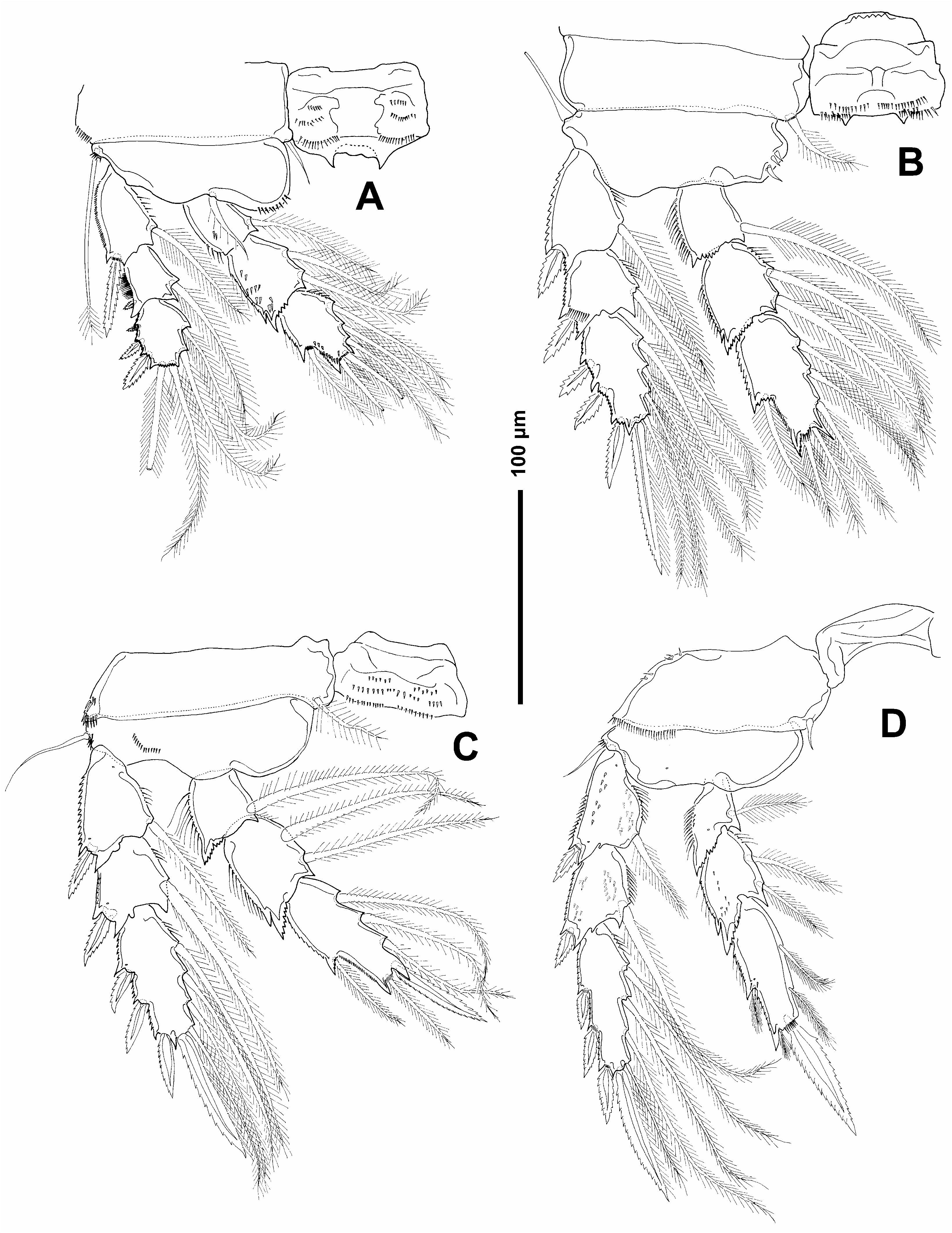
Legs 1–4 biramous ( Figure 8A–D View Figure 8 ), with three-segmented protopods and threesegmented rami. Intercoxal sclerite present on legs 1–4, ornamented with patches of spinules on legs 1–3 and pair of processes on legs 1 and 2. Formula for armature as follows:
Coxae of all legs ornamented with spinule rows laterally as figured. Inner coxal seta plumose in legs 1–3 and reduced and naked in leg 4. Except on leg 2, basal seta of all legs long and naked. Posterior surface of legs 1–4 ornamented with flattened epicuticular scales, arranged in irregular, overlapping rows ( Figure 10D View Figure 10 ). Lateral margins of exopodal segments with minute serrations; lateral margins of endopodal segment with row of setules.
Leg 5 ( Figure 6D View Figure 6 ) with elongated free segment. Three terminal setae, two of them plumose. Few minute spinules on both sides of free segment. Adjacent seta on body somite plumose. Leg 6 ( Figure 6D View Figure 6 ) represented by seta on genital area.
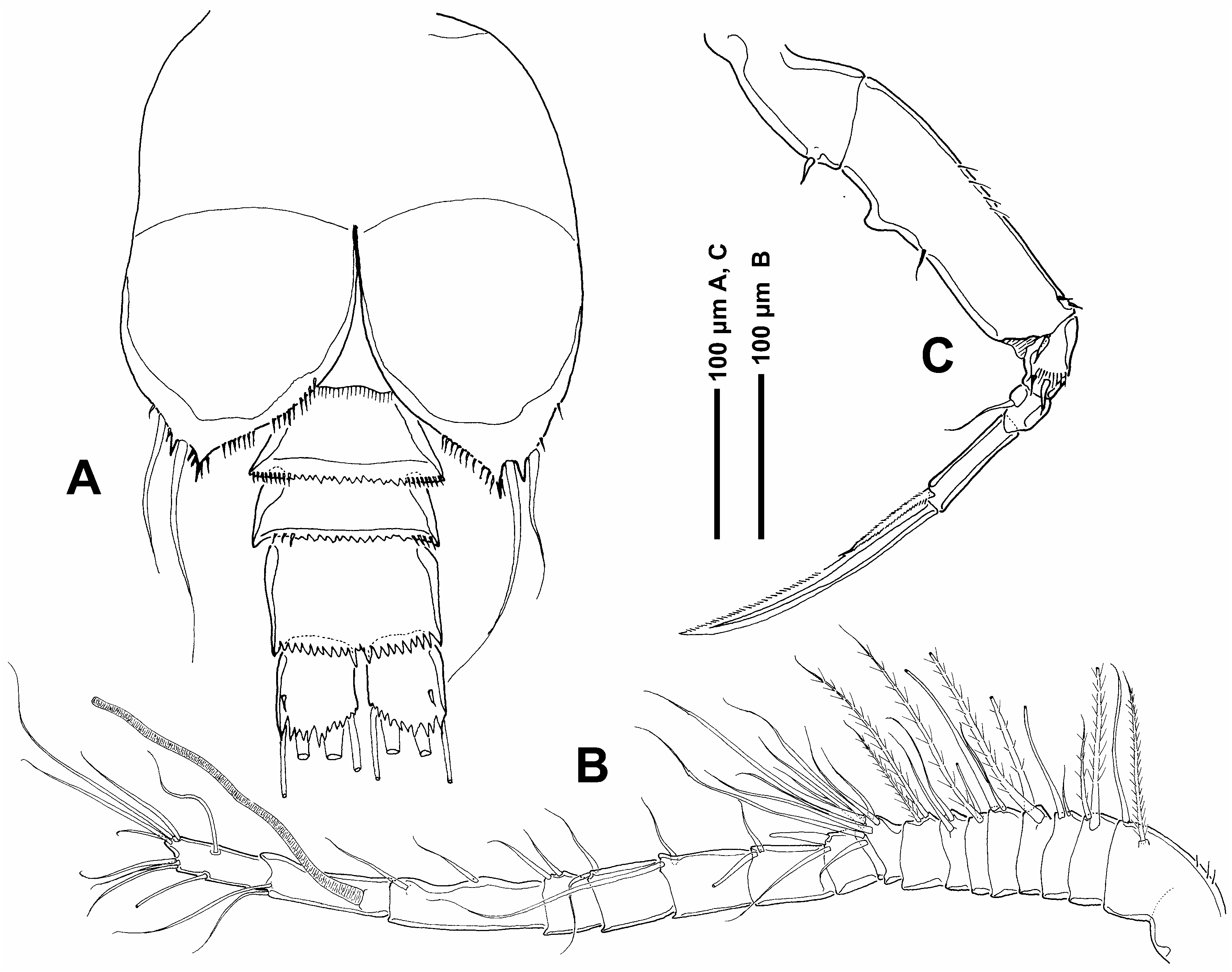
Male. Only one adult male specimen but seriously damaged. Body cyclopiform, slightly more slender than in female, with oval-shaped cephalothorax and cylindrical urosome. Urosome five-segmented, comprising pedigerous somite 5, genital somite and three free abdominal somites. Posterior margins of all somites ornamented with hyaline frills with more or less serrated free margins. Genital somite slightly longer than wide; bearing genital apertures postero-laterally on ventral surface ( Figure 9A View Figure 9 ). Appendages as in female except antennule, maxilliped and leg 6. Leg 5 expected to be different to that of the female but in this specimen it has not been observed due to the fact that it was not complete.
Antennule ( Figure 9B View Figure 9 ) 18-segmented, geniculate; segmental fusion pattern as follows: 1(I), 2(II), 3(III), 4(IV), 5(V), 6(VI), 7(VII), 8(VIII), 9(IX–XII), 10(XIII), 11(XIV), 12(XV), 13(XVI), 14(XVII), 15(XVIII), 16(XIX–XX), 17(XXI–XXIII), 18(XXIV– XXVIII). Geniculation located between segments 16(XIX–XX) and 17(XXI–XXIII). Segments 1–8 each with two setae; segment 9 with eight setae; segment 10–16 each with two setae; segment 17 with two setae plus an aesthetasc; segment 18 with nine setae. Segment 10 (XIII) reduced, partly overlapped by distal expansion of compound segment 9 (IX–XII). Maxilliped ( Figure 9C View Figure 9 ) five-segmented, similar to that of female but second segment with medial proximally directed thorn-like process.
Leg 6 ( Figure 9A View Figure 9 ) forming large opercular plates closing off genital apertures, armed with two smooth setae, ornamented with rows of fine spinules.
Etymology
The name of the species, astroidicola , is a combination of Astroides , the host name, and - icola from the Latin meaning ‘‘inhabiting’’, alluding to the relationship between the copepod and the coral.
Remarks
Eleven valid species of Asterocheres Boeck, 1859 have been reported as possessing a 21- segmented antennule in the female. Three of them, A. suberitis Giesbrecht, 1897 , A. violaceus Claus, 1889 , and A. minutus Claus, 1889 , were collected from NE Atlantic and Mediterranean coasts and another eight from other areas, namely A. bulbosus Malt, 1991 from Hong Kong, A. jeanyeatmanae Yeatman, 1970 from Chesapeake Bay, A. tenuicornis Brady, 1910 from Antarctica, A. reginae Boxshall and Huys, 1994 from Belize, A. flustrae Ivanenko and Smurnov, 1997 from the White Sea, A. lunatus Johnsson, 1998 from Brazil, A. urabensis Kim, 2004 from the Pacific Coast of Panama, and A. hirsutus Bandera et al., 2005 from Antarctica. Except for A. urabensis and A. hirsutus , all of these species are characterized by having a relatively short siphon which does not extend beyond the insertion of the maxillipeds ( Boxshall and Huys 1994). Asterocheres urabensis , A. hirsutus , and the new species, A. astroidicola , possess a longer oral cone. In the first two species it reaches to the insertion of leg 1 and in the latter species the oral cone enlarges nearly to the intercoxal plate of leg 2. However, in a detailed comparison of A. astroidicola and the remaining species with a 21-segmented antennule, a number of additional differences can be discussed. Firstly, A. bulbosus , A. violaceus , and A. minutus have a one-segmented mandibular palp in contrast to the two-segmented mandibular palp present in the new species. As for A. tenuicornis , it differs from A. astroidicola by its very elongated caudal rami which is almost six times longer than wide.
From the point of view of body shape, A. reginae and A. jeanyeatmanae differ from the new species by their dorso-ventrally flattened prosome. Moreover, A. astroidicola can be distinguished from A. reginae by having one additional seta on the inner lobe of the maxillule and from A. jeanyeatmanae by having one distal additional seta on the free segment of the fifth leg. The extremely large inner lobe of the maxillule, at least four times longer than outer lobe, of A. astroidicola and A. hirsutus serves to separate them from the remaining four species, A. flustrae , A. suberitis , A. lunatus , and A. urabensis , which have a much smaller endite in the maxillule. Asterocheres suberitis and A. lunatus possess four terminal setae on the inner lobe of the maxillule in contrast with the five setae present in A. astroidicola . Asterocheres flustrae has a six-segmented maxilliped instead of the five-segmented maxilliped present in A. astroidicola . The caudal rami is 2.5 times longer than wide in A. hirsutus , whereas in A. astroidicola it is only slightly longer than wide.
Concerning hosts, except for A. suberitis and A. tenuicornis whose hosts are unknown, A. flustrae lives in association with the bryozoan Flustra foliacea L. and A. minutus and A. violaceus live in association with an echinoderm. The remaining species of this group, except for A. urabensis , are symbionts on sponges. Asterocheres urabensis and A. astroidicola are associated with scleractinian coral, Pocillopora damicornis (L.) and Astroides calycularis (Pallas, 1766) , respectively. These two species are very similar but they can be distinguished by the following features: (1) the endopodal claw of the antenna; (2) the length of the oral cone; (3) the shape of the inner lobe of the maxillule; (4) the armature of the free segment of leg 5. The claw of the antenna is longer than the entire endopod in A. urabensis , while in the new species it is smaller and the oral cone is longer in A. astroidicola . The inner lobe of the maxillule is less than three times longer than the palp in A. urabensis , while that of A. astroidicola is more than four times longer and more than three times wider than the palp. The free segment of leg 5 has three smooth setae in A. urabensis and adjacent small seta on body somite 5 whereas A. astroidicola possesses two plumose setae and one smooth seta on leg 5 and a long adjacent seta on body somite 5 which reaches to the end of the free segment.
Acontiophorus ( Brady, 1880) Acontiophorus scutatus ( Brady and Robertson, 1873)
( Figure 11 View Figure 11 )
Solenostoma scutatum Brady and Robertson, 1973 . Acontiophorus scutatus Brady, 1880 ; Thompson, 1883; Claus, 1889; Canu, 1891, 1892,
1894; Thompson, 1883?, 1887; Giesbrecht, 1895, 1897, 1899; Norman and Scott,
1906; Sars, 1915, 1918; Hansen, 1923; Gotto, 1993. Acontiophorus angulatus Thompson, 1888 .
Material examined
BEIM ( COP 518 View Materials ), five females and two copepodids, associated with the scleractinian Astroides calycularis, Tarifa Island, 36 u 019N, 5 u 379W, 10–20 m depth, 8 November 1996 ; BEIM ( COP 535 View Materials ), six females, three males and two copepodids, associated with the scleractinian Astroides calycularis, Tarifa Island, 36 u 019N, 5 u 379W, 10–20 m depth, 14 July 1999 ; BEIM ( COP 525 View Materials ), one female, associated with the scleractinian Astroides calycularis, Tarifa Island, 36 u 019N, 5 u 379W, 10–20 m depth, 22 September 1999 .
Remarks
The specimens from Tarifa differ slightly from those drawn by Giesbrecht (1899) in the second antenna, the maxillule, and the fourth leg. The setae ornamentation of the last endopodal segment of the second antenna is slightly dissimilar since the medial seta is shorter and has setules and the innermost seta is stronger than Giesbrecht’s specimens. On the maxillule of our specimens the four setae of the endite are ornamented: the two outermost setae are densely plumose and of the two innermost setae, one is barbed and the other plumose laterally only. With respect to the legs, the only difference is in the length of the basis seta of leg 4 which is shorter in the specimens found in Tarifa.
Host
This species has been found free and caught either by surface-net at night ( Brady 1880), or by dredging ( Brady 1880; Thompson 1883). It was also discovered among seaweeds such as Laminaria saccharina and Sargassum ( Sars 1915; Gotto 1993). The only invertebrate host known is the sponge, Spongelia fragilis var. ramosa . However, according to Canu (1892), this siphonostomatoid is unusually associated with sponges and ascidians. This is the first time that this species has been found associated with Cnidaria.
General distribution
Species widely distributed in the North Atlanthic Ocean: Faroes ( Hansen 1923), Norway ( Sars 1915, 1918), UK ( Brady 1880; Brady and Robertson 1873; Norman and Scott 1906), France ( Canu 1892), and Strait of Gibraltar (present paper). Thompson’s record (1887) of this species in Madeira is dubious ( Giesbrecht 1899; Hansen 1923). It is also present in the Mediterranean Sea: Adriatic Sea ( Claus 1989) and Tyrrhenian Sea ( Giesbrecht 1899), but the record from New Zealand is probably wrong ( Hansen 1923).
No known copyright restrictions apply. See Agosti, D., Egloff, W., 2009. Taxonomic information exchange and copyright: the Plazi approach. BMC Research Notes 2009, 2:53 for further explanation.
|
Kingdom |
|
|
Phylum |
|
|
Class |
|
|
Order |
|
|
Family |
|
|
Genus |
Asterocheres astroidicola
| Conradi, Mercedes, Bandera, M Eugenia & López-González, Pablo J. 2006 |
Solenostoma scutatum
| Brady and Robertson 1973 |
Acontiophorus scutatus
| Brady 1880 |