
Atlantisina acantha, Berning & Harmelin & Bader & Cibio, 2017
|
publication ID |
https://doi.org/ 10.5852/ejt.2017.347 |
|
publication LSID |
lsid:zoobank.org:pub:41385EAB-F391-468D-89CA-F7A574F820AB |
|
DOI |
https://doi.org/10.5281/zenodo.3850624 |
|
persistent identifier |
https://treatment.plazi.org/id/8F0594E7-42C5-429C-8DBC-0AA244256C4F |
|
taxon LSID |
lsid:zoobank.org:act:8F0594E7-42C5-429C-8DBC-0AA244256C4F |
|
treatment provided by |
Carolina |
|
scientific name |
Atlantisina acantha |
| status |
gen. et sp. nov. |
Atlantisina acantha gen. et sp. nov.
urn:lsid:zoobank.org:act:8F0594E7-42C5-429C-8DBC-0AA244256C4F
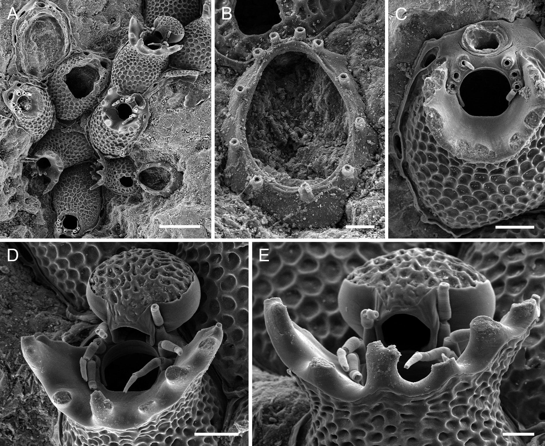
Fig. 10 View Fig A–E, Table 10
Diagnosis
Frontal shield with a reticulate pattern of raised ridges encircling round to polygonal depressions, distolaterally raising to form a huge flaring collar that is equipped with several pointed mucrones and that surrounds at least the proximal half of the orifice; lateral walls reduced proximally and laterally, septular pores therefore small and very elongated, the slightly raised distal pore large and suborbicular. Orifice orbicular, condyles very short and not thickened distally, orifice margin with six spines. Ectooecium covering slightly more than lower half of ooecium, endooecial surface as frontal shield but with narrower and more irregularly shaped elongate depressions. Ancestrula with 11 or 13 spines, opesia rather oval, not pyriform.
Etymology From the Greek Ἀκάνθα (Eng.: thorny) for its thorn-bearing peristome.
Material examined
Holotype
CANARY ISLANDS: 1 ovicellate colony with an ancestrula on a pebble, together with an immature colony, Stn 12 ( MNHN-IB-2014-71 ).
Paratype
CANARY ISLANDS: 1 damaged colony on a rock fragment, Stn 12 (MNHN-IB- 2014-72).
Description
Colony encrusting, unilaminar, forming small patches and bi- to triserial ribbons ( Fig. 10A View Fig ). Zooecia oval, separated by deep grooves and a thin ridge. Frontal shield convex, surface with a reticulate pattern of raised ridges encircling round to polygonal depressions ( Fig. 10C View Fig ), imperforate except for some four small marginal pores hardly visible in frontal view or in older zooecia; suboral area occupied by a tall, thickly calcified and flaring collar encircling almost two-thirds of orifice and sloping abruptly towards orifice and laterally towards first or second pair of spines, crest of collar serrated by up to seven large, pointed mucrones of different shape and height, longest ones distalmost, pointing distally; smooth gymnocystal calcification comprising distal part of collar clearly demarcated from proximal reticulate pattern of cryptocystal-type frontal shield by a wavy line, with wave peaks at bases of mucrones ( Fig. 10A View Fig , D–E); lateral walls well developed only in distal part of zooecium, narrow laterally, (disto) lateral septular pores comparatively small, transversely oval to extremely narrow and elongate, area surrounding pores therefore reduced; distal ooecial pore large, suborbicular, surrounded by a broad nodular area ( Fig. 10C View Fig ).
Orifice orbicular, widest at about mid-distance, proximal border fairly straight, proximal fourth delimited by a pair of very short, blunt condyles that parallel orifice margin ( Fig. 10C View Fig ); distolateral margins equipped with six whip-like spines with thick bases, arranged in two groups of three with a distinct distal gap; all six spines present in ovicellate zooids with distal pair almost incorporated into proximolateral ooecium wall.
Ovicell hyperstomial, ooecium barely resting on distal zooid’s frontal shield, a compressed sphere, the only observed one broader than long, with a short tubular peristome wedged in between distalmost pair of spines and opening at distal orifice margin, ooecial aperture orbicular, acleithral ( Fig. 10D View Fig ); ectooecium covering a little more than lower half; exposed endooecium extensive, hemispherical, surface topography similar to that of frontal shield but with smaller elongated depressions.
Ancestrula longer than wide (ca 370 µm long, 290 µm wide), smooth gymnocyst very narrow and similarly steeply sloping all around, cryptocyst practically absent ( Fig. 10B View Fig ); opesia extensive (ca 280 µm long, 200 µm wide), oval, somewhat narrowing distally, surrounded by 11 or 13 spines, in which distal four are situated slightly closer together; first generation autozooid budded distally or distolaterally ( Fig. 10A View Fig ).
Remarks
Atlantisina acantha gen. et sp. nov. has the most impressive suboral structure in this genus. A vertical outgrowth of the frontal shield forms a huge flaring collar around the proximal half, or even two-thirds, of the orifice, occasionally levelling only at the distal pair of spines. Moreover, this massively calcified collar is equipped with several pointed and occasionally branching mucrones, the largest lateral ones pointing distally and reaching beyond the zooid border.
Atlantisina acantha gen. et sp. nov. is morphologically close to A. lionensis gen. et sp. nov. and A. gorringensis gen. et sp. nov., particularly in having both a broad and tall suboral structure with pointed mucrones, and also the same reticulate pattern on the frontal shield and the exposed endooecium. However, the suboral structure forms a flared collar in A. acantha gen. et sp. nov., while it is planar and laterally more reduced in A. lionensis gen. et sp. nov. These two species also differ in the size of certain characters, particularly in the ooecium width, which is clearly larger in A. acantha gen. et sp. nov. with a ratio of ooecium length/width <1, while it is> 1 in A. lionensis gen. et sp. nov. Unfortunately, the number of measurements that could be taken of A. acantha gen. et sp. nov. were insufficient for statistical comparisons. Atlantisina gorringensis gen. et sp. nov. furthermore differs from A. acantha gen. et sp. nov. in having distinctly smaller zooids and orifices, and the suboral crest always terminates at the proximal pair of oral spines.
Another peculiarity in A. acantha gen. et sp. nov. are the gymnocystal lateral margins, which are, in contrast to all other species of Atlantisina gen. nov., often reduced to a thin band surrounding very narrow and elongated septular pores ( Fig. 10C View Fig ). The distal suborbicular pore through which the ooecium is budded is, on the other hand, comparatively large. Atlantisina acantha gen. et sp. nov. also differs from most other congeners in its ancestrula ( Fig. 10B View Fig ), which has 11 to 13 mural spines instead of the usual nine (only A. gorringensis gen. et sp. nov. has 12 spines).
Ecology
In the holotype, the only mature colony available for study, an ovicell is formed very early in astogeny (presumably in the seventh zooid, see Fig. 10A View Fig ), showing characters of a spot colony (cf. Bishop 1989). As the other species of Atlantisina gen. nov., A. acantha gen. et sp. nov. forms small patches and bi- to triserial colonies, which encrust small rocks and occur at around 660 m depth.
Distribution
Atlantisina acantha gen. et sp. nov. occurs sympatrically with A. inarmata gen. et sp. nov. off NW Gran Canaria (Canary Islands).
No known copyright restrictions apply. See Agosti, D., Egloff, W., 2009. Taxonomic information exchange and copyright: the Plazi approach. BMC Research Notes 2009, 2:53 for further explanation.