
Austrognathia glandifera, Achatz, Johannes G. & Sterrer, Wolfgang, 2015
|
publication ID |
https://doi.org/ 10.11646/zootaxa.3955.2.5 |
|
publication LSID |
lsid:zoobank.org:pub:A38F87D7-C55C-4172-831B-D665AFBEF6C6 |
|
DOI |
https://doi.org/10.5281/zenodo.5631765 |
|
persistent identifier |
https://treatment.plazi.org/id/FB11986F-FE73-FF98-FF5B-FD7B70C1F960 |
|
treatment provided by |
Plazi |
|
scientific name |
Austrognathia glandifera |
| status |
sp. nov. |
Austrognathia glandifera View in CoL n. sp.
( Figs. 1–3 View FIGURE 1 View FIGURE 2 View FIGURE 3 ; Table 2 View TABLE 2 )
Diagnosis. Small, fairly slender Austrognathia (mean length 730 µm, width 97.5 µm; index 7.47; see Table 2 View TABLE 2 ). Basal plate 6.60 µm long, 22.60 µm wide (index 0.29), with conspicuous median and lateral lobes. Jaws 21.2 µm in length; dorsal tooth row with 7 short, even teeth; ventral row with 4–8 longer, uneven-sized teeth. Conuli large and blunt (30.75 µm long by 22.42 µm wide; index 1.37), with capitulum enveloping two thirds of the conulus (capitulum length 20.17 µm; index 1.62).
Type material. Holotype: USNM 1270621, one adult from Hong Kong (sample #2) in squeeze preparation.
Type locality. Sample HK 2 from Hong Kong (see Table 1 View TABLE 1 ).
Other material examined. Four adults and two juveniles from sample #2, one adult from sample #9. Semithin and ultrathin sections of two mature specimens.
Etymology. Referring to the acorn-shape of mature conuli (from Latin glans = acorn).
Description. Organization ( Fig. 2 View FIGURE 2 A). Adults measure 730 µm in length (600–800 µm), and 97.50 µm in width at U55 (body index 7.47), with a short rostrum (index 0.94). The epidermis of the tail region is studded with adhesive glands.
Sclerotized pharyngeal parts ( Figs. 1 View FIGURE 1 A–D; 2C–F). The basal plate is 6–8 µm long and 20–24 µm wide (index 0.29). It has well-defined lobes, with the lateral pair pointing medio-rostrally. Caudally the basal plate is set with up to 15 teeth of which the largest 3 are each flanked by a pair of smaller teeth; even smaller ones separate the three groups. Jaws are 20–22 µm long, with a strong terminal tooth and two rows of teeth: a dorsal row of up to 7 even teeth, and a ventral row in which a larger tooth is followed by up to 8 uneven teeth.
Male system ( Figs. 2 View FIGURE 2 A, B, G, H, I; 3B, C, F, G). The dorsal testis contains 5–8 mature conuli; one specimen had but a single conulus located in the penis. Mature conuli range from 27 µm to 35 µm in length (mean 30.75 µm), and 18 µm to 28 µm in width (mean conulus index 1.34). They are plump and blunt, with the semi-circular capitulum reaching 20.17 µm in length, which is two-thirds of the conulus length (capitulum index 1.62). The pointed end of the conulus is usually enveloped in a matrix (i.e., the sperm cytoplasm), and is often bilobed, an indication of the peculiar conulus composition along two longitudinal axes. The cingulum is inconspicuous but long, barely protruding caudally past the capitulum. A granular layer below the capitulum is conspicuous in the conuli ( Fig. 2 View FIGURE 2 G, H, I). The male copulatory organ consists of a glandular part and an epidermal penis that measures 30 µm in length and 15 µm in width. The glandular part (pg) is ~70 µm long; the spermatic duct runs through it from the dorsal to the ventral side, where it proceeds into the ejaculatory duct of the penis (p). The cells constituting the glandular part are filled with vesicles, which are refractive in live observation ( Fig. 2 View FIGURE 2 B) and electron-dense by transmission electron microscopy ( Fig. 3 View FIGURE 3 F, G). A group of cells, which lie adjacent to the penis, contain spherical vesicles that measure up to 1 µm ( Fig. 3 View FIGURE 3 B, F, G). The vesicles in the more anterior part (ag) are potato-shaped and are more electron-dense when smaller, and larger in size the further posteriorly and ventrally they are positioned (pg), starting with ~ 200 nm in diameter and reaching up to 1 µm ( Fig. 3 View FIGURE 3 B, F, G). The cells of the penis contain numerous electron-dense vesicles (~ 150 nm) and rods, which measure up to 5 µm in length and 200 nm in width. The rods are especially numerous around the gonopore ( Fig. 3 View FIGURE 3 C). Some obliquely striated longitudinal muscles run along the glandular and epidermal part of the penis ( Fig. 3 View FIGURE 3 F).
Female system ( Fig. 3 View FIGURE 3 A, D). The ovary including the mature egg extends from about U25 to U66; the single mature egg can be 340 µm long and 65 µm wide, and often wraps around the bursa, which lies immediately behind it, at U57 to U65.
The bursa is ~50 µm long and consists of a cellular wall (4–5 layers) and a syncytial, multinucleated interior. Even though a distinction into an anterior and posterior part is not conspicuous in its form it is by its content. The posterior part (bp) often contains large electron-dense material of undefined shape, whereas the anterior part (ba) contains filamentous material ( Fig. 3 View FIGURE 3 A, D). Overall the content of the bursa is nearly devoid of cell organelles, e.g. mitochondria. The origin and function of the electron-dense material is not known. On the anterior end the cells of the bursal wall are highly intermingled, but in an unorganized fashion ( Fig. 3 View FIGURE 3 E). On the dorsolateral side the bursa is enclosed by parenchymal cells and connects to the body wall at the posterior end through a vagina that lacks a lumen ( Fig. 3 View FIGURE 3 A, D, E).
Remarks. Members of the genus Austrognathia are mainly distinguished by the dimensions and proportions of their sperm (conuli). The mean sperm index of the 10 described species ranges from 2.79 (in A. australiensis ) to 1.71 (in A. hymanae ); none has a sperm index as low as 1.37. Mature sperm in 5 species are considerably smaller (from 8.0 µm in A. nannulifera to 22.8 µm in A. hymanae ) than in A. glandifera , and considerably larger in another four (from 36.73 µm in A. singatokae to 52.50 µm in A. macroconifera ); only the conuli of A. christianae (31.00 µm) and A. riedli (34.29 µm), two closely related species, are of comparable length but both are significantly more slender (conulus index 1.72 viz. 1.73). The consistency in shape and dimensions of 12 conuli measured in five specimens also suggests this species to be new.
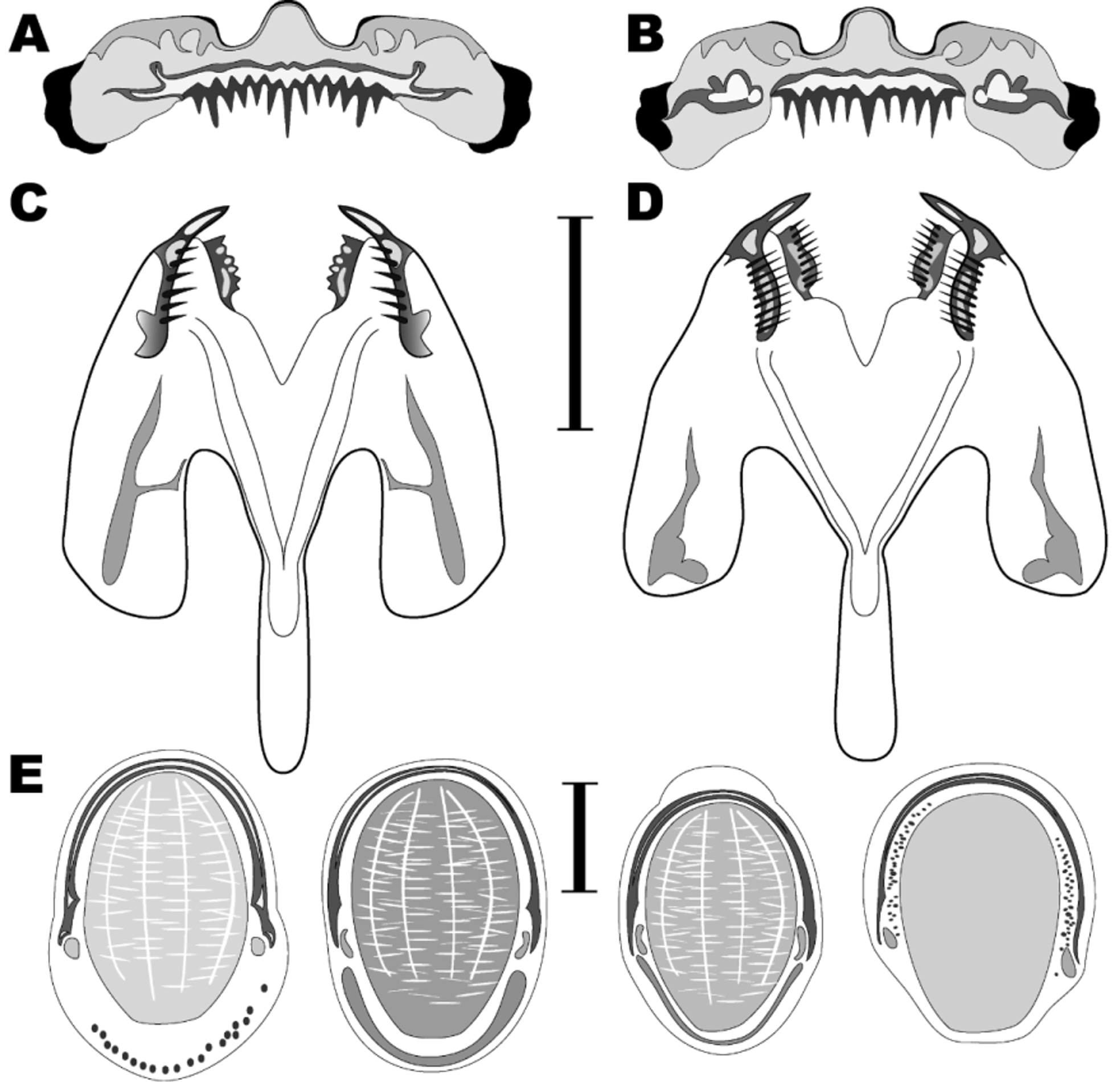
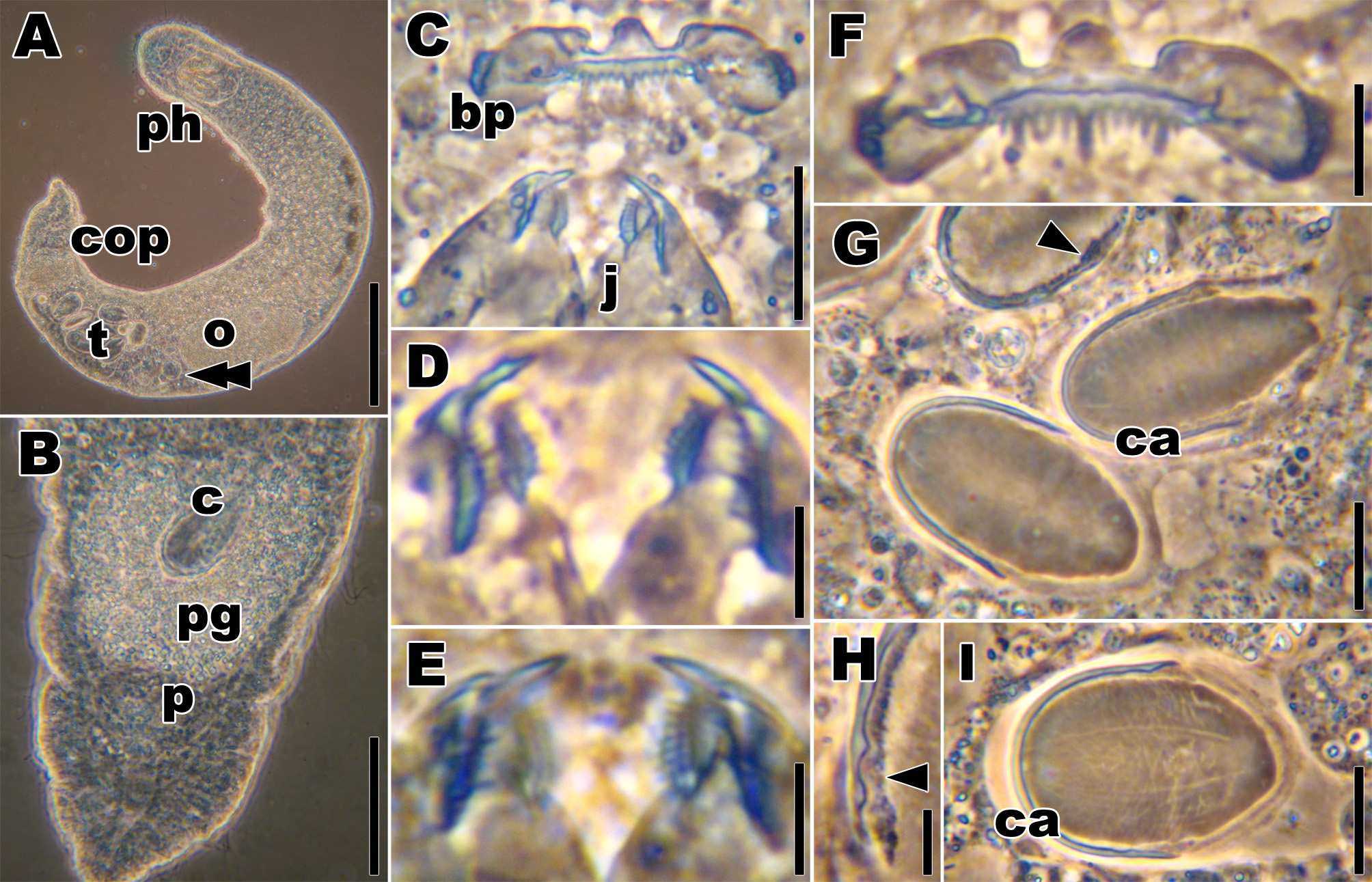
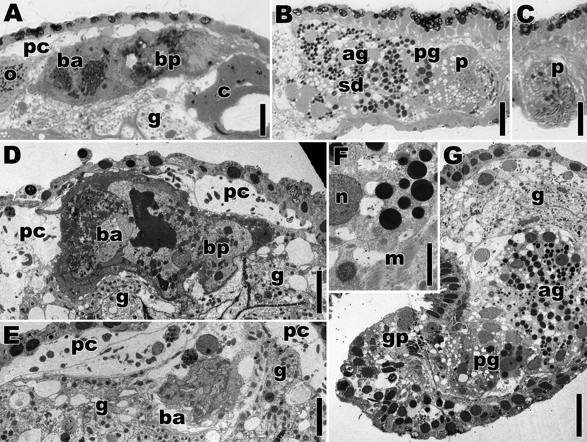
TABLE 2. Morphometric data (in µm) for Austrognathia glandifera n. sp.
| Hong Kong | Mean | StDev | Max | Min | n |
|---|---|---|---|---|---|
| Body length of adults | 730.00 | 89.07 | 800 | 600 | 4 |
| Body width of adults | 97.50 | 5.00 | 100 | 90 | 4 |
| Body index of adults | 7.47 | 0.57 | 8.00 | 6.67 | 4 |
| Rostrum index of adults | 0.94 | 0.22 | 1.10 | 0.79 | 2 |
| Jaw length | 21.20 | 0.84 | 22 | 20 | 5 |
| Basal plate length | 6.60 | 0.89 | 8 | 6 | 5 |
| Basal plate width | 22.60 | 1.52 | 24 | 20 | 5 |
| Basal plate index | 0.29 | 0.03 | 0.33 | 0.26 | 5 |
| Sperm length | 30.75 | 2.34 | 35 | 27 | 12 |
| Sperm width | 22.42 | 2.71 | 28 | 18 | 12 |
| Sperm index (sp l/w) | 1.37 | 0.19 | 1.78 | 1.20 | 12 |
| Capitulum length | 20.17 | 2.48 | 27 | 18 | 12 |
| Capit. index (sp l/cap l) | 1.62 | 0.18 | 1.84 | 1.43 | 5 |
| USNM |
Smithsonian Institution, National Museum of Natural History |
No known copyright restrictions apply. See Agosti, D., Egloff, W., 2009. Taxonomic information exchange and copyright: the Plazi approach. BMC Research Notes 2009, 2:53 for further explanation.