
Bairdoppilata alcyonicola Maddocks, 1969
|
publication ID |
https://doi.org/ 10.11646/zootaxa.5175.3.1 |
|
publication LSID |
lsid:zoobank.org:pub:44FB9C3D-3188-4BFB-BDB8-C1324729A396 |
|
DOI |
https://doi.org/10.5281/zenodo.7003516 |
|
persistent identifier |
https://treatment.plazi.org/id/03FE6B50-FFF9-FFAF-ECD6-AF436AD918A0 |
|
treatment provided by |
Plazi |
|
scientific name |
Bairdoppilata alcyonicola Maddocks, 1969 |
| status |
|
Bairdoppilata alcyonicola Maddocks, 1969 View in CoL
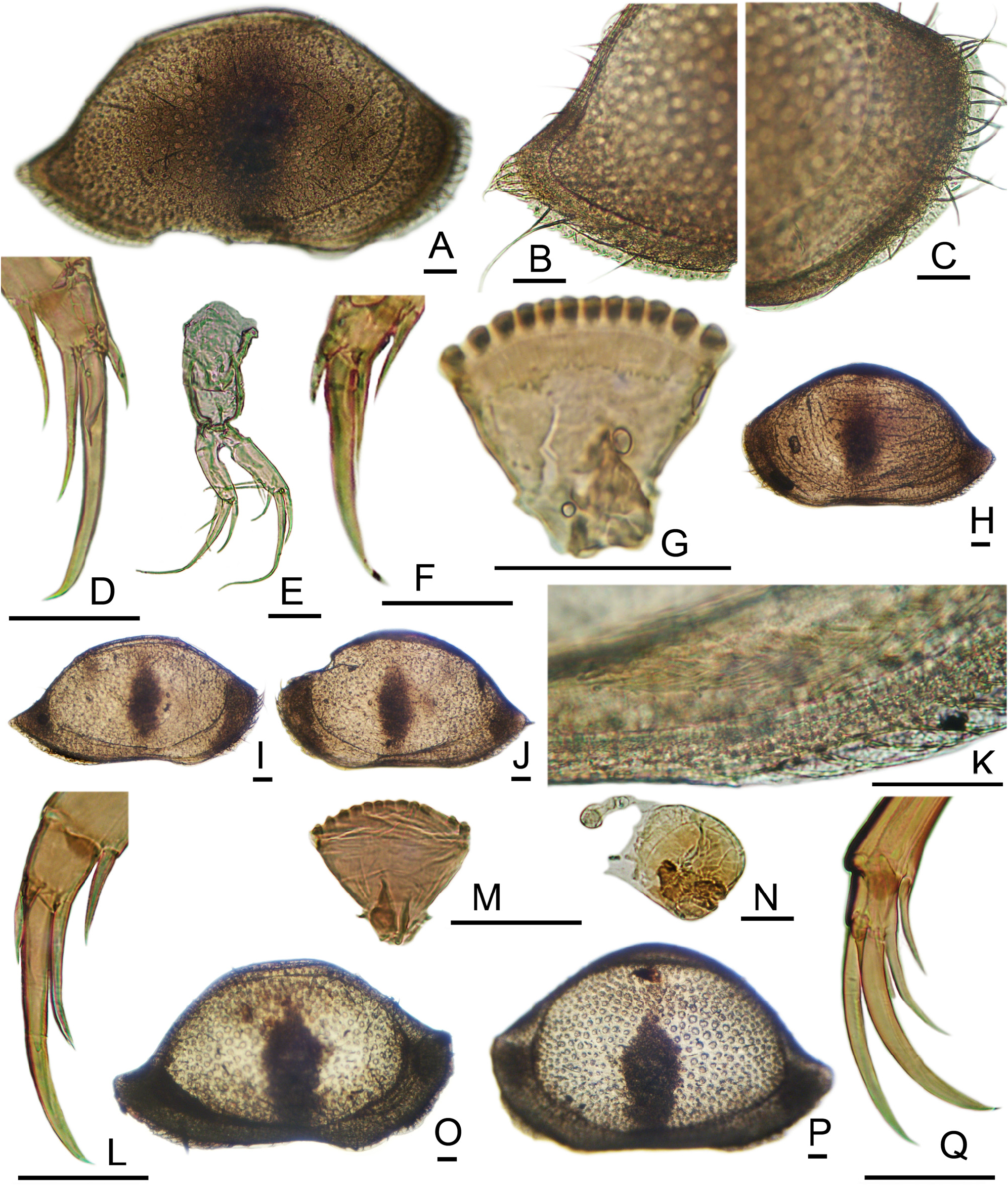
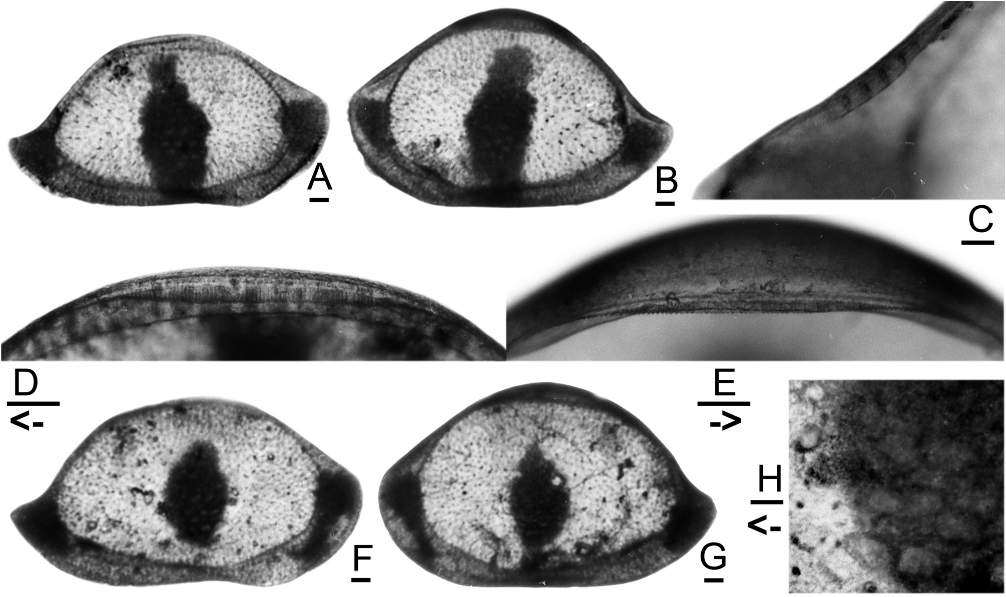
( Figures 1A–K View FIGURE 1 , 2A–E View FIGURE 2 )
1969 Bairdoppilata (Bairdoppilata) alcyonicola Maddocks : 71, figs. 36A–H, 37A–H, 38A–C.
1995 Bairdoppilata alcyonicola Maddocks. —Maddocks, p. 216, pl. 6, figs. 1–4; pl. 14, figs. 7–8.
Material Examined: Five juvenile specimens and several subfossil carapaces from sands of the fringing reef, Nosy Be, Madagascar.
Dimensions: Specimen 168J: LVL 0.706 mm, LVH 0.404 mm; RVL 0.698 mm, RVH 0.373. Specimen 523J: RVL 0.703 mm, RVH 0.399. Specimen 524J: LVL 0.719 mm, LVH 0.419 mm. Specimen 764W: LVL 0.970 mm, LVH 0.590, RVL 0.930, RVH 0.530 mm. Specimen 2629WF: LVL 0.930 mm, LVH 0.590 mm, RVL 0.950 mm, RVH 0.520 mm. Specimen 3089WM: LVL 0.880 mm, LVH 0.540 mm, RVL 0.900 mm, RVH 0.490 mm. A height: length scatter plot for adult valves was published by Maddocks (1995, Graph 1).
Esophageal Valve: The plate is flat and thin ( Fig. 1G View FIGURE 1 ) The evenly curved posterior perimeter carries 12 low, mound-like teeth, of equal size and evenly spaced, which project upward (dorsally) and very slightly posteriorly. The two corner teeth are no larger than the others, have an indistinct or no secondary cusp (not compound), and are set apart from the others by only a small gap. A solitary guide pin (setule) rises at a shelf outside each corner from the ventral brush below. The anterolateral scroll is asymmetrical, with a triangular tab.
Anatomical Remarks: The adult carapace has a somewhat irregular texture produced by numerous tiny puncta, which are not aligned in rows and are separated by broader muri ( Maddocks 1995, Pl. 6, figs. 1–4; Pl. 14, figs. 7–8). The caudal process may be slightly thickened, but there is no horizontal bar. Delicate posteroventral marginal denticles may be present. The opaque pattern consists of a broad, irregular vertical streak across the central region, which almost reaches the ventral margin, narrows above mid-height, and does not reach the dorsal margin ( Figs. 2A–B View FIGURE 2 ). Small opaque spots are also present at the anterodorsal and posterodorsal corners.
The A–1 carapace is thin–walled with delicately impressed, shallow, round puncta ( Figs. 1A–C View FIGURE 1 ). The opaque pattern consists of an elongate central spot over the AMS, a long band along the anteroventral margin, and a spot filling the posteroventral angle. Marginal denticles are not well developed on the juvenile LV, but the RV has broad anterior and posterior fringes. The fused zone (infold) is narrow. The anterior and posterior vestibules are deep and connected through the ventral region. The inner lamella is very weakly calcified and not as wide as in adults. About 6 large sensilla are located at the caudal angle ( Fig. 1B View FIGURE 1 ). In the vestibule of a molting A–1, folds of new cuticle with attached marginal and carapace sensilla are stored ( Fig. 1H View FIGURE 1 ).
In juveniles the anterodistal antennal claw is represented by a short, tapering anlage. In a molting individual one can see the tip of the adult anterodistal claw being withdrawn from this A–1 anlage ( Figs. 1D, F View FIGURE 1 ).
No known copyright restrictions apply. See Agosti, D., Egloff, W., 2009. Taxonomic information exchange and copyright: the Plazi approach. BMC Research Notes 2009, 2:53 for further explanation.