
Batillipes algharbensis, Santos & Rubal & Veiga & da Rocha & Fontoura, 2018
|
publication ID |
https://doi.org/ 10.5852/ejt.2018.425 |
|
publication LSID |
lsid:zoobank.org:pub:AB5C3414-92EC-4CE3-8963-880E07648D11 |
|
DOI |
https://doi.org/10.5281/zenodo.3816375 |
|
persistent identifier |
https://treatment.plazi.org/id/005ABE3C-1A30-4E9C-8C29-2AD0F8AA3B3B |
|
taxon LSID |
lsid:zoobank.org:act:005ABE3C-1A30-4E9C-8C29-2AD0F8AA3B3B |
|
treatment provided by |
Valdenar |
|
scientific name |
Batillipes algharbensis |
| status |
sp. nov. |
Batillipes algharbensis View in CoL sp. nov.
urn:lsid:zoobank.org:act:005ABE3C-1A30-4E9C-8C29-2AD0F8AA3B3B
Figs 2–3 View Fig View Fig , Table 1 View Table 1
Diagnosis
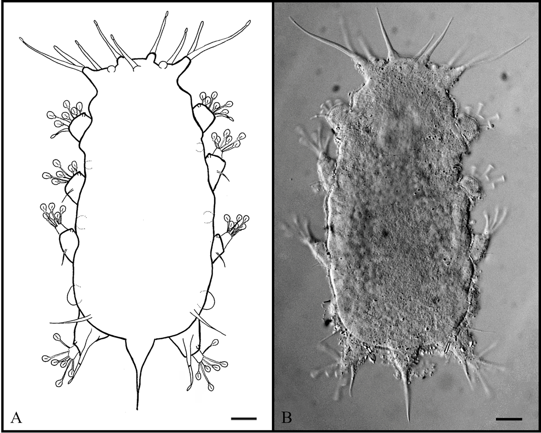
Batillipes with distinct head, separated from the body by a neck constriction followed by lateral processes. Primary clavae very thin and undivided. Strongly developed secondary clavae present. Cephalic cirri with spatula-like distal tips. Sensory spines on all legs. Sensory spine on leg I with spatula-like distal tip separated from basal portion by a van der Land’s organ. On fourth feet, middle toes 3 and 4 of different lengths, with toe 3 longer than 4. A dorsal papilla present at distal extreme of all legs. Blunt lateral body projections present between all leg pairs: faint between legs I–III and very well-developed between legs III–IV. The caudal apparatus consists of a sharp and long spine inserted in a slightly elongated swollen base. Dorsal cuticle coarsely punctated. Rosette-like female gonopore.
Etymology
The name refers to the region where the species was found, the Portuguese region of Algarve, from the Arabic ‘ Al Gharb ’ (Al = the + Gharb = West); algharbensis = inhabiting Algarve.
Material examined
Holotype
PORTUGAL: ♀ adult, collected at Meia-Praia Beach, Lagos, Algarve, 37°07′01″ N, 8°38′37″ W, the only adult found, mounted in glycerol (slide C. VII-88). GoogleMaps
Paratype
PORTUGAL:1 four-toed larva, collected at Meia-Praia Beach, Lagos,Algarve, 37°07′01″ N, 8°38′37″ W, mounted in glycerol (slide C. VII-89).
Additional material
SPAIN: 9 specimens (8 ♀♀ and 1 six-toed juvenile), collected in Galicia (NW Spain), at Rias of O Barqueiro (43°44′37″ N, 7°39′06″ W – 43°45′15″ N, 7°41′22″ W) and Foz (43°33′47″ N, 7°15′19″ W – 43°34′11″ N, 7°14′41″ W) (slides GAL. I-9 – GAL. I-17).
Description
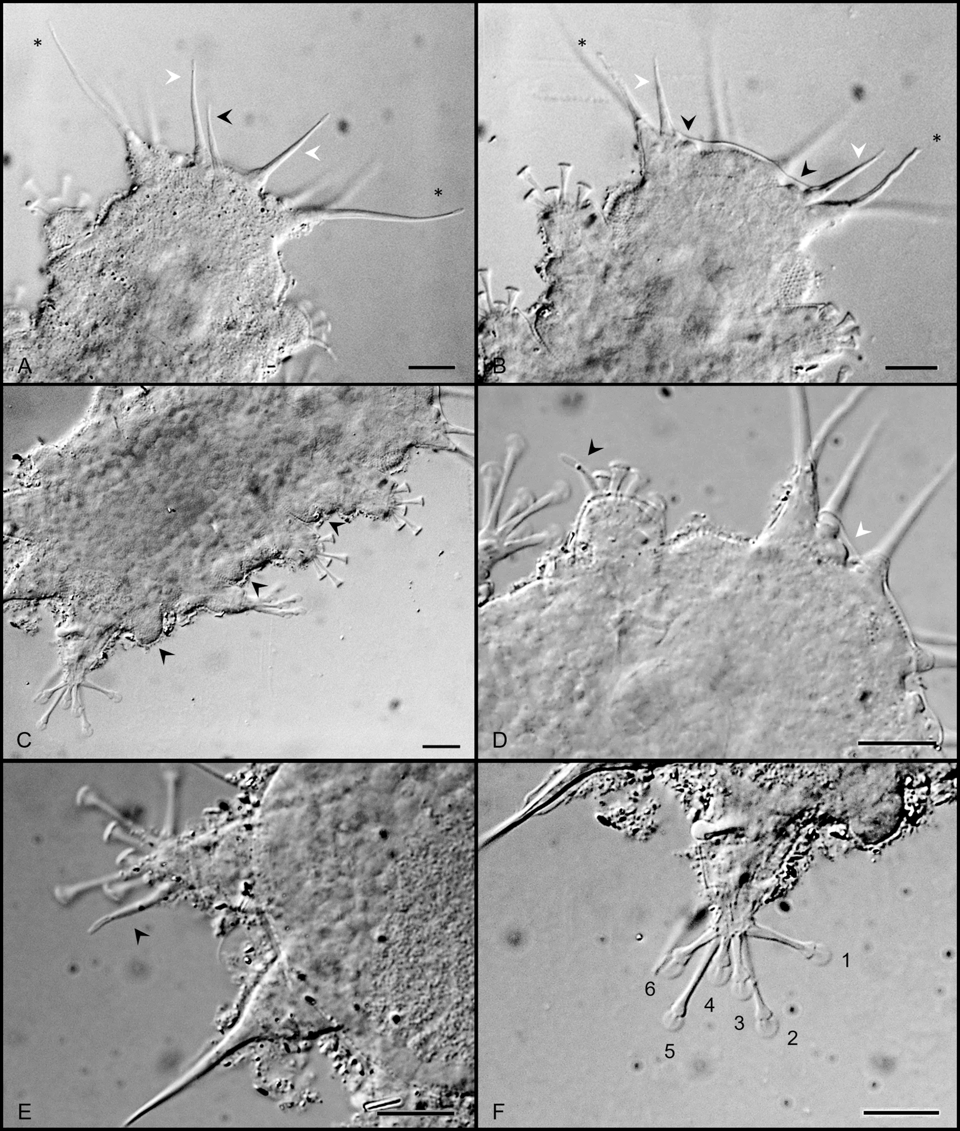
Holotype, female with a body length 121.1 µm (150.6 µm including the caudal apparatus) and 57.9 µm wide between the third and fourth pair of legs ( Fig. 2 View Fig A–B; Table 1 View Table 1 for morphometrics). Eye spots not observed in mounted specimens. Trapezoid head ( Fig. 3 View Fig A–B) separated from the body by a neck constriction followed by lateral processes (body projection 1). Internal cephalic cirri inserted dorsally, bearing cirrophores (cirrophores ca 3.4 µm long) ( Fig. 3A View Fig ). External cephalic cirri, with indistinct cirrophores, inserted more ventrally, near the common pedestal bearing lateral cirri A and primary clavae ( Fig. 3B View Fig ). The median cirrus with a cirrophore (ca 3.5 µm long). Lateral cirri A located dorsally in relation to the unconstricted primary clavae ( Fig. 3A View Fig ). The primary clavae, with a van der Land’s organ at the base, particularly thin (thinner than lateral cirri) and sharpened at the tip ( Fig. 3B View Fig ). In the frontal edge of the head, conspicuous and strongly developed dome-shaped secondary clavae ( Fig. 3B, D View Fig ) also present (major diameter 3.8 µm). All cephalic cirri, have long spatula-like swollen tips ( Fig. 3 View Fig A–B). Ovoid pharyngeal bulb 14 µm long and 16.6 µm wide. Ventral mouth opening in a protruded circular cone. Stylets and placoids not visible after slide mounting.
Blunt ventro-lateral body projections (body projections 2 to 4, respectively from legs I to IV) present between all leg pairs: very small and difficult to see between the first three pairs of legs and welldeveloped between legs III–IV ( Fig. 3C View Fig ). The caudal apparatus is constituted by a long and sharp spine inserted in a slightly elongated swollen base ( Figs 2 View Fig A–B, 3E).
Sensory spines present on all legs, decreasing in length from leg I to leg III. First ( Fig. 3D View Fig ) and fourth ( Fig. 3E View Fig ) leg sensory organs with spatula-like tips similar to the ones of cephalic appendages and both divided into two parts, separated by a van der Land’s organ. Basal and distal parts are respectively 4.8 µm and 2.5 µm long for the sensory organ on legs I and 12.6 and 7.6 µm long for the sensory organ
on legs IV. The large spatula-like tips of sensory organs on legs IV are 3.7 µm long. The entire fourth leg sensory organ is 20.2 µm long. Sharply pointed cirri E with small cirrophores present.
Telescopic legs with toes having the distal stalk enlarged distally (about 2.1 µm wide), ovoid suction discs (diameter 2.9 µm measured on toes of leg IV) with conspicuous braces. On the first three pair of legs, toe 2 is the shortest (considering toe 1 the most cephalically), toes 3 and 5 are the longest and toes 1, 4 and 6 are medium sized. On feet of the fourth pair of legs, the middle toes 3 and 4 are of different lengths with toe 3 longer than toe 4; toes 2 and 5 are the longest, and toes 1 and 6 of intermediate size ( Fig. 3F View Fig ). A dorsal papilla (dark when observed under PCM) is present at the distal extreme of each leg.
Dorsal cuticle uniformly and coarsely punctated, about 9–10 pillars / 10 µm (each pillar about 1.4 µm high), with barely visible transversal folds. Ventrally the cuticle punctation is more delicate. Body with a considerable amount of adherent detritus, mainly near the lateral body projections.
Rosette-shaped gonopore separated from the anus by a weakly defined groove. Other details of the gonoporal apparatus are not visible.
Differential diagnosis
Only one known species of Batillipes , B. tubernatis , originally described from the Northeast Atlantic Ocean ( Scotland, Germany and England), shares with the new species the same toe arrangement pattern in the fourth feet, characterized by having toes 3 and 4 of different lengths and toe 3 longer than 4. In the original description, the caudal region of B. tubernatis is described as being round without appendages ( Pollock 1971). However, this character can be very variable and should be used with caution in species comparisons ( Gallo D’Addabbo et al. 2000). Actually, in the emended description of B. tubernatis by McKirdy (1975), based on specimens from Florida (Gulf of Mexico), USA, (hereby designated B. tubernatis sensu McKirdy 1975 ), a strong swollen-based caudal spike was present in about 80% of the examined specimens.
Batillipes algharbensis sp. nov. can be clearly distinguished from B. tubernatis by the presence of ventrolateral body projections that are very well-developed between legs III–IV (lacking in all morphotypes of B. tubernatis ). Moreover, in the new species, the primary clava is very thin and sharp-tipped, while in B. tubernatis it is short, thicker and tube-shaped. The dorsal cuticle is coarsely uniformly punctated in the new species (9–10 pillars / 10 µm) and finely punctated (about 14 pillars / 10 µm) in B. tubernatis whereas exhibiting apunctate areas in specimens from Florida. In addition, secondary clavae are conspicuous dome-shaped in the new species and indistinct in B. tubernatis . Finally, in B. tubernatis sensu McKirdy toe discs are quadrate with a slightly indented distal edge while in B. algharbensis sp. nov. toe discs are ovoid.
Body shape and caudal apparatus of B. algharbensis sp. nov. are very similar to B. spinicauda Gallo D’Addabbo, Sandulli & de Zio Grimaldi, 2005 , justifying a comparison between the two species. In addition to a different toe arrangement pattern in the fourth feet (Toe 3 = Toe 4 Ĕ Toe 1, Toe 6 in B. spinicauda ), the new species differs from B. spinicauda in having undivided primary clavae, blunt body projections between legs III and IV (conical in B. spinicauda ), toes in feet of legs I–III inserted at the same level (tarsus are oblique in B. spinicauda ), and by lacking lateral papilla (malleolus) on leg IV.
Associated species
Batillipes lusitanus sp. nov., B. pennaki , B. phreaticus and H. greveni .
Remarks
The four-toed larva (morphometry in Table 1 View Table 1 ) is similar to the adult. Specimens collected in Galicia (NW Spain) and previously identified by Veiga et al. (2009) as Batillipes sp. 1 with similarities with B. spinicauda also exhibit the unique characters of B. algharbensis sp. nov. ( Table 1 View Table 1 ). Therefore, they are assigned to B. algharbensis sp. nov.
| N |
Nanjing University |
| W |
Naturhistorisches Museum Wien |
| C |
University of Copenhagen |
| O |
Botanical Museum - University of Oslo |
No known copyright restrictions apply. See Agosti, D., Egloff, W., 2009. Taxonomic information exchange and copyright: the Plazi approach. BMC Research Notes 2009, 2:53 for further explanation.
|
Kingdom |
|
|
Phylum |
|
|
Class |
|
|
Order |
|
|
Family |
|
|
Genus |