
Boholina munaensis, Boxshall, Geoff A. & Jaume, Damià, 2012
|
publication ID |
https://doi.org/ 10.5281/zenodo.279542 |
|
DOI |
https://doi.org/10.5281/zenodo.5693710 |
|
persistent identifier |
https://treatment.plazi.org/id/03E287D0-7014-AC65-FF25-FB68B2533BE5 |
|
treatment provided by |
Plazi |
|
scientific name |
Boholina munaensis |
| status |
sp. nov. |
Boholina munaensis n. sp.
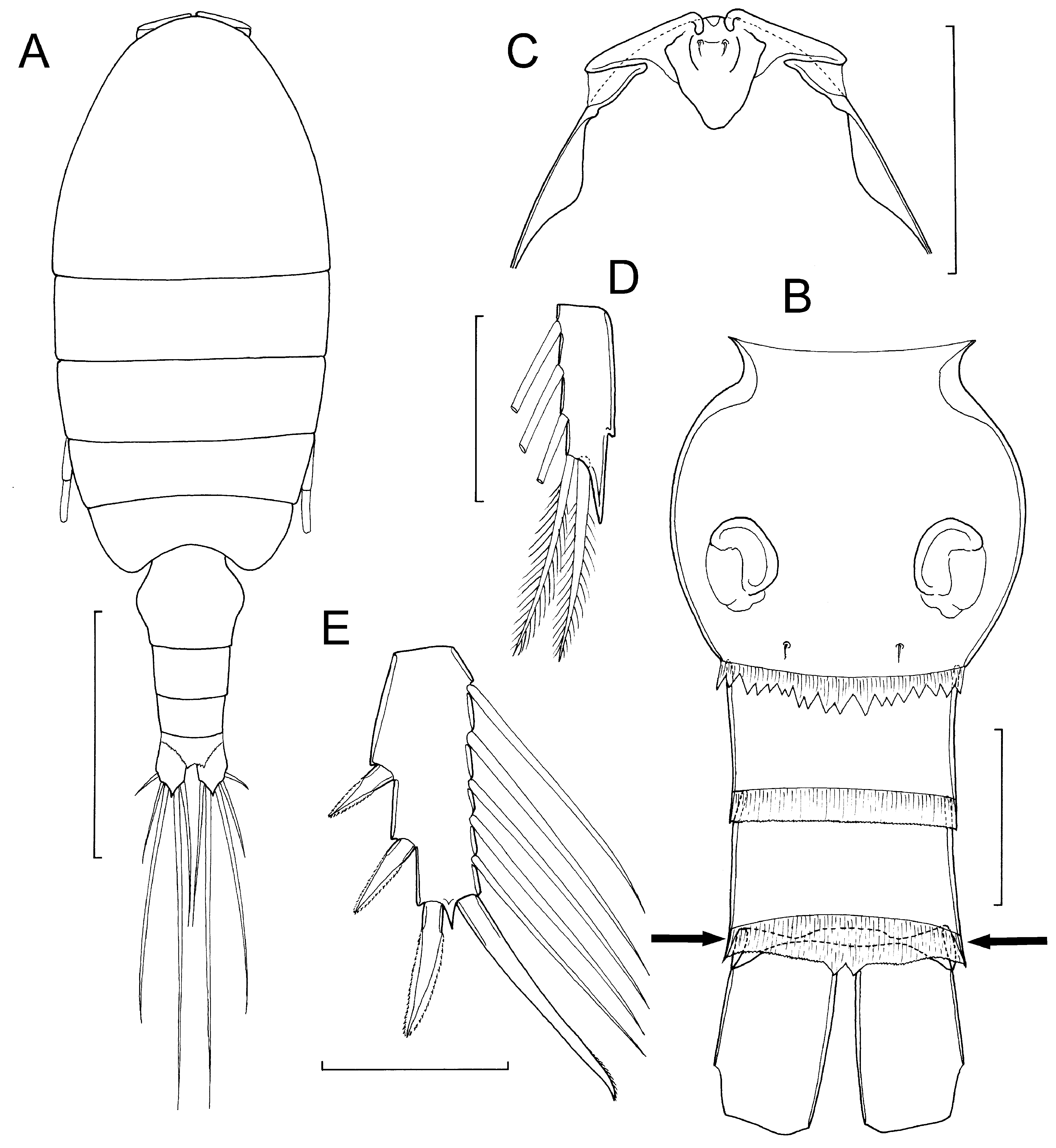
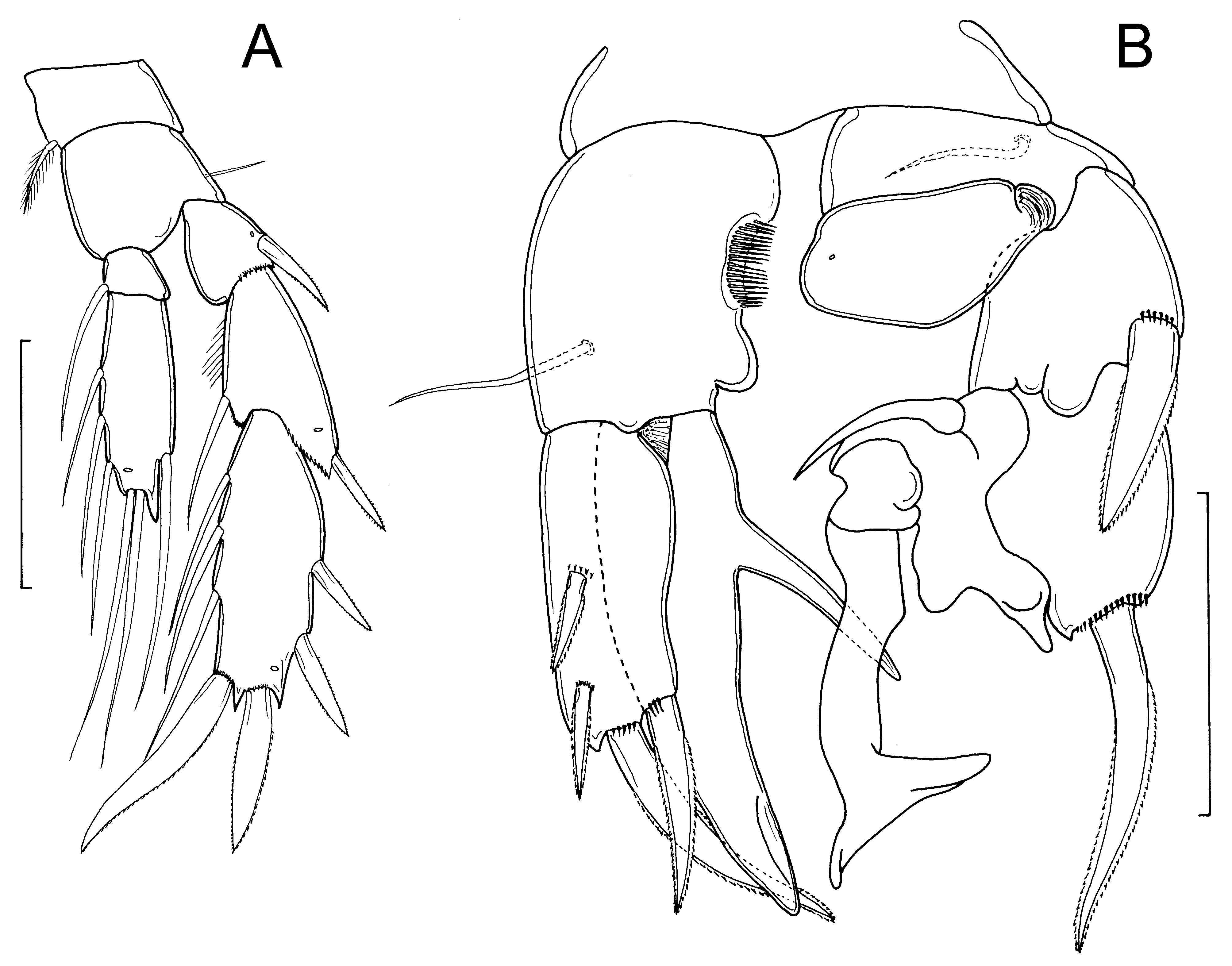
( Figs. 11–12 View FIGURE 11 View FIGURE 12 )
Type material. Holotype female, 2 paratype females, 2 paratype males, collected from Lawou cave spring in Walengkabola village, Muna Island, Indonesia located at 5º 10.950’S 122º 34.720’E; salinity 1.8 ppt, on 13 September 2007 by G.A. Boxshall and D. Jaume. Registration numbers: holotype female [ MZB.Cru Cop.107], 2 paratype females and 1 paratype male [ MZB.Cru Cop. 108] in Museum Zoologicum Bogoriense; 1 paratype male in Natural History Museum, London [ BMNH 2011.1178].
Additional material. 13 copepodid stages with same collection data: tentatively identified as B. munaensis n. sp.
Etymology. The name of the new species is based on its type locality, Muna Island in Indonesia.
Description of adult female. Body ( Fig. 11 View FIGURE 11 A) length ranging from 0.70 to 0.77 mm, with mean of 0.74 mm (based on 3 specimens). Prosome 5-segmented; postero-lateral corners of second and third pedigerous somites forming acute posteriorly-directed points; postero-lateral corners of posterior double-somite rounded, symmetrical. Ratio of prosome to urosome length about 2.2:1. Urosome 4-segmented as in B. parapurgata . Genital doublesomite ( Fig. 11 View FIGURE 11 B) symmetrical, with strongly convex lateral margins widest about at mid-length; posterior border ornamented with finely serrate hyaline membrane dorsally and strongly but slightly irregularly dentate hyaline membrane ventrally ( Fig. 11 View FIGURE 11 B); genital openings paired, more widely separated on mid-ventral surface than in B. parapurgata ; 1 pair of sensillae present on ventral surface near posterior margin of double-somite. First and second free abdominal somites subequal in length; first with finely serrated hyaline membrane all around posterior margin, second with posterior margin hyaline membrane expanded mid-ventrally and mid-dorsally, to form double-pointed flap functioning as pseudoperculum concealing anal opening. Hoop of integument representing anal somite, irregular in thickness (arrowed in Fig. 11 View FIGURE 11 B). Caudal rami as for B. parapurgata .
Frontal margin of dorsal cephalic shield forming elaborate transverse crest of thickened integument; continuous with weakly developed, tapering rostrum ( Fig. 11 View FIGURE 11 C).
Antennules, antennae, maxillules, maxillae and maxillipeds all as in B. parapurgata . Legs 1–4 as for B. parapurgata except leg 1 with distinct notch on outer margin of third endopodal segment ( Fig. 11 View FIGURE 11 D), and leg 4 with terminal spine on third exopodal segment ( Fig. 11 View FIGURE 11 E) 68 μm long, about 1.9 times longer than adjacent distolateral spine (36 μm) and about same length as segment (= 68 μm).
Leg 5 ( Fig. 12 View FIGURE 12 A) biramous, with 3-segmented exopod and 2-segmented endopod, intercoxal sclerite smooth and unornamented. Setal formula as in B. parapurgata . Basis of leg 5 with acute process located on posterior surface near base of exopod. Exopod longer than endopod: tip of endopod reaching almost to level of proximal outer spine on third exopodal segment; distal endopodal segment 2.6 times longer (39 μm) than wide (16 μm). Outer spines on exopod each with serrate membrane bilaterally. Terminal spine on exopod with serrate membrane externally and finely serrate membrane internally, inner distal spine (46 μm) about 1.4 times longer than terminal spine (34 μm), both shorter than segment (56 μm).
Description of adult male. Body 0.68 mm in length (based on 1 complete specimen). Prosome 5-segmented, as in female: postero-lateral corners of second and third pedigerous somites forming acute posteriorly-directed points, as in female. Ratio of prosome to urosome length about 2.3:1. Urosome 5-segmented. Genital somite slightly asymmetrical, with single gonopore opening posterolaterally on left side; genital and first to third free abdominal somites similar in length, although often variably telescoped within preceding somite; each with hyaline membrane around posterior border, except membrane lacking ventrally on genital somite. Anus opening terminal, located between caudal rami, concealed beneath pseudoperculum formed by hyaline membrane on third free abdominal somite. Caudal rami as in female.
Antennules as in male B. parapurgata ; antenna to maxillipeds and legs 1–4, as described above for female B. parapurgata .
Leg 5 ( Fig. 12 View FIGURE 12 B) strongly asymmetrical; coxae and intercoxal sclerite fused to form common base. Left leg biramous: basis with slender outer basal seta located on posterior surface; exopod 3-segmented; first segment with bilaterally serrate outer spine 35 μm in length; second segment modified, bearing slightly curved spine, about 56 μm long on outer margin; third segment highly transformed bearing several rounded processes, one modified setal element, and a long process terminating in a T-shaped distal expansion ( Fig. 12 View FIGURE 12 B); endopod an unarmed lobe, about 1.9 times longer (40 μm) than wide (21 μm). Right leg biramous, basis armed with slender outer basal seta located on posterior surface and ornamented with medial comb of spinules in mid-margin; inner distal corner of basis forming rounded lobe; exopod unsegmented with 2 spines on outer margin; proximal spine (18 μm) shorter than distal spine (23 μm long); curved apical spine 47 μm in length, subapical inner spine 37 μm long; endopod forming an elongate lobe, about 81 μm long, tapering distally, bearing strong, spiniform process originating medially on inner surface ( Fig. 12 View FIGURE 12 B).
Remarks. The new species shares with B. crassicephala the presence of four well developed spines on the exopod of the male right leg 5, but can be distinguished from it by the relative lengths of these spines and by the presence of a large spinous process on the medial surface of the endopod of the right leg 5. In the female leg 5, the two distal spines on the exopod are more dissimilar in size than in B. crassicephala . The inner distal spine is 1.4 times longer than the outer terminal spine in the new species but only 1.1. times longer in B. crassicephala . In addition B. munaensis has pointed corners on the tergites of the second and third pedigerous somites in both sexes, as in B. parapurgata , whereas these corners are rounded in B. crassicephala according to Fosshagen & Iliffe (1989).
It is interesting to note that Boholina tends to occur in cave pools. In Bohol the two species co-occurred in the pools in San Vincente Cave ( Fosshagen & Iliffe 1989). On Muna, a dense population of B. munaensis was found in Lawou, a cave spring with a water depth of up to 100 cm and a salinity of only 1.8 ppt. Villagers were washing clothes there every time we visited and the air smelled of soap and detergent yet the copepods were abundant. Interestingly, the pool in San Vicente cave on Bohol where B. purgata and B. crassicephala were originally found, was also used for clothes washing and the water was described as “milky and opaque from an overload of detergents” ( Fosshagen & Iliffe 1989). Other crustaceans occurred in these caves including an atyid shrimp of the genus Caridina H Milne Edwards, 1837 (Wowor, pers. comm.), a cymothoid isopod parasitic on the Caridina , and a melitid amphipod.
| MZB |
Museum Zoologicum Bogoriense |
No known copyright restrictions apply. See Agosti, D., Egloff, W., 2009. Taxonomic information exchange and copyright: the Plazi approach. BMC Research Notes 2009, 2:53 for further explanation.