
CAECILIIDAE, Rafinesque, 1814
|
publication ID |
https://doi.org/ 10.1111/j.1096-3642.2012.00838.x |
|
persistent identifier |
https://treatment.plazi.org/id/03DB87B7-FFF6-FFB5-FC4F-96A5FB31664D |
|
treatment provided by |
Marcus |
|
scientific name |
CAECILIIDAE |
| status |
|
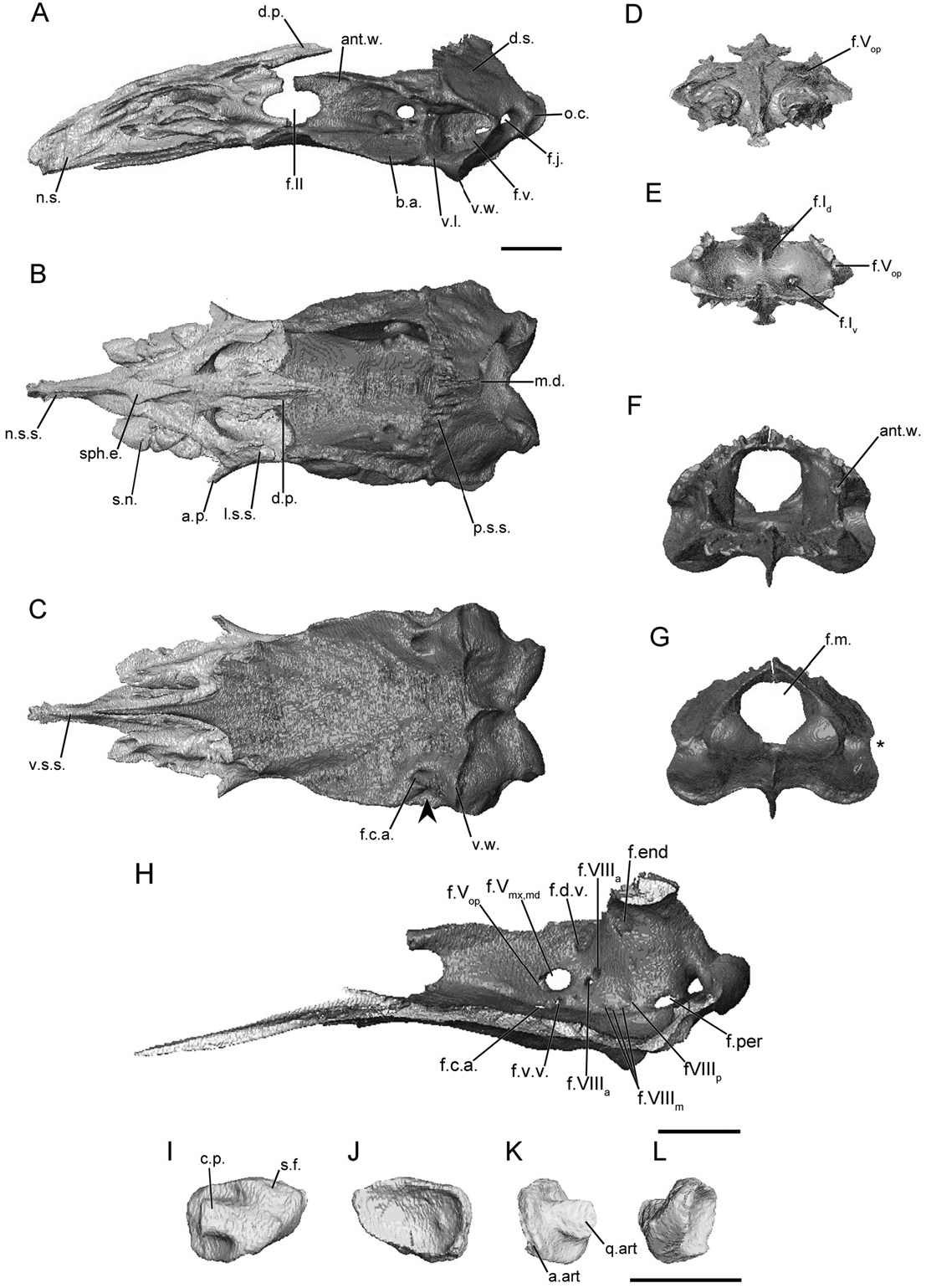
CAECILIIDAE View in CoL ( FIGS 2, S2–S 3 View Figure 3 )
The main body of the sphenethmoid of caeciliids accounts for less than half of the total length of the element ( Fig. 2A). The lateral wall of the main body is short and is capped by a broad sutural surface for the reception of a large anterolateral lappet of the parietal. The anterolateral corner of the dorsal surface is slightly expanded; however, an ossified anterolateral process is absent ( Fig. 2B). A broad dorsomedial process is present and has a large dorsally exposed region ( Fig. 2B). The exposure is bounded by the nasals, frontals, and parietals. The process does not reach the level of the antotic wall of the os basale in Cae. tentaculata ( Fig. 2A), but does so in Cae. volcani and Oscaecilia ochrocephala ( Figs. S2A and S 3A View Figure 3 ). The ·
posterior margin of the lateral wall is quite deeply incised by the anterior margin of the oval optic foramen ( Fig. 2A). The posterior margin of the floor of the sphenethmoid is almost completely divided by a deep medial incision. Otherwise the floor is extensive and broad, and its posterior margin is only slightly concave. The ventral surface of the main body bears a pair of grooves that form the dorsal portion of a canal for the palatal ramus of the facial nerve ( Fig. 2C).
The nasal region is divided by a moderately long, ossified nasal septum ( Fig. 2A) terminating just posterior to the external naris. It is relatively uniform in height throughout its length in the species of Caecilia ( Fig. 2A), but it tapers dramatically in O. ochrocephala ( Fig. S3A View Figure 3 ). The broad dorsal sutural surface along the main body of the sphenethmoid extends onto the nasal septum ( Fig. 2B). The surface tapers in width distally, but terminates relatively more broadly than those of several other species. Sola nasi are absent.
The foramina in the anterior wall, which transmit the dorsal and ventral branches of the olfactory nerve ( Maddin, 2011) are paired, as they are in the majority of species examined here ( Fig. 2D, E). The foramina of each pair are located close to either side of the midline of the sphenethmoid. The dorsal foramina lead to canals just beneath the sutural surface of the nasal septum, exiting almost half way along the length of the nasal septum. The ventral foramina also extend into short canals before exiting into the nasal capsule. An anterolateral foramen transmitting the ophthalmic branch of the trigeminal nerve ( Maddin, 2011) pierces the base of the anterolateral expansion of the sphenethmoid.
In dorsal view the anterior portion of the antotic wall of the os basale runs parallel to the contralateral antotic wall ( Fig. 2B). Posteriorly it is bowed laterally at the level of the antotic foramina. In anterior view the antotic wall is vertical ( Fig. 2F). The dorsal sutural surface is extremely thin, but expands posteriorly over the antotic foramina, and is continuous with that along the anterior margin of the otic capsule. The anterior margin of the antotic wall is incised by the posterior margin of the oval optic foramen ( Fig. 2A). There are two foramina present in each antotic wall ( Fig. 2H). Both trunks of the trigeminal nerve, as well as the facial nerve and ventral vein, pass through the large antotic foramen ( Maddin, 2011). A dorsal vein exits through a much smaller, posterodorsal foramen. This is referred to here as Pattern 6.
The dorsal surface of the otic-occipital complex is strongly inclined posteroventrally. The surface tapers slightly in width towards the midline. A broad region is exposed dorsally posterior to the parietal, and it also bears an extensive sutural surface along its anterior margin that receives the parietal ( Fig. 2B). The dorsal surface extends laterally, creating a thin shelf dorsal to the fenestra vestibuli for the species of Caecilia ( Fig. 2A), but not for O. ochrocephala ( Fig. S3A View Figure 3 ). The fenestra vestibuli is small and ovoid longitudinally and orientated roughly horizontally. In posterior view the fenestra vestibuli deeply incises the lateral margin of the otic-occipital complex for the species of Caecilia ( Figs 2G, S2G), but does not for O. ochrocephala ( Fig. S3G View Figure 3 ). In lateral view the occipital condyle is extremely well developed, protruding far posterior to the otic capsule. A large jugular foramen is present in the base of the occipital condyle, and is visible in lateral view. The foramen magnum is diamond-shaped.
The medial wall of the otic capsule contains seven or eight foramina ( Fig. 2H). The endo- and perilymphatic foramina, and the smaller foramina for the anterior and posterior branch of the vestibulocochlear nerve, are present in their typical locations, as described in Maddin (2011). Variably within the family three or four small foramina form an arc along the ventral margin of the medial wall of the otic capsule and these conduct the medial branch of the vestibulocochlear nerve.
The floor of the os basale of caeciliids is concave dorsally, rather than flat. The anterior margin tapers continuously to a point, and terminates beneath the nasal septum of the sphenethmoid. A central ridge corresponds to the medial incision of the floor of the sphenethmoid, and the main body of the sphenethmoid overlies much of the remainder of the anterior portion of the floor. A pair of weak depressions is located between the antotic walls. This corresponds to the location of the paired cerebral hemispheres. A deeper depression is located posteriorly, between the otic capsules. This corresponds to the location of the hypophysis of the brain ( Maddin, 2011), and is particularly well defined in caeciliids ( Fig. 2B, F). Lateral to the antotic wall the floor expands dramatically into well-developed basicranial articular surfaces ( Fig. 2A, B). In ventral view the lateral margin of the floor is constricted slightly, just posterior to the basicranial articulation (arrowhead; Fig. 2C). A welldeveloped, wing-like projection is present under each otic capsule ( Fig. 2A, C), demarcating the boundaries of the attachment site for the m. longus capitis ( Bemis et al., 1983). Posteriorly the floor of the os basale terminates at a rounded point, which almost reaches the ventral margin of the foramen magnum.
A foramen that leads to a canal for the carotid artery perforates the floor of the os basale anterior to the otic capsule ( Fig. 2C). The canal coursing anteriorly from this foramen bifurcates into lateral and medial canals, which terminate at foramina located on the lateral and medial sides of the antotic wall, respectively. Both foramina occur at roughly the equivalent positions on either side of the antotic wall, ventral to the large antotic foramen ( Fig. 2H).
The footplate of the stapes of caeciliids is markedly elongated anteroposteriorly, with its long axis orientated horizontally ( Fig. 2I, J). The footplate is closely applied to the anterior margin of the fenestra vestibuli. The articulation between the footplate and margin of the fenestra vestibuli has previously been described as a synchondrosis ( de Jager, 1939; Wever, 1975). The remainder of the margin of the footplate does not contact the cartilaginous joint with the fenestra vestibuli, but it closely approaches its margins. The medial surface of the footplate is concave. The columellar process extends anterodorsally from the lateral surface of the footplate. It is long and slightly depressed dorsoventrally, and greatly expanded distally into a broad articular surface that contacts the quadrate ( Fig. 2K). The lateral margin of the columellar process is reduced to a thin ridge ( Fig. 2L). There is no stapedial foramen present.
No known copyright restrictions apply. See Agosti, D., Egloff, W., 2009. Taxonomic information exchange and copyright: the Plazi approach. BMC Research Notes 2009, 2:53 for further explanation.
|
Kingdom |
|
|
Phylum |
|
|
Class |
|
|
Order |
|
|
Family |