
Caligus ogawai, Venmathi Maran & Ohtsuka & Shang, 2012
|
publication ID |
https://doi.org/ 10.12782/sd.17.2.201 |
|
persistent identifier |
https://treatment.plazi.org/id/701FA908-D23A-FFBB-17FA-7FC74DCDF830 |
|
treatment provided by |
Felipe |
|
scientific name |
Caligus ogawai |
| status |
sp. nov. |
Caligus ogawai sp. nov.
( Figs. 5 View Fig , 6 View Fig , 7 View Fig )
Material examined. Holotype: Ovigerous ♀ (KMNH IvR 500,501), Nagasu , off Shodo Island, Seto Inland Sea, Kagawa Prefecture, Japan (St. 3), 7 October 2008 . Paratype: 1 adult but not ovigerous ♀ (KMNH IvR 500,502), Suonada , Seto Inland Sea, Japan (St. 9), 28 October 2010 . Allotype: adult ♂ (KMNH IvR 500,503), off Hirado Island , Nagasaki Prefecture, Japan (St. 10), 29 October 2010 ( Table 1) .
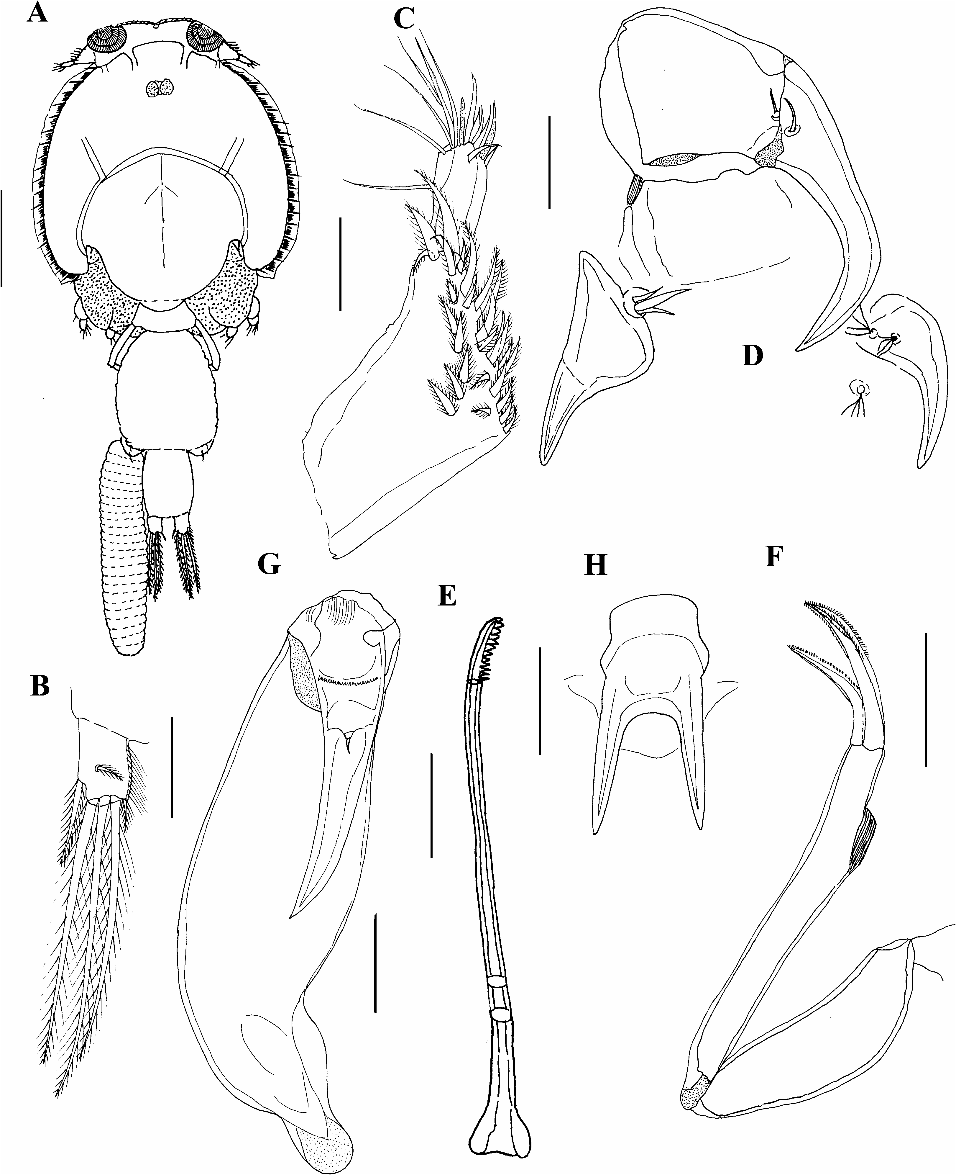
Description. Female (based on holotype). Body ( Fig. 5A View Fig ) 4.65 mm long excluding caudal setae. Cephalothorax suborbicular, slightly longer than wide, 2.53× 2.23 mm. Fourth pediger partly concealed by cephalothorax, wider than long, 0.38, 0.40×0.57, 0.58 mm. Genital complex slightly tapering anteriorly, longer than wide, 1.09× 0.90 mm. Abdomen longer than wide, 0.64× 0.46 mm. Caudal ramus ( Fig. 5B View Fig ) longer than wide, 0.15× 0.12 mm, with 3 long terminal setae, 2 short subterminal setae, and 1 seta located at midlength. Egg string, uniseriate, containing 25 eggs.
Antennule ( Fig. 5C View Fig ) 2-segmented; proximal segment distinctly longer than distal, armed with 27 setae; distal segment armed with subterminal seta on posterior margin and 11 setae plus 2 aesthetascs around apex. Antenna ( Fig. 5D View Fig ) 3-segmented; proximal segment small, unarmed, produced into small dentiform process postero-medially; middle segment subrectangular with 1 small seta distally; distal segment drawn out into curved claw bearing 1 small seta proximally. Postantennal process ( Fig. 5D View Fig ) weakly curved, bearing 2 basal papillae each, with 4 setules; another similar papilla located on nearby sternite. Mandible ( Fig. 5E View Fig ) styliform with 12 marginal teeth subapically. Maxillule ( Fig. 5D View Fig ) comprising dentiform posterior process and anterior papilla bearing 3 short setae. Maxilla ( Fig. 5F View Fig ) slender, 2-segmented; distal segment slender, bearing subterminal hyaline membrane on outer edge and 2 large, curved elements ornamented with serrate hyaline membrane. Maxilliped ( Fig. 5G View Fig ) indistinctly 3-segmented; proximal segment robust, unarmed; middle segment flap-like, indistinct; distal subchela comprising incorporated endopodal segment plus distal claw, armed with minute seta at midlength. Sternal furca ( Fig. 5H View Fig ) with proximal subquadrate box and long, acutely pointed tines.
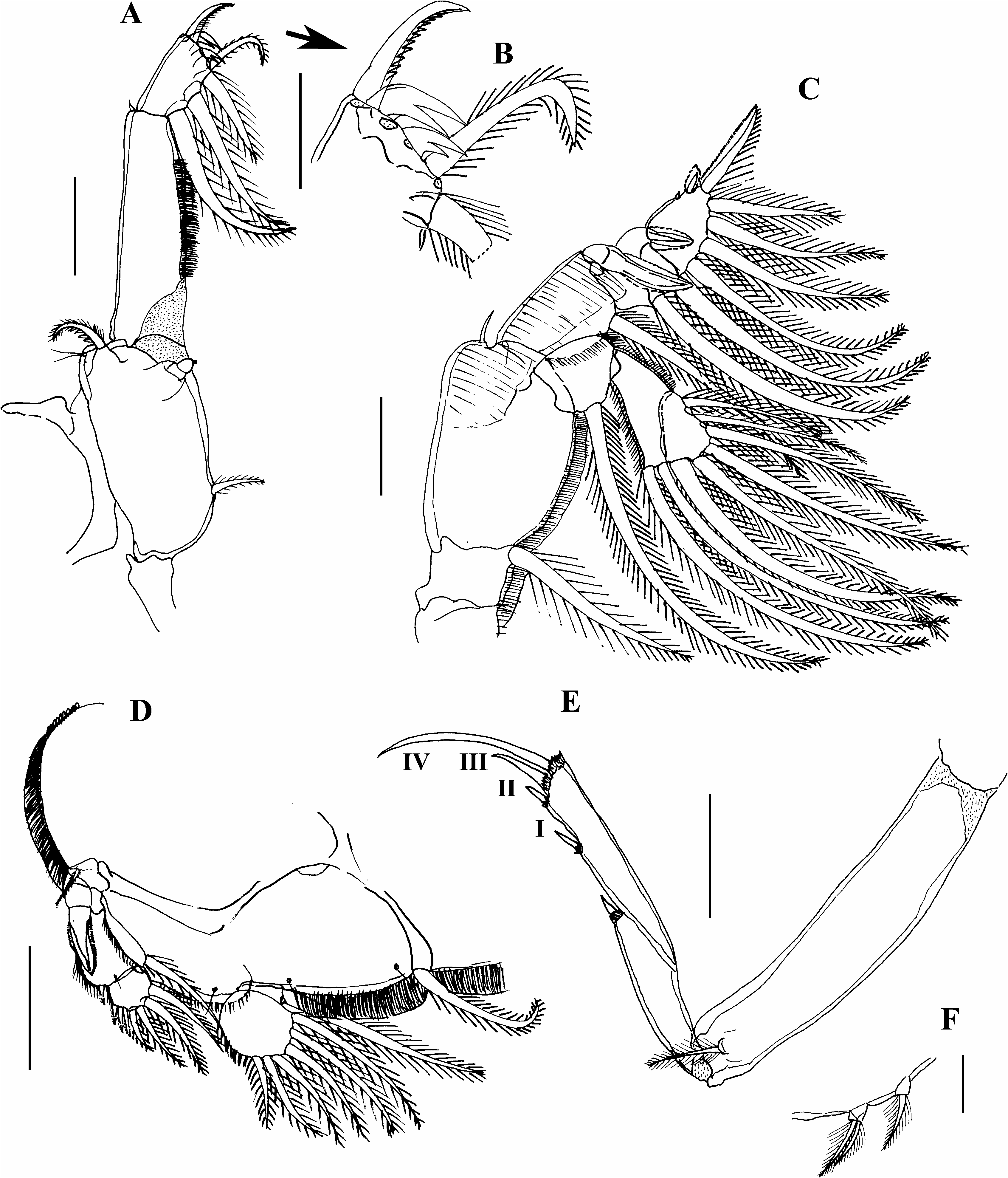
Armature on rami of legs 1–4 summarized in Table 3.
Leg 1 ( Fig. 6A View Fig ) protopod consisting of coxa with papilla on outer margin carrying 2 setules, and long, plumose outer (basal) and short, plumose inner (coxal) setae; vestigial endopod tipped with setules; middle 2 of 4 terminal elements on distal margin of second exopodal segment bifid ( Fig. 6B View Fig ). Leg 2 ( Fig. 6C View Fig ) with coxa having large, plumose inner seta; basis with small, naked outer seta; both outer and medial edges of basis fringed with marginal membrane; similar membrane present dorsally on outer margin of first segment of exopod; distal segment of exopod with very small proximal outer spine. Leg 3 ( Fig. 6D View Fig ) protopod with small, plumose outer seta on posterior marginal membrane. Leg 4 ( Fig. 6E View Fig ) with 2-segmented exopod; all outer exopodal spines with pecten at base, outer terminal spine longer than adjacent spines. Leg 5 ( Fig. 6F View Fig ) represented by 2 papillae at posterolateral corner of genital complex, these being tipped with 2 and 1 plumose setae, respectively.
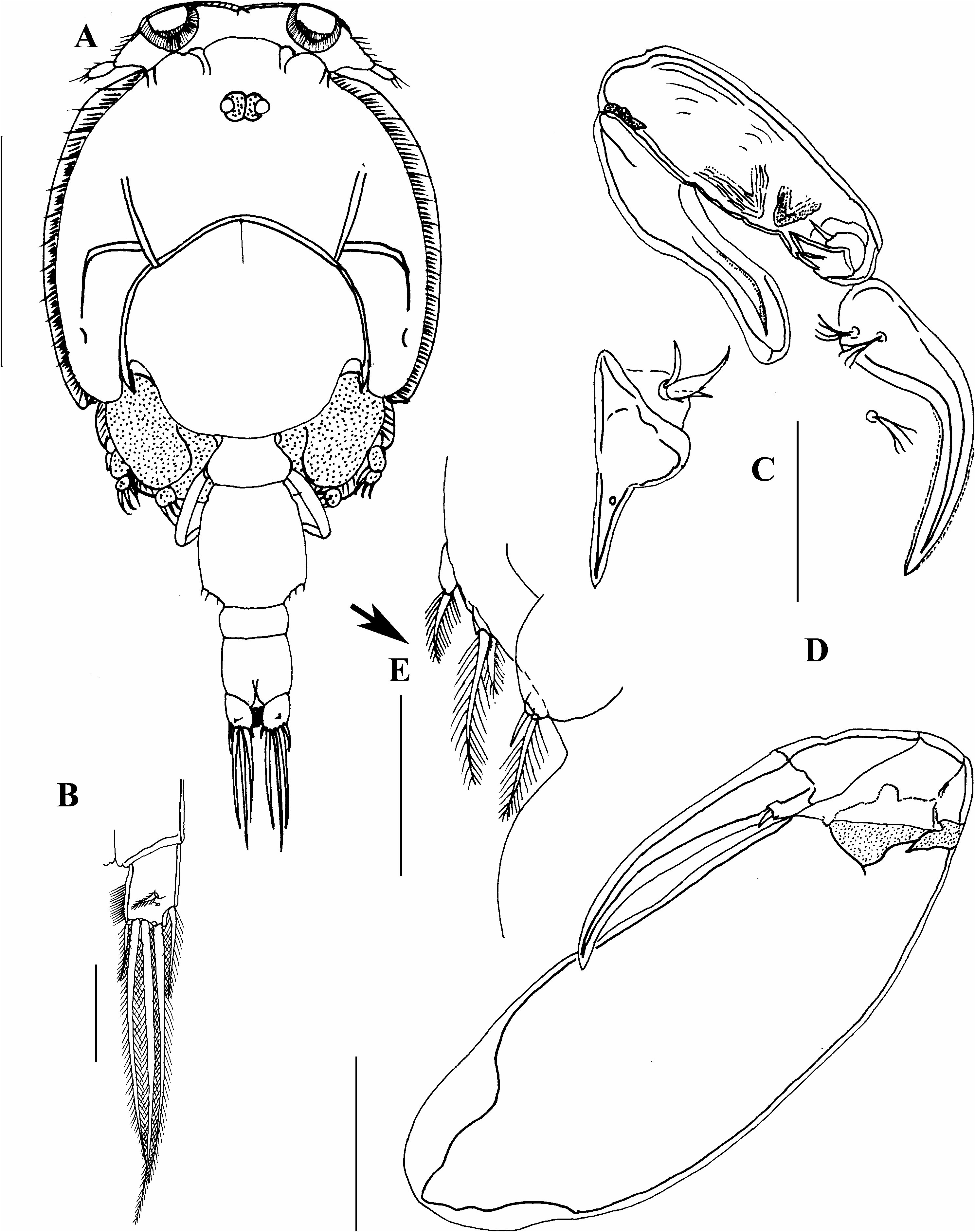
Description. Male, allotype: Body ( Fig. 7A View Fig ) 3.65 mm long excluding caudal setae. Cephalothorax suborbicular, longer than wide 2.18× 1.90 mm wide. Fourth pediger free, 0.21× 0.43 mm, wider than long. Genital complex 0.62× 0.56 mm, vase-shaped, longer than wide, with legs 5 and 6 projecting slightly. Abdomen 0.50× 0.34 mm, 2-segmented, 1st segment small, 2nd segment longer than wide. Caudal ramus 0.17× 0.12 mm, longer than wide, setae as in female ( Fig. 7B View Fig ).
Antenna ( Fig. 7C View Fig ) 3-segmented; proximal segment slen- der, unarmed; middle segment largest, armed with 2 slightly corrugated adhesion pads distally, distal segment armed with basal seta and 2 sclerotized plates. Postantennal process ( Fig. 7C View Fig ) generally as in female, curved, ornamented with hyaline membrane, bearing 2 basal papillae, with 4 setules each, another similar papilla located on nearby sternite. Maxillule ( Fig. 7C View Fig ) distally pointed, with small pore at mid length. Maxilliped ( Fig. 7D View Fig ) robust as in female and bearing 1 seta midlaterally. Leg 5 ( Fig. 7E View Fig ) as in female, represented by 2 papillae at posterolateral corner of genital complex, one bearing 1 small seta and another papilla bearing 1 long and 1 small seta. Leg 6 ( Fig. 7E View Fig ) located near insertion of abdomen, represented by papilla tipped with 1 long seta and 1 small spine.
Remarks. The female of the new species is characterized by the following features: (1) the dentiform process of the antennary proximal segment is tiny and pointed; (2) the maxilliped has one minute seta at the midpoint of the subchela; (3) the sternal furca is long and broad with sharp tines; and (4) the leg 4 exopod is armed with I-0; I,III (IV) spines ( Fig. 6E View Fig ), with the terminal spine markedly longer than the other spines. The male is characterized by having the following features: (1) a simple maxilliped; (2) a 2-segmented abdomen; and (3) leg 6 bearing a long seta and spine. The new species is closely similar to C. savala Gnanamuthu, 1948 in the general body structure and sternal furca but, differs in the tiny, pointed dentiform process of the antennary proximal segment (larger in C. savala ). Also, the maxilliped has a minute seta at the middle region of the subchela in C. ogawai , while in C. savala it has a proportionally longer seta located distal to the midlength of the subchela ( Gnanamuthu 1948; Pillai 1985). Leg 4 is armed with an equal number of spines in C. savala , but the new species differs in having them arranged as a series of smaller spines leading to a long terminal spine ( Fig. 5E View Fig ).
The new species is similar to the following congeners in the leg 4 armature of the female (I-0; I,III): Caligus latigenitalis Shiino, 1954 ; C. arii Bassett-Smith, 1898 ; C. dactylus Ho, Lin and Chang, 2007 ; C. hobsoni Cressey, 1969 , and C. rufimaculatus Wilson, 1905 , but can be differentiated from them as follows: from C. latigenitalis by the abdomen (rounded vs rectangular in the new species) and sternal furca (simple tines vs tines with flanges in the new species); from C. arii by the abdomen (larger and quite long vs small and rectangular in the new species) and the postantennal process (absent vs present in the new species); from C. hobsoni by the abdomen (2-segmented vs 1-segmented in the new species); and from C. rufimaculatus by the sternal furca (short, blunt tines vs long, pointed tines with flanges in the new species) ( Cressey 1969; Izawa and Choi 2000; Suàrez Morales et al. 2003; Ho and Lin 2004a).
Concerning the male characteristics, C. latigenitalis is again similar to the present new species, but the former markedly differs from the latter in the structure of the maxilliped. In C. latigenitalis , the maxilliped has a myxal area on the stout corpus comprising three unequal protuberances ( Izawa and Choi 2000), but it is simple with no protuberances in the new species ( Fig. 7D View Fig ). All these congeners here considered have been found exclusively as parasites on teleosts, except for C. latigenitalis and C. rufimaculatus , which have also been reported from plankton in Japan ( Venmathi Maran et al. 2012a) and Mexico (Suàrez Morales et al. 2003), respectively.
Etymology. This new species is named in honor of retired Prof. Kazuo Ogawa (University of Tokyo); currently at Meguro Parasitological Museum, in recognition of his major contributions to marine parasitology.
No known copyright restrictions apply. See Agosti, D., Egloff, W., 2009. Taxonomic information exchange and copyright: the Plazi approach. BMC Research Notes 2009, 2:53 for further explanation.
|
Kingdom |
|
|
Phylum |
|
|
Class |
|
|
Order |
|
|
Family |
|
|
Genus |