
Coeliccia macrostigma Laidlaw, 1918
|
publication ID |
https://doi.org/ 10.11646/zootaxa.4184.1.5 |
|
publication LSID |
lsid:zoobank.org:pub:2312F650-EB9C-457E-B3AD-71BC3984A30A |
|
DOI |
https://doi.org/10.5281/zenodo.6087469 |
|
persistent identifier |
https://treatment.plazi.org/id/03B7F636-FFAF-FFE4-4CB6-8FC9FDB0D46C |
|
treatment provided by |
Plazi |
|
scientific name |
Coeliccia macrostigma Laidlaw, 1918 |
| status |
|
Coeliccia macrostigma Laidlaw, 1918 View in CoL
Figs. (5, 6, 7, 14, 15, 16, 23, 24, 29, 30, 35, 36, 41, 42, 47, 48, 53, 54, 59, 60, 65, 68, 72)
Coeliccia macrostigma Laidlaw 1918: 225 View in CoL –227, Figs. 5, 6 View FIGURES 5 – 13 (original description both sexes from Baram, female specimen collected day before male in 1910, accidently destroyed after description);— Laidlaw 1920: 333;— Laidlaw 1932: 35 – 36;— Lieftinck 1954: 48 (part, Sarawak);— Kimmins 1970: 180 (notes on holotype);—Thompson & van Tol 1993: 69 (Sungai Ingei, Brunei);— Orr 2001: 187 (site 24, Brunei);— Orr 2003: 39, 78, 79 (part).
Material. Holotype. Ƌ, Baram , [ Sarawak], Borneo, 20 x 1910, in BMNH.
Other material. In collection Dow unless otherwise noted. Sarawak, Malaysia. 1 Ƌ, dry peat swamp forest near Sungai Dabai , Marudi, Miri division, 5 ix 2011, leg. RAD, in RMNH ; 1 ♀, Mentawei Boundary Trail (kerangas forest), Gunung Mulu National Park, Limbang division, 13 ii 2006, leg. J. Simun.
Belait district, Brunei. 2 Ƌ, peat swamp forest near camp, Sungai Ingei area , ulu Belait, 23 ii 2014, leg. RAD; 1 Ƌ, Sungai Ingei Kanan, same area, 24 ii 2014, leg. RAD; 4 Ƌ (2 in RMNH), kerapa forest near Sungai Ingei, same area, 3 iii 2014, leg. RAD; 2 Ƌ, 1 ♀, same location and date, leg. Minoh ; 1 Ƌ, 1 ♀, swamp forest mosaic, same area, 6 iii 2014, leg. RAD; 1 Ƌ, same location, 9 iii 2014, leg. Minoh ; 2 Ƌ (one missing most of abdomen), “guarding small pools in swamp”, Badas stream, 16 xii 1995, leg. A.G. Orr, in coll. Orr ; 1 Ƌ, peat swamp forest, Kuala Belai Road , 2 iii 2013, leg. RAD; 7 Ƌ (including BRU 13 View Materials _PCD42, used for the description below) , 2 ♀ (one in tandem with one of the males), same location, 4 iii 2013, leg. RAD; 3 Ƌ , same location, 21 iii 2013, leg. RAD; ♀ ( BRU 13 View Materials _PCD10, used for the description below) , same location, 24 iii 2013, leg. RAD; 2 Ƌ, 1 ♀ (in tandem with one of the males) , same location, 27 v 2013, leg. RAD; 3 Ƌ, peat swamp forest, sawmill site by Sungai Belait, 5 iii 2013, leg. RAD; 2 Ƌ, small stream at same location, 5 iii 2013, leg. RAD; 1 Ƌ , same location, 1 vi 2013, leg. RAD; 7 Ƌ (1 in RMNH) , 1 ♀ (in RMNH), peat swamp forest beside Sungai Belait, 3 iii 2013, leg. RAD; 3 Ƌ, peat swamp forest adjacent to “sand beach”, Sungai Belait, 25 v 2013, leg. RAD; 5 Ƌ , 1 ♀, same location, 28 vi 2013, leg. RAD; 2 Ƌ, in peat swamp forest adjacent to burnt sand ridge, Lumut Pipeline Road, 6 iii 2013, leg. RAD; 4 Ƌ , 3 ♀, peat swamp forest, same area, 8 iii 2013, leg. RAD; 1 Ƌ, same habitat and area, 13 iii 2013, leg. RAD; 5 Ƌ (1 in RMNH), peat swamp forest accessed from Labi Road ca 17-18 km from Labi, 11 iii 2013, leg. RAD. Tutong district, Brunei. 1 Ƌ, 1 ♀, Telsai [Telisai], 13 xii 1983, leg. Harman, in CUMZ.
Description of male (based on BRU13_PCD42). Head: Labium pale cream except hooks of labial palps, which are black. Labrum shining black. Mandible bases shining black, sub-rectangular pale area in lower anterior corner. Clypeus and most of lower part of genae shining black, anteclypeus with obscure grey areas, pale blue band running from eye margin to clypeus and narrowly for short distance above clypeus ( Fig. 6 View FIGURES 5 – 13 ). Antenna with top part scape and base pedicel white, remainder dark brown and black. Frons and vertex mostly black, short whitish stripe based on outer edge of lateral ocellus, directed towards rear of but not reaching antenna base, small pale mark at eye margin ( Fig. 5 View FIGURES 5 – 13 ). Ocelli white. Whitish, elongate oval, transverse postocular spots. Underside of head ( Fig. 7 View FIGURES 5 – 13 ) black with large bluish-white marks at eye margins, just extending onto genae, narrowing towards point of attachment with prothorax, narrowly connected (right) or just separated (left) from postocular markings.
Thorax: Prothorax with propleuron entirely pale except narrowly black in upper rear corner. Upper part of notopleural projection clearly present ( Fig. 24 View FIGURES 23 – 28 ), pale, fused to lower lateral part of anterior pronotal lobe. Posterior and anterior pronotal lobes black. Anterior carina of anterior pronotal lobe slightly lower than main part, which is rounded at top in lateral view, sloping to rear. Middle lobe mostly pale, black centrally and to rear on dorsum, in lateral view rounded and dome-like ( Fig. 24 View FIGURES 23 – 28 ). Posterior pronotal lobe simple, collar-like, raised along free margin. Synthorax with mesepisternum black with large blue antehumeral markings ( Fig. 35 View FIGURES 35 – 40 ), extending from short distance to rear of mesostigmal plate to just beyond apex of antealar triangle, occupying more than half width of mesepisternum near prothorax, outer margin following mesepleural suture for most of length but diverging from it abruptly shortly before level of apex of antealar triangle, inner margin running approximately parallel to mid-dorsal carina except ca semicircularly but irregularly excised centrally. Mesepimeron black except narrowly blue above interpleural suture for some distance and tiny pale mark in upper corner adjacent to antealar carina ( Fig. 36 View FIGURES 35 – 40 ). Metepisternum largely blue with black stripe running from antealar carina most of way towards spiracle and small black area below interpleural suture immediately to rear of extension of blue above suture. Metepimeron pale. Venter of synthorax pale. Mesinfraepisternum black except lower corner adjacent to metepisternum, metinfraepisternum entirely pale. Legs with coxae and trochanters almost entirely pale, femur mostly pale with black stripe along extensor surface, black stripe along outer flexor surface in distal ca 2/3 of anterior femur only, tibia mostly pale, dark along flexor surface, tarsi pale with obscure darker areas, claws brown. Wings with arc situated slightly distal to Ax 2. Fw with 16 Px, Hw with 15 (left) or 17 (right) Px. Three postquadrilateral cells in all wings. R4 at (Hw) or very slightly proximal (Fw) to Sn. Pt black with narrow, almost complete, white margin, almost rhomboidal, covering one underlying cell entirely and ca 1/4 to 1/3 of another.
Abdomen: S1 whitish, black in narrow apical annulus including posterior carina and area behind, broadly at base on dorsum, narrowing apically. S2 pale laterally, dark brown dorsally, this widest apically; faint narrow pale stripe centrally on dorsum basally and pair of faint obscure pale marks subapically. S3–7 pale lower laterally, becoming brown dorsally, this darkening on successive segments so black by apical part S7, pale basal annulus, interrupted dorsally, on each segment, bright on S3, faint on others, very faint and obscure paler subapical area on S3–5, apical dark annulus. S8 mostly pale blue lower laterally, otherwise black ( Figs. 47, 48 View FIGURES 47 – 52 ). S9 almost entirely pale blue, except small but broad-based black dorsal subtriangular marking and narrow wedge from base along lower ca 3/4 segment length. S10 pale blue except narrow, short basal black dorsal triangle and narrowly apically. Cerci mostly blue laterally and dorsally in outer half, mostly black interiorly and ventrally. In dorsal view ( Fig. 59 View FIGURES 59 – 64 ), a broad epiproct just visible, cercus with basal tooth visible, expanded on inner margin at ca half-length, this expansion rounded and curving gently out to apex which is situated on outer margin, which is approximately straight from base to apex, a distinct ridge running diagonally forwards from apex almost to inner margin. In lateral view ( Fig. 60 View FIGURES 59 – 64 ) subrectangular, upper margin directed rather abruptly downwards towards apex, which is slightly hooked downward, rounded, central tooth with apex at ca half-length, large. Paraprocts mostly pale in visible parts, black at tips, relatively short, tips at ca level of those of cerci; in ventral view narrowing gently after base along outer margin, then curving gently out again before turning in just before apices, which are rounded and not quite pointing at each other, with excised appearance in apical half of inner margin ( Fig. 65 View FIGURES 65 – 67 ). In lateral view paraprocts narrow after base, running approximately parallel to cercus until just before apices, where turned inward and upward. Genital ligula typical for the membranipes -group.
Measurements (mm): Abdomen without anal appendages 35.5, cerci ca 0.75, Hw 22.
Description of female (BRU13_PCD10). As male except as noted. Head: Pale markings on mandible bases more extensive ( Fig. 15 View FIGURES 14 – 22 ), obscure grey marking centrally on anteclypeus. Pale marking between median and each lateral ocellus, another running diagonally rearwards from eye margin to almost behind antenna base, parallel to but not in line with that running from lateral ocellus toward antenna base ( Fig. 14 View FIGURES 14 – 22 ). Postocular markings separated from markings on underside of head ( Fig. 16 View FIGURES 14 – 22 ).
Thorax: upper cervical spur poorly developed, subtriangular, lower cervical spur well developed, broad, overlapping propleuron but well separated from notopleural projection, rounded. Very large lower part of notopleural projection ( Figs. 29, 30 View FIGURES 29 – 34 ), in form of near vertical rounded flap, concave to front, upper part large but less prominent, fusing smoothly with lower lateral part of anterior pronotal lobe. Anterior carina of anterior pronotal lobe approximately as high as rounded top of main part, which slopes at rear. Middle pronotal lobe strongly dome-like in lateral view. Posterior pronotal lobe with short lapels, dorsally flattened, apex below level of highest part of middle lobe, in dorsal view straight sided interiorly at base, rounded toward apex ( Fig. 29 View FIGURES 29 – 34 ); horn narrow, longer than middle lobe measured centrally, slanted gently forward in lateral view ( Fig. 30 View FIGURES 29 – 34 ). Middle lobe only black narrowly centrally ( Fig. 29 View FIGURES 29 – 34 ). Synthorax with mesostigmal plates bearing fringe of long setae on low ridge at rear, depressed at front and obscured, but appear small. A few long, erect, setae on quarter of mesepisternum behind prothorax. Antehumeral markings similar to male but separated into two at position of excision in male; rearward section narrowly separated from mesepleural suture for whole length. Fw 17 Px, Hw 16 Px. Pt greyish brown.
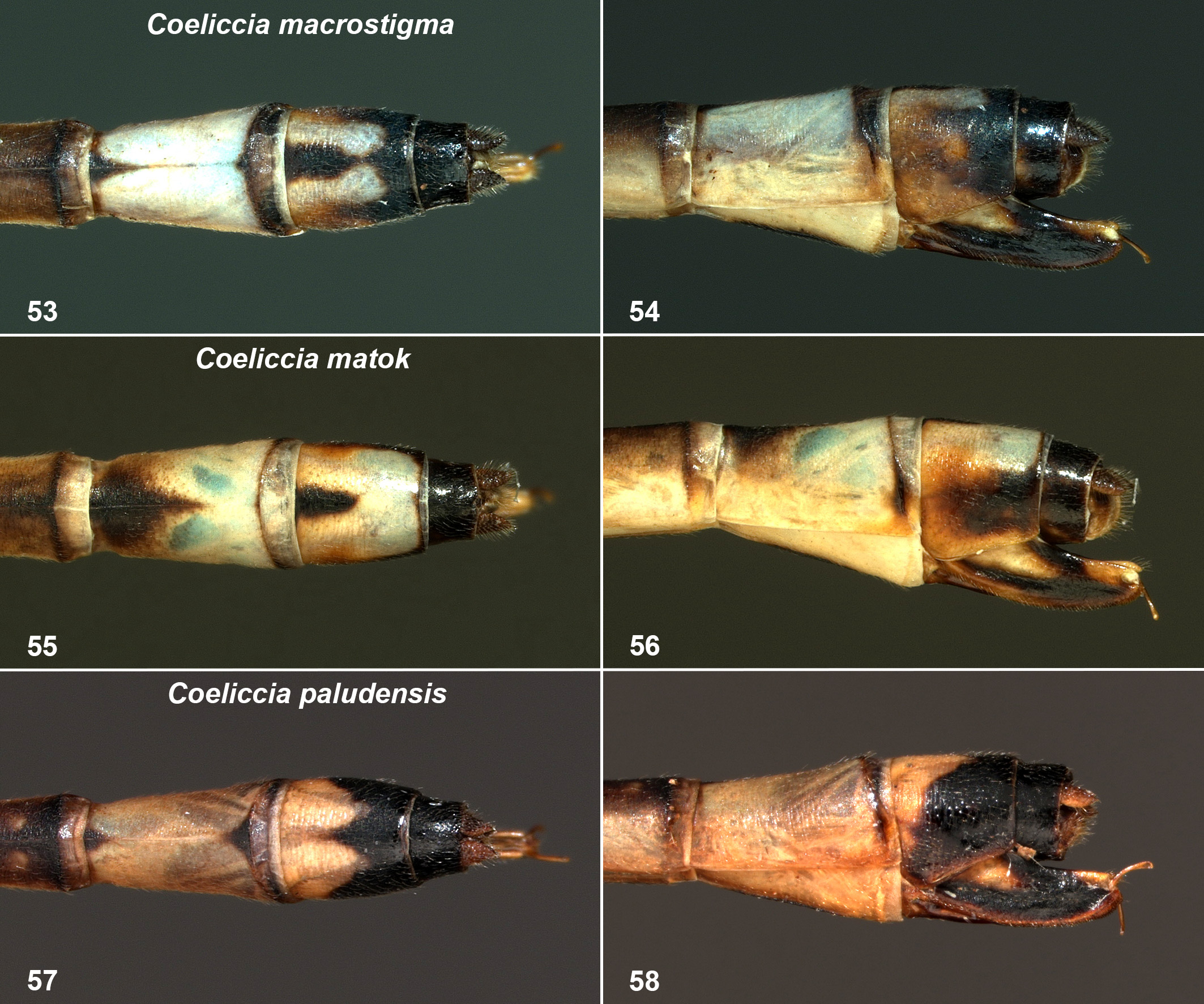
Abdomen: Coloration overall similar to male but slightly less dark above. S8 pale blue except subtriangular basal dark brown dorsal mark, apex produced as line extending to posterior carina. S9 black apically and dorsally, laterally brown in basal two-thirds, merging with pair of elongate subrectangular blue dorsal marks ( Figs. 53, 54 View FIGURES 53 – 58 ). S10 black with small brown basal lateral area. Cerci shorter than S10, dark brown. Ovipositor mostly black, basal pale patch, brown along lower margin, pale mark dorsally near apex, extending well beyond cerci.
Measurements (mm): Abdomen with anal appendages or ovipositor 33, Hw 22.3.
Variation. Males. There is considerable variation in the exact shape of pale markings on the mandible bases. In a few individuals the markings on the vertex are as in the female described, or intermediate. The postocular markings are frequently disconnected from the markings on the underside of the head, which are sometimes further subdivided. R4 is sometimes well proximal to Sn, occasionally slightly distal to it. One individual has two postquadrangular cells in the left Hw. The pale border of Pt is sometimes less complete, and sometimes only one underlying cell is covered. There is some variation in the obscure pale marks on S2. The basal annuli on S3–7 are frequently bright all the way to S6, occasionally to S7. There is sometimes a pair of small pale dorsal apical markings on S8, very occasionally connected to the lateral pale colouration. The basal parts of the marking on S9 vary considerably in length and occasionally have a rearward spur lower laterally, the exact shape of the dorsal part also varies. The cerci are occasionally black; the basal tooth is sometimes not visible and might actually be absent in a few individuals (removal of the paraprocts would be necessary to check this). The position of the central tooth of the cercus appears to vary to some extent, occasionally appearing slightly more distal or more apical than in the male described and illustrated. The paraprocts are sometimes a little shorter relative to the tips of the cerci in lateral view.
Females. The extent of pale colouration on the mandible bases is variable, sometimes the entire lower twothirds pale. The postocular markings are sometimes connected to the pair of markings on the underside of the head. In one female the mark between left lateral ocellus and median ocellus is smaller, in another that on the corresponding position on the right side is absent. In one specimen the lower cervical spur is less well developed. In some individuals the lower part of the notopleural projection is more strongly concave to the front, giving the impression of being strongly folded forward, so that the separation from the lower cervical spur is less; in one specimen it is hardly concave or folded forward at all on one side. There is some variation in the length of the prothoracic horn; at minimum it is subequal to the length of the middle posterior lobe centrally. The horn is sometimes more strongly slanted forward and sometimes curved; in the female from Gunung Mulu National Park it overlaps the anterior pronotal lobe. The antehumeral stripes are sometimes not broken on one side or both, or only very narrowly broken. One individual has three Ax in the left Hw, the same specimen has two postquadrangular cells in both wings on the right side; another has three-and-a-half postquadrangular cells in the left Fw. The colour of the Pt is sometimes dark brown. There is sometimes a brown basal lateral mark on S8. The size of the dorsal basal mark on S8 is variable, and sometimes its apex is only around half segment length. The extent of paler markings on S9 is also variable.
Measurements (mm): Males 15–19 Px in Fw, 13–16 in Hw; abdomen without anal appendages 31–38; Hw 20–22. Females 14–17 Px in Fw, 14–16 in Hw; abdomen without anal appendages and ovipositor 31.5–35.5; Hw 21–22.5.
Remarks. Coeliccia macrostigma is a distinctive species, easily distinguished from all other members of the membranipes -group. The male can be separated from all species except C. nigrohamata , C. matok , C. octogesima , C. resecta and C. paludensis by the antehumeral stripes with an excision on the inner side, as opposed to either linear or crescentic without excision ( C. nemoricola and allies and C. membranipes ), or expanded so as to cover almost the entire mesepisternum ( C. cyaneothorax ). There is an as-yet-undescribed species in Sarawak and Brunei, occurring in different habitats, that has variable antehumeral markings which sometimes approximate those of C. macrostigma , but that differs clearly in the anal appendages. The male is distinguished from those of C. nigrohamata , C. matok , C. octogesima , C. resecta and C. paludensis by the simple shape of the excision in the antehumeral stripes and the form of the male anal appendages. The female is distinguished from all species except C. paludensis by the very large lower part of the notopleural projections, without any nipple-like projection, from C. paludensis in that the lower part of the notopleural projection forms a vertical or almost vertical rounded flap, concave to front, whereas in C. paludensis it is almost flat at the top, and by the shape of the anterior pronotal lobe. Lieftinck (1954) gives the distribution of this species as west and northwest Borneo and the habitat as “Lowland forests of Sarawak and western Borneo”; these statements appear to be extrapolation based on the types and a series of C. nigrohamata misidentified as C. macrostigma in RMNH, from the Singkawang area of northwest Kalimantan, collected in the 1930s. Unfortunately Lieftinck’s statements are wildly inaccurate. The male and female from Baram (clearly the Baram river area and almost certainly the section between the mouth of the river and Long Lama, where the peat swamp habitats are, or were), the male from Marudi and the female from Gunung Mulu National Park are the only records actually of this species outside of Brunei, which on the basis of the data available now appears to have a restricted range in the lower Baram area of Sarawak and Brunei’s Belait district, with one record from the Telsai area in Tutong district, immediately adjacent to Belait district. All records for which details of habitat are known are either from peat swamp forest or from kerapa forest (intermediate between peat swamp forest and kerangas forest). Peat swamp and kerapa habitats in the lower Baram area are mostly already lost and what remains is mostly extremely degraded; searches in peat swamp forest in the Marudi area (quite possibly where the types were collected since this area was readily accessible in 1910, when Marudi was known as Claudetown) in 2011 yielded only a single specimen. The only other recent specimen from Sarawak is a female from a part of Gunung Mulu National Park very close to the border with Brunei. At some locations in Belait district the species is abundant.
The female specimen described by Laidlaw (1918), was collected in “Baram” on 19 October 1910, the day before the holotype male. Unfortunately this female specimen was, as Laidlaw (1918: 227) notes, destroyed while in his possession, but not before he had made the descriptive notes used in the paper; he states that “the evidence that they belong to the same species is not conclusive”, but the description he gives broadly agrees with the female of C. macrostigma as described above. In particular Laidlaw writes: “The structure of the prothorax is very remarkable. A curious crescentic projection stands out on either side of the middle lobe attached to the prothorax by its convex border. When looked at obliquely from above, each of these projections shows like the moon at the end of the first quarter, but when viewed directly from above each shows as a single outstanding spur.” This description clearly refers to the notopleural projections, and appears to agree with the females described above. The description of the holotype in Laidlaw (1918), although insufficiently detailed and inadequately illustrated, agrees with more recently collected males; moreover, direct comparison of the holotype with these specimens has confirmed that they are the same species.


No known copyright restrictions apply. See Agosti, D., Egloff, W., 2009. Taxonomic information exchange and copyright: the Plazi approach. BMC Research Notes 2009, 2:53 for further explanation.
|
Kingdom |
|
|
Phylum |
|
|
Class |
|
|
Order |
|
|
Family |
|
|
Genus |
Coeliccia macrostigma Laidlaw, 1918
| Dow, Rory A. 2016 |
Coeliccia macrostigma
| Orr 2003: 39 |
| Orr 2001: 187 |
| Tol 1993: 69 |
| Kimmins 1970: 180 |
| Lieftinck 1954: 48 |
| Laidlaw 1932: 35 |
| Laidlaw 1920: 333 |
| Laidlaw 1918: 225 |