
Cyanogomphus angelomachadoi, Pinto, Ângelo Parise & Almeida, Marcus Vinícius Oliveira De, 2016
|
publication ID |
https://doi.org/ 10.11646/zootaxa.4078.1.6 |
|
publication LSID |
lsid:zoobank.org:pub:3D0D79C2-2583-47F4-BC01-5CF347614B44 |
|
DOI |
https://doi.org/10.5281/zenodo.6077749 |
|
persistent identifier |
https://treatment.plazi.org/id/61BDDFBA-202E-41CD-AA6D-2E65CD171CF4 |
|
taxon LSID |
lsid:zoobank.org:act:61BDDFBA-202E-41CD-AA6D-2E65CD171CF4 |
|
treatment provided by |
Plazi |
|
scientific name |
Cyanogomphus angelomachadoi |
| status |
sp. nov. |
Cyanogomphus angelomachadoi View in CoL sp. nov.
LSID urn:lsid:zoobank.org:act:61BDDFBA-202E-41CD-AA6D-2E65CD171CF4 ( Figures 1 View FIGURE 1 , 2 View FIGURE 2 A–B, 3A, 4A–B, 6A–C, 7A–C, 8A–B, 9C, 10A–B, 11A–B, 12)
Cyanogomphus View in CoL sp. nov. — De Almeida et al. (2013): 418 (mention of the material described here from “Parque Nacional da Serra do Cipó”).
Material examined (4♂, 2♀). Holotype ♂. BRAZIL. Minas Gerais State: Jaboticatubas municipality, Parque Nacional da Serra do Cipó ( PNSC),, light trap using a white sheet with mixed mercury vapor lights (250W) at Córrego das Pedras stream (19°22’17”S, 43°36’03”W, 766 m a.s.l.), 12.XII.2011, A.P.M. Santos & D.M. Takiya leg. ( DZRJ 1449); 1♀ Paratype, same data as holotype but 6 meter-long Malaise trap crossing Córrego das Pedras stream, 9–13.XII.2011, A.P.M. Santos, D.M. Takiya, M.L. Monné & R.R. Cavichioli leg. ( DZRJ 1448); 1♂ Paratype, [Lagoa Santa municipality], Estr[ada] B[elo] Horizonte, Serra do Cipó, Km 46–47 [19°33’40.56”S, 43°54’47.21”W, 664 m a.s.l.], Exc[ursão] 221, Col[eta] 30, 01.XII.1963, N.[D.] Santos, [J.P.] Machado & Borges leg. ( MNRJ); 1♂ Paratype, Serra do Salitre municipality, river at riparian Forest, 13.I.2014, J.E. Santos Junior leg. ( ABMM); São Paulo State: 1♂ Paratype, Brotas municipality, Fazenda Santa Lúcia da Boa Vista (22°12’38.88”S, 48°14’35.88”W, 485 m a.s.l.), 02.XI.2001, F.A.A. Lencioni leg. (RWG, not examined). Additional specimen. BRAZIL. Minas Gerais State: 1♀, Serra do Salitre municipality, river at riparian forest, II.2014, J.E. Santos Junior leg. ( ABMM). Specimens in DZRJ collected under ICMBIO/SISBIO licenses numbers 14591-8 and 23620-6.
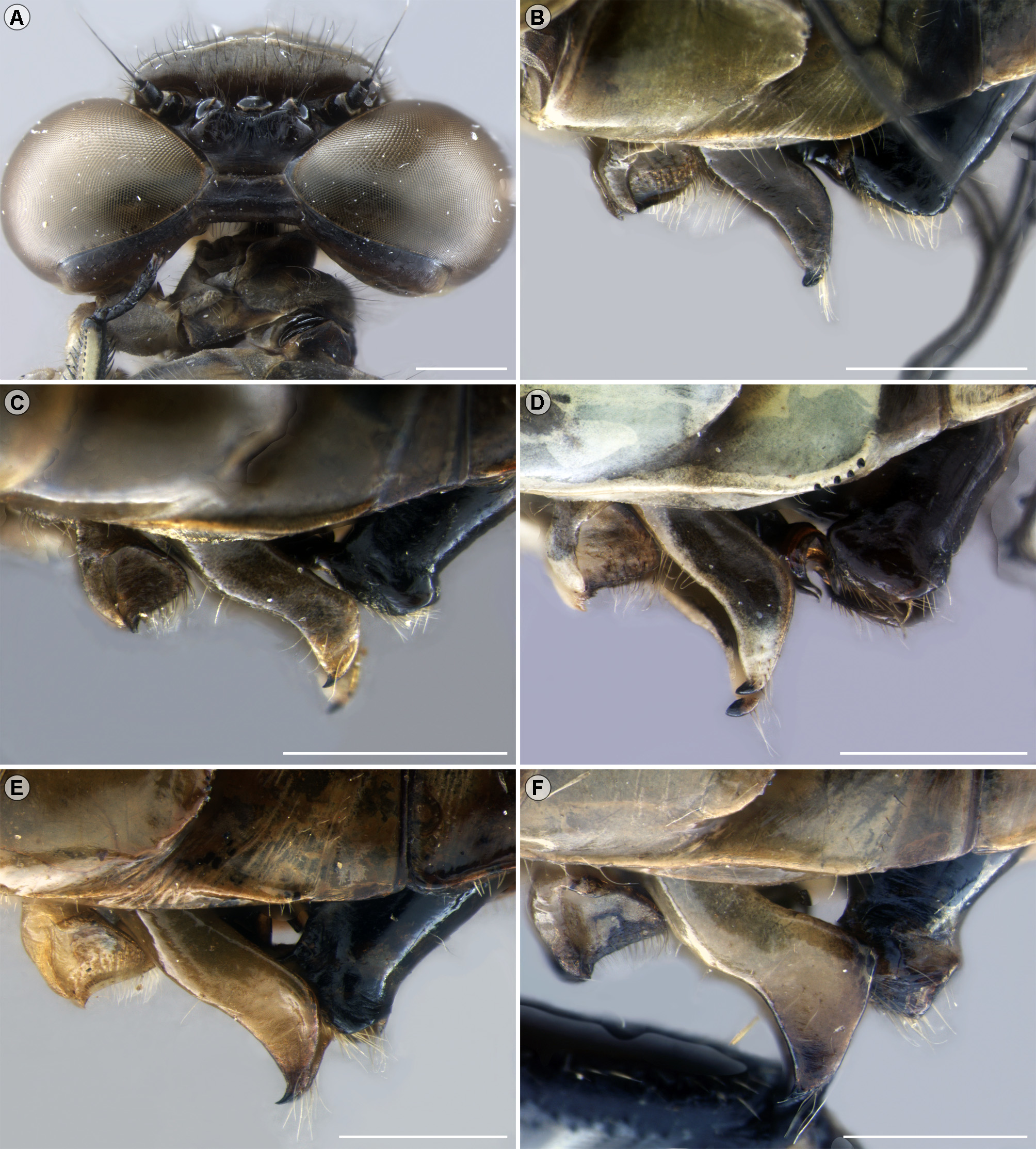
Description of male holotype. Head ( Fig. 4 View FIGURE 4 A). Labium greenish-yellow, visible parts of maxilla and mandible greenish-yellow, except dark-brown to glossy black movable hook, extreme apex of maxillary outer lobe and mandibular incisor lobe. Genae brown. Labrum greenish-yellow, with a narrow straight dark brown to black stripe at ventral margin, one lateral margin with a narrow dark brown line from ventral margin to level of clypeolabral suture, clypeolabral suture light brown connected to a small mesal rounded spot over labrum occupying about half of its total height. Anteclypeus brownish-yellow, clypeus greenish-yellow. Frons yellowishgreen, darker than remainder of face, with dark brown bar at posterior 0.4, extended laterally. Antenna, vertex and occiput dark brown to black, except yellowish membranous part of scape and area close to postocellar suture noticeably lighter than adjacent areas of cranium. Posterior region of cranium (“postgena” plus “occiput”) gradually lightening ventrally with irregular dark areas, especially close to postoccipital suture. Ventral margin of labrum with a wide concavity almost mesal, giving to sclerite a slight asymmetric shape; ante- and postfrons division carinated, dorsal margin of frons sinuous in anterior view with a small mesal concavity, anterior margin regularly convex and with a shallow mesal furrow in dorsal view. Posterior portion of vertex with a ridge (postocellar ridge sensu Belle 1973) slightly projecting anteriorly over lateral ocelli, occiput (occipital plate) in dorsal view with a distinct ridge along its entire width ( Fig. 4 View FIGURE 4 A), area posterior to ridge distinctly higher than anterior, posterior margin of occiput with scattered setae, not forming a distinct fringe.
Thorax. Prothorax greenish-yellow, lightening ventrally to basisternum with dark brown spots or stripes on anterior margin of anterior lobe, an ill-defined spot on dorsum of median lobe and distinct stripes surrounding central pale area of posterior lobe. Synthorax ( Figs 1 View FIGURE 1 A–B) largely pale, patterned with dark brown spots and greenish-yellow stripes dorsally, almost entirely greenish-yellow laterally; dorsal carina dark on anterior third, then with narrow yellowish line; mesepisternum with first and second pale antehumeral stripes distinct, first antehumeral stripe joined to pale mesothoracic collar, to second antehumeral stripe at about half length, and at level of antealar sinus ( Figs 1 View FIGURE 1 A–B); pale areas enclosing three large dark spots, a triangular dark brown mesal spot from collar to antealar sinus, at maximum half pleurite width, a barely defined rounded brown spot from mesinfraepisternum to about of posterior 0.4, and an ellipsoid dark brown spot posteriorly; a narrow brown stripe parallel to the pale mesopleural suture (humeral) beginning at 0.2 height of epimeron and extending to near antealar sinus, enlarged posteriorly; mesepisternum with a dark brown stripe parallel to mesopleural suture darkened and wider ventrally close to mesinfraepisternum ( Figs 1 View FIGURE 1 A–B); metepisternum with a barely defined brown stripe dorsally from metapleural antealar process to intersegmental suture, remainder of synthorax greenish-yellow lightening to light yellow ventrally, with irregular ill-defined dark areas laterally ( Fig. 1 View FIGURE 1 B). Legs, coxa and trochanter yellowish-brown, ventral and posteroventral surfaces of femur light yellow, anteroventral and dorsal surfaces dark brown to black in pro- and mesothoracic legs, in metathoracic leg yellowish-brown with irregular dark areas and distal 0.1 black; tibia black with dorsal surface yellow to brownish-yellow ( Fig. 3 View FIGURE 3 A); tarsus black, pretarsal claws dark brown with distinct supplementary inferior tooth. Proximal portion of trochanter (1 Tr sensu Snodgrass 1935) armed with a row of small black spurs only in the mesothoracic leg, distal portion (2 Tr) armed on all legs, most numerous on mesothoracic leg. Femur with rows of numerous small black spurs, metathoracic femur slight convex anteriorly in lateral view, anteroventral surface of prothoracic tibia armed with 10–12 spurs (including scale-like spurs of tibial comb), 11–12 on mesothoracic, 14–15 on metathoracic, posteroventral surface with 14–15 on prothoracic, 14–15 on mesothoracic, 13–14 on metathoracic, usually at least two times longer than intervening space. Tibial and tarsal spurs modified, basally enlarged ( Fig. 3 View FIGURE 3 A).
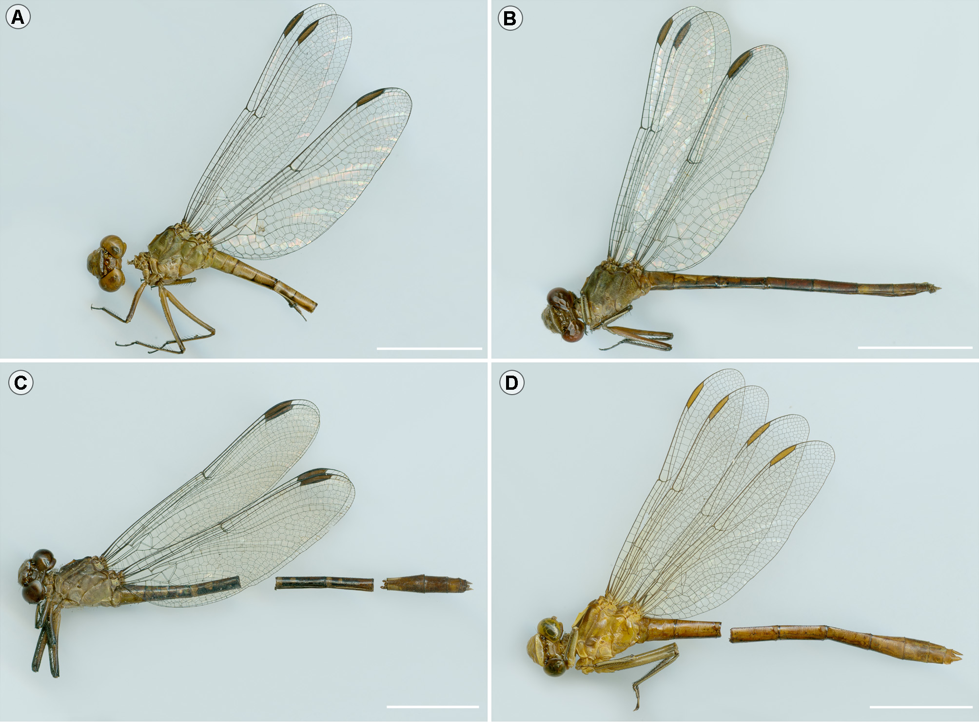
Wings ( Fig. 2 View FIGURE 2 A). Membrane hyaline; venation dark-brown to black; pt brown, contrasting with black of marginal veins; one basal subcostal crossvein in all wings, membranula strongly reduced. Venation as follows: 12–13 Ax in Fw, 9–10 Ax in Hw, Ax2 the 5th in all wings; 9 Px all wings; 8–9 postsubnodals (including brace vein) in Fw, 8 in Hw; 5–6 bridge crossveins in Fw, 4 in Hw; arc at or slightly distal to second Ax; sectors of arc not stalked, origin located 0.5 basal of arc in all wings; 3–4 crossveins in the space between arc and RP-midfork in Fw, 2–3 in Hw; Rspl indistinct in all wings; discoidal triangles, supratriangles and subtriangles not crossed in four wings; space between CuP-crossing and PsA with one crossvein in all wings; Fw discoidal field with two rows of cells up to level of nodus, beyond that three and increasing to 9–11 cells at wing margin; Hw discoidal field divergent, with 4–5 rows of two cells, followed by 1–2 rows of three cells, then broadening to 9–11 cells towards wing margin; Mspl indistinct in the four wings; anal loop rectangular with four cells; seven paranal cells from wing base to apex of subtriangle in Fw, five in Hw; anal triangle distinct, three-celled, with seven strong and short spines along the dorso-proximal margin (AP&AA).
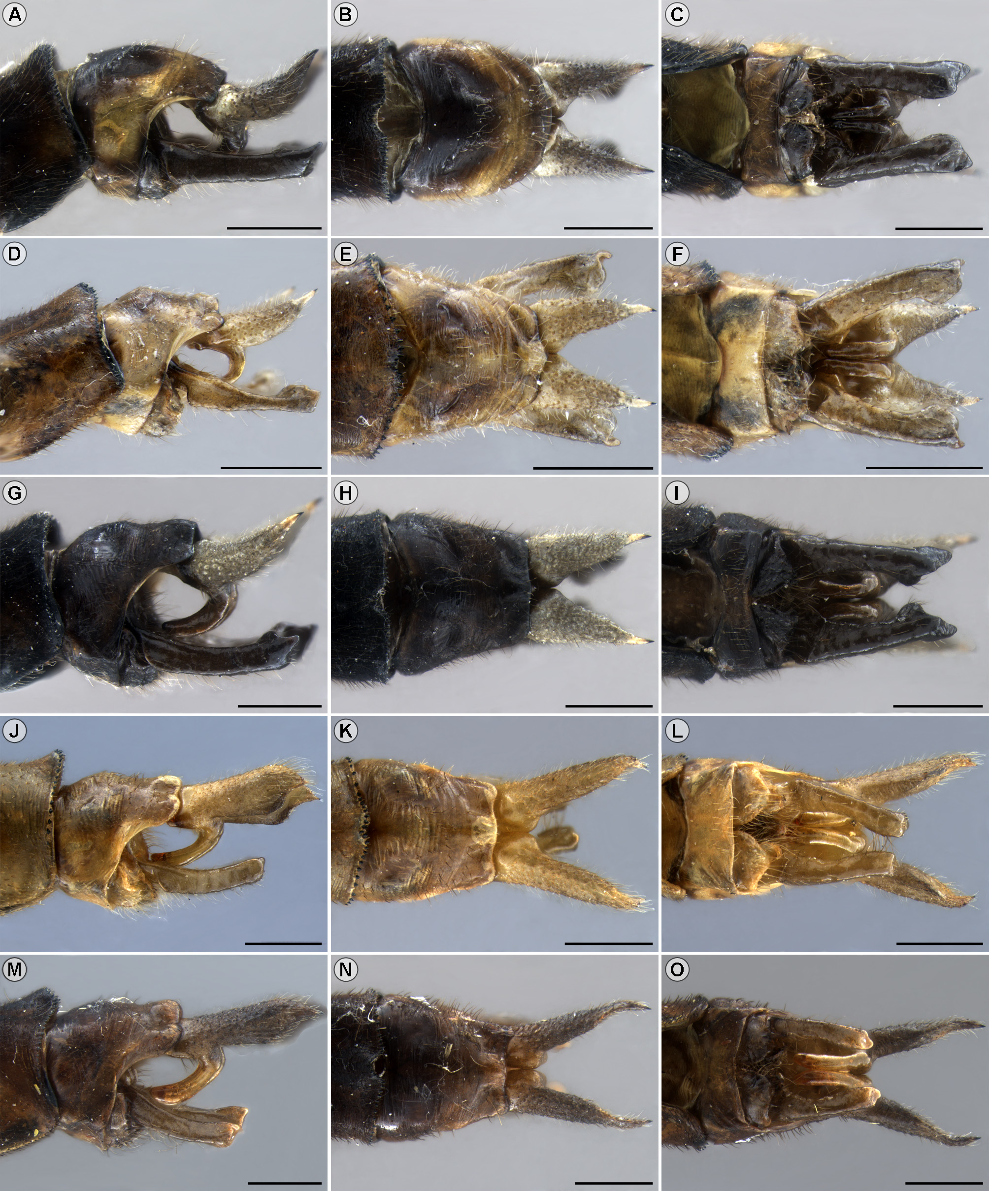
Abdomen ( Fig. 10 View FIGURE 10 A). Dark brown with pale greenish-yellow spots. Tergite of S1 almost entirely greenishyellow expect the dark brown rounded process (swelling of abdominal articulation sensu Asahina 1954) close to posterior carina; S2 with a large pale spot on anterior 0.6, narrowing posteriad to 0.33 of segment height in lateral view, a small pale spot close to posterior carina and a middorsal pale line along entire segment; S3–7 with a pale proximal ring up to transverse carina, darkened in its middle, remainder of segment posteriorly dark brown to black; S8 almost entirely dark brown with an irregular lateroventral pale spot, enlarged at both ends of segment; S9 dark brown with ill-defined lateral lighter areas; S10 dark brown above, with pale lateral spot extended dorsally in a narrow ring at 0.66 length separating a rounded basal spot from the dark posterior end ( Figs 7 View FIGURE 7 A–C, 10A); sternites dark brown to black; ventral carina of S2–8 and anterior 0.5 of S9 greenish-yellow; S1–7 cylindrical, S8–10 distinctly wider than previous segments, maximum width at S8. Auricles rounded, positioned obliquely to S2 longitudinal axis, length about one-third height of S2; posterior margin with two patches of 5–6 minute dark spines; posterior carina of S9 armed with small spines dorsally and with V-shaped concavity. Secondary genitalia typical of the Agriogomphus -complex, anterior lamina not visible in lateral view, in ventral view posterior margin with a wide U-shaped ridge, anterior hamule ( Fig. 4 View FIGURE 4 B) yellowish-brown, quadrangular in lateral view, anterior margin strongly ridged, sickle-shaped, posteriorly prolonged into a concave laminar projection with scattered hairlike setae denser posteriorly, a small ventral V-shaped concavity between anterior ridge and posterior laminar projection; in ventral view posterior margin convergent, the limit between anterior ridge and posterior laminar projection concave in a wide V-shape; posterior hamule ( Fig. 4 View FIGURE 4 B) light brown, except black stout acute anteriorly directed apex, distinctly longer than other structures of secondary genitalia in lateral view, anterior margin slightly sinuous, slightly convex at proximal 0.4 then gently concave, with small deep concavity before extreme distal; two distinct sets of pale hair-like long setae, a basal line with 8–10 setae at anterior margin and a distal tuft with about a dozen setae close to acute apex ( Fig. 4 View FIGURE 4 B); in ventral view regularly curved toward convergent black apexes, somewhat flattened; without a distinct genital lobe. Vesica spermalis ( Fig. 4 View FIGURE 4 B) with V1 black, in lateral view slightly higher than level of anterior hamule, scoop-like process on V1 (median cleft sensu Belle 1988) rounded in front view, strongly concave and covered with hair-like yellow setae internally; see paratype for full description. Cercus ( Figs 7 View FIGURE 7 A–C, 8A–B) greenish-yellow spotted with brown by numerous tubercular setal alveoli proximally, darkening to black apex, ventral branch dark brown ( Fig. 7 View FIGURE 7 A); in lateral view cercus strongly curved at half-length at level of epiproct posteriorly, directed obliquely dorsad ( Figs 7 View FIGURE 7 A, 8A); apex acute and slightly directed posteriorly; posterior margin flattened, forming a distinct rounded keel ( Fig. 7 View FIGURE 7 A); ventral branch strongly curved in lateral view with apex directed anteriorly ( Figs 7 View FIGURE 7 A, 8A); cercus in dorsal view cylindrical, tapering posteriorly to acute apex, slightly divergent ( Fig. 7 View FIGURE 7 B); mesal margin with a rounded basal projection, then gradually tapering, external margin almost straight ( Fig. 7 View FIGURE 7 B); cerci in ventral view touching each other proximally and gradually diverging distally, apexes almost parallel, ventral branch strongly directing parallel toward anteriorly, apex rounded, slightly flattened ( Fig. 7 View FIGURE 7 C). Epiproct dark brown, largely bifid, posteriorly at level of cercus ( Figs 7 View FIGURE 7 A, C, 8A–B); almost straight in lateral view, with a dorsal and ventral ridge at proximal 0.25, a small tubercle-like rounded process on dorsal margin at ca. distal 0.70 ( Figs 7 View FIGURE 7 A, 8A–B); laterodistal projection forefinger-like, apex extending obliquely upward, posterior margin slightly concave ( Figs 7 View FIGURE 7 A, 8A–B); in dorsal view slightly convergent, dilated at about posterior 0.70 arms surrounding a deep quadrangular concavity (hereafter distal concavity, Fig. 8 View FIGURE 8 B); in dorsolateral view with a strong tubercle-like process anterior to the concavity ( Fig. 8 View FIGURE 8 B); in ventral view almost straight, laterodistal projection distinctly posterior to internal margin, posterior margin slightly sinuous ( Fig. 7 View FIGURE 7 C).
Measurements. Total length (including caudal appendages) 40.5; abdomen length (excluding caudal appendages) 29.2; head maximum width 5.7; Fw length 25; Hw length 23.5; Fw maximum width 5.6, in Hw 7.2; pt length 2.8 in Fw, 2.9 in Hw; length of metathoracic femur 6.8; metathoracic tibia 5.4; length of S9+ 10 in lateral view 3.3; total length of cercus in lateral view 1.4.
Description of female paratype. Similar to holotype, only the differences described here. Head. Genae greenish-yellow. Dark brown to black stripe at ventral margin of labrum prolonged laterally to dorsal 0.5. Frons uniformly light brown, darker than remainder of face. Vertex and occiput light brown, the latter slightly paler. Posterior region of cranium (“postgena” plus “occiput”) light brown with irregular pale areas. Ante- and postfrons division slightly carinated. Occiput (occipital plate) behind carina clearly swollen, posterior margin sinuous with a mesal concavity.
Thorax. Prothorax with posterior lobe entirely greenish-yellow. Synthorax with dark areas slightly paler than in holotype ( Figs 1 View FIGURE 1 E–F, 11A); pale area over dorsal carina larger. Legs with dark areas on anteroventral and dorsal surfaces more extensive; tibia dark brown to black with dorsal surface yellow ( Fig. 11 View FIGURE 11 A). Anteroventral surface of prothoracic tibia armed with 11 spurs, 12–13 on mesothoracic, 16 on metathoracic, posteroventral surface of prothoracic tibia with 14, 16 on mesothoracic, 13–15 on metathoracic, usually more than two times longer than intervening space.
Wings ( Fig. 2 View FIGURE 2 B). Venation as follows: 14 Ax in Fw, 10 in Hw, 9 Px in Fw, 10–11 in Hw; 10–11 postsubnodals (including brace vein) in Fw, 9–10 in Hw; 7 bridge crossveins in Fw, 4–6 in Hw; arc at or slightly distal to second Ax; 4 crossveins in space between arc and RP-midfork in Fw, 2 in Hw; Fw discoidal field increasing to 10–11 cells at wing margin; Hw discoidal field with 4–6 rows of two cells, followed by 1–2 rows of three cells, then broadening to 11 cells toward wing margin; anal loop barely defined with 6 cells; 6–7 paranal cells from wing base to apex of subtriangle in Fw; anal wing base rounded without distinct anal triangle and strong spines along of the dorso-proximal margin (AP&AA).
Abdomen (only S1–4). Largely pale, greenish-yellow with irregular dark brown transverse stripes and irregular spots, transverse carina ill defined ( Fig. 11 View FIGURE 11 A). S2 yellowish lateroventrally in anterior 0.5, light brown at level of transverse carina (not carinated) and dark brown at posterior field, S3–4 with a dark brown stripe on posterior 0.8 adjacent to ventral carina, enlarging and darkening posteriorly, a transverse dark brown stripe at level of transverse carina and at posterior field, and a dorsolateral oval dark brown spot not connected dorsally, similar to PD spot of Walker (1912).
Measurements. Head maximum width 5.7; Fw length 25.8–26.0; Hw length 24.4–24.5; Fw maximum width 5.7, in Hw 7.2; pt length 2.8 in Fw, 3.0 in Hw; length of metathoracic femur 6.3; metathoracic tibia 5.
Variation in the male paratypes. The general appearance in size and color of the male from Lagoa Santa (MNRJ) is very similar to that of the holotype, though slightly paler, while that from Serra do Salitre (ABMM) is smaller and distinctly darker with first and second humeral stripes clearly defined and limited by dark ground color ( Figs 1 View FIGURE 1 C–D, 10B). They agree with the holotype except as follows (characters observed in one or both specimens): Head. Labrum with the black distal stripe almost restricted to ventral margin, mesal dark spot longer, occupying 0.66 of its total length (height). Anteclypeus greenish-yellow. Frontoclypeal suture with a distinct brownish stripe widening dorsolaterally over frons. Thorax. Prothorax with dark brown areas better defined. Synthorax with larger and clearly better defined dark areas; mesepisternum at level of junction between first and second pale antehumeral stripes only slightly paler than anterior and posterior dark areas, pale areas not enclosing dark spots, showing a regular gomphid pattern with defined mesepisternal stripes ( Figs 1 View FIGURE 1 C–D); metepisternal dark stripe better defined. Legs, coxa and trochanter greenish-yellow; tibia dark brown to black with dorsal surface yellow. Anteroventral surface of prothoracic tibia armed with 10–11 spurs, 11–12 on mesothoracic, 14–18 on metathoracic, posteroventral surface 13–14 on prothoracic, 13 on mesothoracic, 14–16 on metathoracic. Wings ( Fig. 10 View FIGURE 10 B). Membrane hyaline, very slightly smoked with light brown. Venation as follows: one Hw lacking basal subcostal crossvein; 11–13 Ax in Fw, 10 Ax in Hw, Ax2 the 4th in one Fw and one Hw; 7–10 Px in Fw, 7–8 in Hw; 8–9 postsubnodals (including brace vein) in Fw, 9–10 in Hw; 5–7 bridge crossveins in Fw, 5 in Hw; arc at or 0.3 proximal to second Ax; 3–5 crossveins in the space between arc and RP-midfork in Fw, 2–3 in Hw; Fw discoidal field 9–12 cells at wing margin; Hw discoidal field with 2–5 rows of two cells, followed by 1–2 rows of three cells, then broadening to 9–13 cells towards wing margin; anal loop barely defined with five cells; 6 paranal cells from wing base to apex of subtriangle in Fw, 4 in Hw; 6–9 spines along of the dorso-proximal margin (AP&AA). Abdomen. S2 with the large pale spot with irregular margin; S7 with a pale spot entirely greenish-yellow; S8 with a round pale spot at anterior 0.33; S10 distinctly paler than former segments with ill-defined paler areas. Posterior margin of auricles with irregular rows containing 8–14 minute spines, 5 setae at the line of basal hair-like setae on anterior margin of posterior hamule. Vesica spermalis (based on paratype from Lagoa Santa, Figs 6 View FIGURE 6 A–C). Sclerotized parts dark brown to black; ventral surface of V1 with two processes, a small rounded one at proximal third and a large strong scoop-like process at distal third (median cleft sensu Belle 1988), the latter laminar in lateral view, in anteroposterior view with converging rounded apexes and covered with hair-like yellow setae internally ( Figs 6 View FIGURE 6 A, C); V2 tubular, lacking a defined H; V3 globose in ventral view, dorsal surface (process ‘f’ of Pfau 2011) with a rounded, slightly sclerotized distal process ( Figs 6 View FIGURE 6 B, h-f) prolonged over joint between V3 and V4 (J v3-v4), ventrally with a distinct proximal sclerite ( Figs 6 View FIGURE 6 A, p-ps), and a membranous-like hood distally over V4, tapering into a tongue-like apex ventrally ( Figs 6 View FIGURE 6 A, “mh”); V4 cornet-like, with a pair of lateroproximal sclerotized projections ( Figs 6 View FIGURE 6 A-B, “lsp”), curved anteriorly in lateral view ( Fig. 6 View FIGURE 6 A), pair of long distal flagella somewhat forcipate in ventral view ( Fig. 6 View FIGURE 6 B, d-fl), in lateral view as long as V4 length ( Fig. 6 View FIGURE 6 A). Cercus brownishyellow to dark brown to black, ventral branch light brown to dark-brown. Epiproct brown to almost black.
Measurements. Abdomen length (excluding caudal appendages) 27.5–29.2; head maximum width 6.0 (specimen from Serra do Salitre not measured, eyes smashed); Fw length 23.4–25.0; Hw length 22.2–23.8; Fw maximum width 5.1–5.2, in Hw 6.4–6.7; pt length 2.6–2.7 in Fw, 2.6–2.8 in Hw; length of metathoracic femur 6.0–6.3; metathoracic tibia 4.6–4.8; length of S9+ 10 in lateral view 2.8; total length of cercus in lateral view 1.2–1.3.
Larva. Unknown.
Diagnosis. The wide cornet-like distal portion of V4 ( Figs 6 View FIGURE 6 A–B), upturned tapered apex of cylindrical cercus ( Figs 7 View FIGURE 7 A–C, 8A–B), distal concavity from posterior 0.7 of epiproct of males ( Fig. 8 View FIGURE 8 B) and short subgenital plate (ratio between length of plate and S9 <0.4) with a wide concavity of females ( Figs 9 View FIGURE 9 C), will separate Cyanogomphus angelomachadoi sp. nov. from Tibiagomphus (V4 distal portion narrow, apex of cercus posteriorly directed, epiproct dorsally smooth, lacking concavity, and female subgenital plate large and with a narrow mesal incision, Figs 6 View FIGURE 6 F–G, 7J– O, 8G–J, 9E).
The dark mesal part of the mesepisternum close to the dorsal carina ( Fig. 1 View FIGURE 1 ), epiproct with a larger distal concavity at posterior 0.70 ( Fig. 8 View FIGURE 8 B) and dark abdomen with pale spots ( Figs 10 View FIGURE 10 A–B, 11A–B) distinguish this species from C. comparabilis (mesal part of mesepisternum pale, epiproct with distal concavity reduced to a small posterodorsal area, abdomen almost uniform in color, Figs 7 View FIGURE 7 E, 8F, 10C). Cyanogomphus angelomachadoi sp. nov. is similar in general appearance to C. waltheri , with which the smallest specimens might be confused. Cyanogomphus angelomachadoi sp. nov. can be distinguished by its smaller size (length of Hw 22.2–24.5 vs. 25.8–31.6 for C. waltheri ); general paler color ( Figs 10 View FIGURE 10 A–B, 11A–B), with large pale areas connected on mesepisternum ( Figs 1 View FIGURE 1 A–B, E–F), except the paratype male from Serra do Salitre ( Fig. 10 View FIGURE 10 B), which is distinctly darker with pale areas only partially connected and showing well-defined mesepisternal stripes limited by dark ground color ( Figs 1 View FIGURE 1 C–D), much like some C. waltheri specimens ( C. waltheri pale areas less broad, with first and second antehumeral stripes well defined); dorsal surface of metathoracic tibia yellow to brownish-yellow ( Fig. 3 View FIGURE 3 A, black in C. waltheri , Fig. 3 View FIGURE 3 B), distal concavity on epiproct larger (beginning at <0.76 length of epiproct, Fig. 8 View FIGURE 8 B; while in C. waltheri at> 0.80 length, Fig. 8 View FIGURE 8 D) with laterodistal projection in lateral view longer and slender, forefinger-shaped ( Figs 7 View FIGURE 7 A, 8A; in C. waltheri epiproct laterodistal projection smaller and stout, thumb-shaped, Figs 7 View FIGURE 7 G, 8C); and cercus upright ( Figs 7 View FIGURE 7 A, 8A) will distinguish it from C. waltheri (cercus less curved upright, Figs 7 View FIGURE 7 G, 8C). Both species appear to be allopatric with C. angelomachadoi sp. nov., thus far known as a representative of the Cerrado, and C. waltheri , a representative of the Atlantic Forest ( Fig. 12 View FIGURE 12 ).
Distribution. States of Minas Gerais and São Paulo, Brazil, in the Cerrado biogeographical province ( Fig. 12 View FIGURE 12 ).
Biological and ecological data. Adults were collected at lotic systems, near rivers, streams, and creeks, surrounded by forests (riparian) in rupestrian formations over ironstone outcrops known as “canga”, at 664–766 meters in the core of Serra do Espinhaço mountain range (PNSC and Lagoa Santa sites) and at 1205 meters of elevation in the Serra da Canastra formation (Serra do Salitre site) in the Brazilian Cerrado domain. The pair from PNSC was caught using a Malaise trap and light sheet, non-traditional methods to capture dragonflies ( De Almeida et al. 2013). Despite more than 300 km between sites in PNSC and Lagoa Santa (Espinhaço mountain range) from that in Serra do Salitre (Canastra mountain range), they are similar environmentally, typical of the Brazilian Cerrado in that region; temperature, altitude, vegetation and rock outcrops are common to both regions. However, the paratype from São Paulo was collected at 485 meters in an apparently different environment, a sedimentary basin located in the north of Serra Geral rather than rupestrian formation where other specimens were collected.
Etymology. angelomachadoi (noun phrase in genitive singular formed by a given and surname) specific name in honor of our friend and eminent Brazilian odonatologist Angelo B. M. Machado on the occasion of his 80th birthday.
Remarks. The only bona fide female from PNSC (DZRJ) is damaged, lacking S5–10 ( Fig. 11 View FIGURE 11 A), precluding a full description, while the complete female collected at same site of paratype male from Serra do Salitre (ABMM) is poorly preserved as to general color pattern ( Fig. 11 View FIGURE 11 B), leaving in some doubt an association with this species. It was for this reason excluded from the type series. The Lagoa Santa paratype (MNRJ) is damaged, with head detached, synthorax crushed, and S1–6 varying from slightly to strongly damaged, with the left auricle broken. The male paratype from São Paulo State in RWG collection was not examined; however, it agrees fairly well with other specimens, showing the mesepisternal stripes divided as in the holotype (RWG pers. comm.). The females from Goiás (ABMM) may also be this species (see under Cyanogomphus sp.).


| MNRJ |
Museu Nacional/Universidade Federal de Rio de Janeiro |
No known copyright restrictions apply. See Agosti, D., Egloff, W., 2009. Taxonomic information exchange and copyright: the Plazi approach. BMC Research Notes 2009, 2:53 for further explanation.
|
Kingdom |
|
|
Phylum |
|
|
Class |
|
|
Order |
|
|
Family |
|
|
Genus |
Cyanogomphus angelomachadoi
| Pinto, Ângelo Parise & Almeida, Marcus Vinícius Oliveira De 2016 |
Cyanogomphus
| De Almeida et al. (2013) : 418 |