
Drosichoides
|
publication ID |
https://doi.org/ 10.11646/zootaxa.4497.2.8 |
|
publication LSID |
lsid:zoobank.org:pub:C24797D6-DF05-414C-B3CF-55DAD1676D3F |
|
DOI |
https://doi.org/10.5281/zenodo.5988715 |
|
persistent identifier |
https://treatment.plazi.org/id/03E74547-FFF9-FF90-FF6C-6B2BFA43B3D3 |
|
treatment provided by |
Plazi |
|
scientific name |
Drosichoides |
| status |
|
Drosichoides View in CoL ? haematoptera (Cockerell)
Llaveia haematoptera Cockerell 1919: 272 View in CoL .
Drosichoides haematoptera (Cockerell) View in CoL ; Morrison 1927: 106. Change of combination.
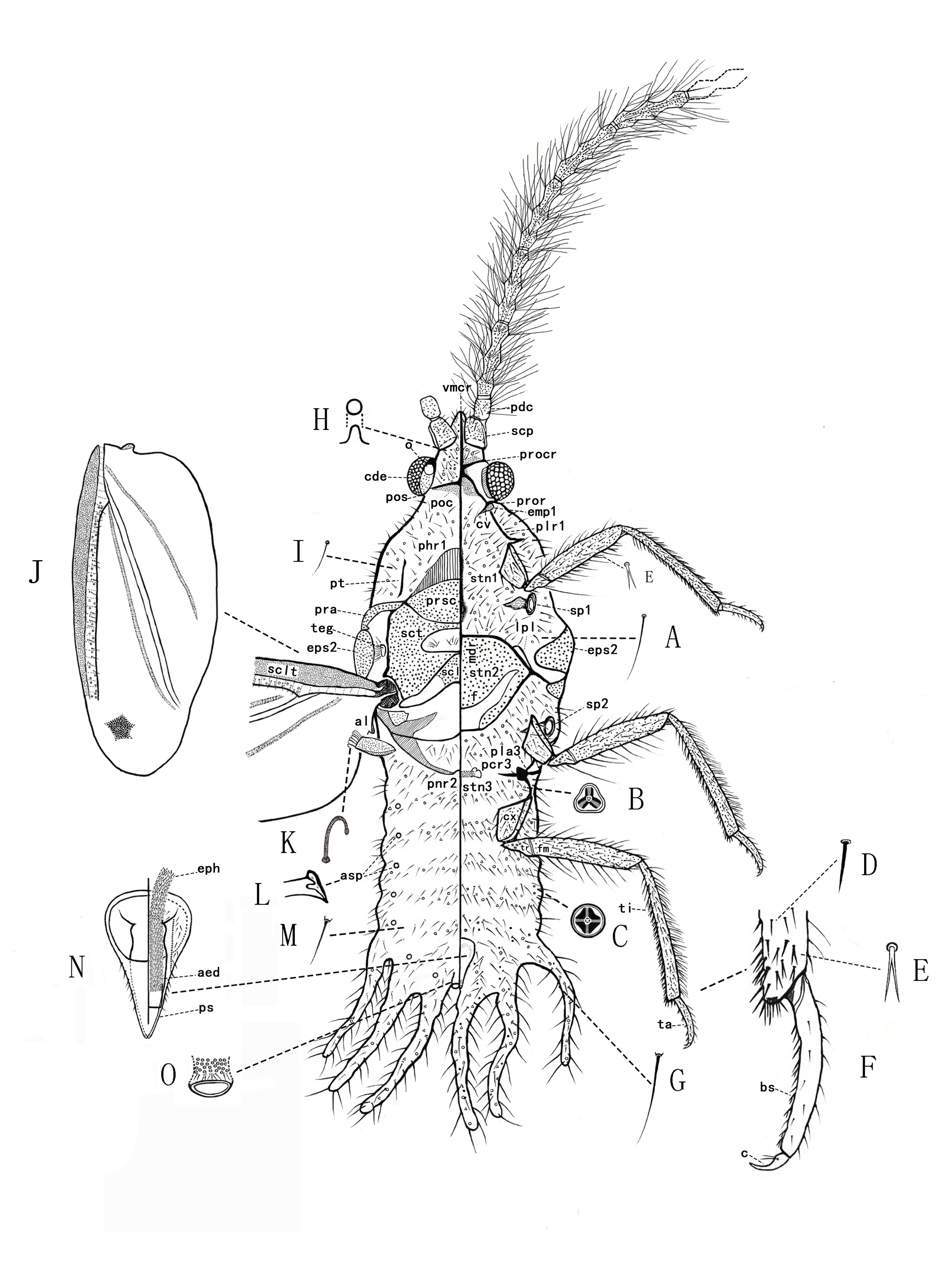
Adult male ( Figs.1 View FIGURES 1–3 and 4 View FIGURE 4 )
Material examined. Two adult males, MALAYSIA: Borneo , Sabah, Keningau district , Mount Trus Madi , Jungle Girl Camp. N5.4430, E116.4512; 1182m; Shi H.L. & Liu Y. light trap, 2016. IV.29 night. GoogleMaps
Unmounted material. Body cylindrical, with abdomen depressed, about 11 mm long (with caudal extensions), 9.5 mm from head to anus, 3.0 mm wide across prealare, and wing span about 21 mm. Body, antennae and legs yellow to light brown; compound eyes dark but becoming crimson after deposition in 75% alcohol for about half a year. Wings large and broad, quite dark except area anterior to subcostal thickening and anal area bright red, with basal diagonal vein nearly attaining wing margin and 2 long diagonal white lines (hyaline folds). Antennae 10- segmented, very long, each segment beyond second segment trinodose and each with 3 whorls of long setae. Abdomen with pairs of long caudal extensions on segments VI, VII and VIII.
Slide-mounted material. Extremely large, 10.7 mm long (with caudal extensions), 9.4 mm from head to anus, and 3.1 mm wide across prealare. Almost all membranous areas on body with numerous setae, longest up to perhaps 490 µm long; also with loculate pores, complex in structure, each 15 µm wide, with 3 ( Fig. 4B View FIGURE 4 ) or 4 (fig. 4C) loculi, present on both surfaces of head, thorax and abdomen including caudal extension, but much less frequent on dorsum of abdomen. Convex pores ( Fig. 4H View FIGURE 4 ) also present on dorsal surface of head and ventral surface of abdominal segments VI–VIII. Antennae all broken but each long, segments beyond pedicel each with 3 whorls of very long setae, of which about 1/2–1/4th with satellite setae. Sclerotised areas without nodulations. Scutum with a membranous area medially. Legs well developed and very setose, many setae spur-like and strongly bifurcated, particularly distally on tibia; tarsi 2-segmented; claws without denticles; claw digitules setose. Abdomen with 3 pairs of long lateral caudal extensions, those on segment VIII longest, becoming gradually shorter on segments VII and VI; absent on segments V–I. Penial sheath ventral, immediately anterior to anus.
Head: triangular in dorsal view, length 1013 µm, width across compound eyes 1538 µm. Dorsally with a welldeveloped postoccipital suture (pos) extending across posterior part of epicranium, and with a triangular postocciput (poc) posteriorly. Dorso-medial part of epicranium apparently entirely membranous (midcranial ridge absent), covered in numerous setae of different lengths (up to 160 µm long) and loculate pores. Laterally with a pair of compound eyes, each 526 µm long, with 180–190 ommatidia. Each compound eye with a narrow, lightly sclerotised, ocular sclerite along dorsal and posterior margins, each with a single ocellus (o) dorsally, each about 189 µm in diameter, on top of a protuberance. Ventrally with a strongly sclerotised series of ridges forming a 5- armed cross, composed of: (i) midcranial ridge (vmcr) anteriorly, extending posteriorly from between antennae on anterior margin of head to (ii) a pair of preocular ridges (procr), which originate from between each antenna and compound eye laterally, and (iii) a pair of posterior preoral ridges (pror) extending laterally to posterior margin of each compound eye. Ventral part of epicranium membranous, apart from a nearly rectangular, sclerotized plate anterior to preocular ridges (procr). Setae present anteriorly between midcranial ridge and antenna, with about 120 setae (each 115–205 µm long); also with loculate pores; lateral area between preocular and preoral ridges without pores and setae. Cranial apophysis distinct as a shallow indentation. Mouth indistinct.
Antennae: broken, at most 7 segments present. Scape (scp) 323 µm long, 374 µm wide, without a sclerotised articulatory socket with head, but with a basal articular process extending posteriorly from each scape laterally and with 20–30 short setae, each about 174–245 µm long; pedicel (pdc) 378 µm long, 245 µm wide, with 15–25 long setae, plus a campaniform sensillum on distal dorsal margin. Each of segments III–VII trinodose, with 3 whorls of long setae; segment III 1045 µm long, 287µm wide, each whorl with 25 or 26 setae (each mostly about 1190–1205 µm long); segment IV 978 µm long, 269 µm wide, each whorl with 16 setae (mostly about 998–1093 µm long); segment V 965 µm long, 259 µm wide; segment VI 1010 µm long, 251 µm wide; segment VII 1004 µm long, 247 µm wide. Based on proportions segments VIII–X to VII on the photo of live male, we estimated the lengths of segment VIII –X about 980 µm, 1000 µm and 2020 µm long respectively.
Thorax. Prothorax: head broadly attached to prothorax, dorsally with no structure representing pronotum, with a pair of diagonal post-tergites (pt), each 605 µm long, slightly broadened at each end. Laterally with a pair of strong cervical sclerites (cv) that articulate anteriorly with posterior end of ocular sclerite and preoral ridge (pror), cv with a proepimeron (epm1). Pleural ridge (plr1) extending dorsally from articulation with coxa. Ventrally, prosternum (stn1) with well-sclerotized median ridge, 890 µm long, broadening posteriorly but with no obvious sternal apophysis at posterior end. Most of membranous areas with setae and loculate pores in groups.
Mesothorax: dorsally, prescutum (prsc) large and approximately oval, 1024 µm long, widest part 446 µm across; mesoprephragma (phr1) large and approximately tongue-like; prescutal ridges short, mesad to each prealare (pra); prescutum without prescutal setae medially. Scutum (sct) with a medium-sized, oblong, membranous area medially just posterior to prescutum, length 986 µm, width 321 µm, with about 25 setae mainly in one anterior and 2 lateral small groups. Scutellum (scl) triangular; scutoscutellar sutures extending from membranous area anteriorly postero-laterally to postalare (pa); each outer angle with an elongate membranous area, about 1/3rd of width of scutellum (scl); without pores and setae. Immediately posterior to scutellum is a large, oval membranous area, bordered posteriorly by the sclerotised mesopostnotum, which is broadly U-shaped, each arm very strongly sclerotized, extending anteriorly to articulate with the mesopleural ridge. Posteriorly, mesopostnotum extends internally under the metathoracic metapostnotum, forming a large mesopostphragma, with a pair of comparatively long, sclerotised mesopostnotal apophyses antero-laterally. Laterally, prealare (pra) elongate, about 518 µm long; tegula (teg) well developed, mesepisternum (eps2) not nodulated. Mesothoracic spiracles (sp1) large, width of peritremes 415µm. Ventrally, basisternum (stn2) large, 1123 µm long, 1746 µm wide; with a distinct, complete but poorly sclerotised median ridge (mdr) (865 µm long), with a line of short setae along it; furca (f) moderately narrow posteriorly, only slightly waisted, with comparatively long arms (each arm about 876 µm long), which diverge strongly.
Metathorax: dorsally, metapostnotum present as a pair of sclerites, with a group of metatergal setae extending across segment. Laterally, Pleural ridge well-developed, precoxal ridge (pcr3) well developed and extending about 556 µm medioventrally, with a moderately deep pleural apophysis (pla3). Posterior spiracles (sp2) large, width of peritremes about 435 µm. Ventrally, metasternum lightly sclerotized.
Wings ( Fig. 4G View FIGURE 4 ): well-developed, each 8.5 mm long, about 3.2 mm wide. Subcostal thickening (sclt) welldeveloped; wing anterior to sclt sclerotized, obliquely truncated well before the apex, rest of wing membranous. A line of circular sensoria (30–32) and fine setae (21–25) extending distally from where radius and cubital veins meet; radial and cubital veins extending close to edge of wings; alar fold present. Alar lobe (al) well developed and sclerotized, and perhaps one alar seta present. Hamulohalteres mainly sclerotized, about 512 µm long, widest point about 263 µm across, narrowing to base; with 6 or 7 hamuli ( Fig. 4K View FIGURE 4 ), each strongly curved with a hooked apex, and about 197 µm long.
Legs: well-developed. Metathoracic legs comparatively longest, prothoracic leg I 4910 µm; mesothoracic leg II 5600 µm; metathoracic leg III 5970 µm. Coxa (cx) lengths: I 513 µm; II 597 µm; III 645 µm; each with 45–50 setae along lateral margins (each 265 µm long). Trochanter (tr) + femur (fm) lengths: I 1625 µm; II 1915 µm; III 2075 µm; trochanter with 30–35 setae, and 1 trochanteral seta about 878 µm long; trochanter (tr) with probably 4 round campaniform sensilla on each surface; femur (fm) with many setae, most shortish (75–105 µm long), but some along ventral and dorsal surfaces up to 190–385 µm; posterior margin of profemur with more than 30 bifurcate setae (bs) ( Fig. 4E View FIGURE 4 ). Tibia (ti) lengths: I 1875 µm; II 2125 µm; III 2250 µm; distal area on each tibia with bifurcate setae (bs) (fig. 4E) laterally and ventrally; with many tibial spurs ( Fig. 4D View FIGURE 4 ) distally, longest each about 63 µm long. Tarsi (ta) ( Fig. 4F View FIGURE 4 ) 2-segmented, proximal segment very short and triangular; lengths: I 656 µm; II 828 µm; III 859 µm; with a tarsal campaniform sensillum; bifurcate setae (bs) ( Fig. 4E View FIGURE 4 ) present along margin; lateral setae all short but some dorsal setae slightly longer; tarsal spurs present on distal end. Claws (c) ( Fig. 4F View FIGURE 4 ) quite long and thin, curved, without a denticle; claw lengths: I 141 µm; II 145 µm; III 150 µm; each with 2 setose digitules shorter than claw.
Abdomen: Caudal extensions absent on segments I–V, short (each 1750 µm long) on VI, longer on VII (each 2065 µm long) and longest on VIII (each 2250 µm long); each extension with setae throughout, mostly each 150– 310 µm long but becoming longer (up to 500 µm) towards extremities. Ventral abdominal setae more abundant and longer than dorsal abdominal seta, but loculate pores appearing fewer than on venter. Ventral setae each about 189– 265 µm long, dorsal setae each about 48–156 µm long. Abdominal spiracles (asp) ( Fig. 4L View FIGURE 4 ) in 7 pairs dorsolaterally, one on each side of segments II–VIII; without a definite circular chitinized collar; but each with an irregularly chitinized tube with opening 45 µm wide, almost slit-like with one or more conical teeth.
Genital segment: Anus ( Fig. 4O View FIGURE 4 ) located between caudal extensions of segment VIII, consisting of a sclerotized ring (153 µm in diameter) with a ring of disk pores and a ring of setae within. Penial sheath (ps) ( Fig. 4N View FIGURE 4 ) heavily sclerotized, positioned ventrally and immediately anterior to anus; penial sheath stout conical, about 809 µm long and 436 µm wide at broadest point, broadest anteriorly, narrowing to a short, blunt, parallel-sided, bifurcated apex posteriorly. Aedeagus (aed) ( Fig. 4N View FIGURE 4 ) mainly lightly sclerotised, cylindrical, lying in a groove along venter of penial sheath, with a very long, strongly setiferous, eversible endophallus (eph) ( Fig. 4N View FIGURE 4 ) retracted into body.
Distribution. Indonesia (Borneo, Java), Malaysia (Borneo).
Remarks. We identified the examined specimen as belonging to the genus Drosichoides by the red anterior vein and costal area, the abdominal spiracles that are slit-like, and the shape of the penis sheath, which narrows gradually to the apex and is hardly constricted before the apex. We considered the species to be D. haematoptera because of its large body (longer than 10 mm) and red thorax ( Cockerell, 1915, 1919; Morrison, 1928; Reyne, 1965). The main difference between our specimens and Morrison’s description is that some pores had 4 loculi instead by 3 loculi. Though the type specimens of Llaveia haematophora and the specimens here studied were collected from Sabah, Borneo, the two collecting sites are nearly 250 km apart, with very different temperatures and condition. Since the microscopical morphology of adult males have no definite morphologically differences for species separation in the genus Drosichoides ( Morrison, 1928) , the identity here as D. haematoptera is likely but not certain, and the species described here may be a new species.
As regards the lengths of caudal extensions, Cockerell (1919) states that the apical extensions are about 3.5 mm long; Reyne (1965) measured the length of caudal extensions on abdominal segments VIII to VI as 2.6 mm, 2.2 mm and 1.8 mm from Borneo and 1.6 mm, 1.2 mm and 0.45 mm from W. Java. Our study, on specimens from Borneo, found them 2.25 mm, 2.07 mm and 1.75 mm long respectively. However, as Reyne (1965) said that there were some differences between the specimens he examined from Java and Borneo, we postulate that the species of Drosichoides from Java and Borneo perhaps represent different species.
Within the Monophlebidae , adult males of four species have been well studied microscopically, namely Drosichoides haematoptera , Drosicha dalbergiae (Green) , Laurencella colombiana Foldi & Watson and Neohodgsonius splendens Foldi. These species share trinodose flagellar segments of the antennae, each with 3 rings of long setae, and an abdomen with more than one pair of caudal extensions (Hodgson and Foldi, 2008; Foldi, 2016). The differences between these species are listed in Table 1.
Notes: The morphological data provided above for Drosichoides haematoptera came from our study, for Drosicha dalbergiae and Laurencella colombiana from Hodgson and Foldi, (2008), and that of Neohodgsonius splendens from Foldi (2016).
...Continued on next page

No known copyright restrictions apply. See Agosti, D., Egloff, W., 2009. Taxonomic information exchange and copyright: the Plazi approach. BMC Research Notes 2009, 2:53 for further explanation.
|
Kingdom |
|
|
Phylum |
|
|
Class |
|
|
Order |
|
|
Family |
Drosichoides
| Wu, Bo-Wen & Wu, San-An 2018 |
Llaveia haematoptera
| Cockerell 1919 : 272 |
Drosichoides haematoptera
| Morrison 1927 : 106 |