
Eidmanacris corumbatai Garcia, 1998
|
publication ID |
https://doi.org/ 10.5281/zenodo.897058 |
|
DOI |
https://doi.org/10.5281/zenodo.6001777 |
|
persistent identifier |
https://treatment.plazi.org/id/AB7EC101-854A-E811-82B4-FF45FDE7FE1D |
|
treatment provided by |
Plazi |
|
scientific name |
Eidmanacris corumbatai Garcia, 1998 |
| status |
|
Eidmanacris corumbatai Garcia, 1998
( Figs. 27–29 View FIGURE 27 View FIGURE 28 View FIGURE 29 )
http://lsid.speciesfile.org/urn:lsid: Orthoptera .speciesfile.org:TaxonName:28455
Eidmanacris corumbatai Garcia, 1998 in Mesa, Sperber & Garcia, 1998: 46 . Type locality: Brazil, state of São Paulo, municipality of Cerrado de Corumbataí.
Eidmanacris corumbatai, Prado & Fontanetti, 2005: 83 –87 (morphology of metanotal gland); Prado, 2006:452 –457 (reproductive behavior); Zefa, Fontanetti & Martins, 2010 (citogenetic note); Souza-Dias, Campos & Nihei, 2015 (note).
Type material examined. Holotype male, allotype. Holotype male labeled: “ Brasil (SP), Cerrado de Corumbataí , 30-IX-95 / A. Mesa, P. García, Camila Cherem, Ejemplares fotografados” . Allotype labeled: “ Brasil (SP), Cerrado de Corumbataí , 10-IX-95, A. Mesa-P. García, C. Cherem ” ( MZSP). Specimens preserved in ethanol 80%.
Other material examined. Total : 4 males and 5 females labeled: “ Brasil, SP, Botucatu , Distrito de Vitoriana, Beira da Estrada no cerrado, 01.xii.1995, F. A.G. Mello—S. Nihei, leg ” ( UBTU). All specimens preserved in ethanol 80%.
Distribution. Brazil, state of São Paulo.
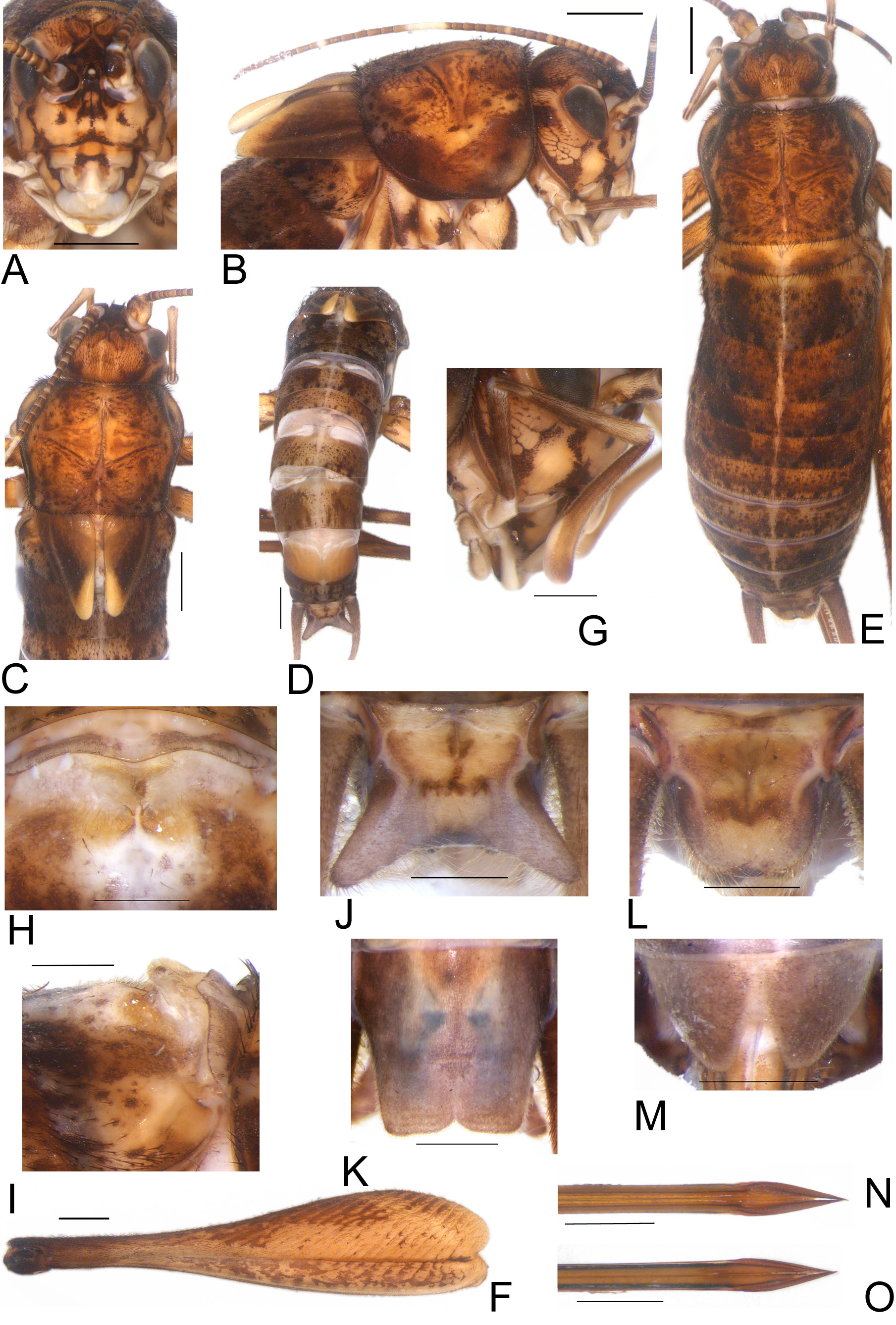
Diagnosis. This species can be distinguished from other Eidmanacris by the following characters: frons with maculae dark brown below antennal scape in frontal view; antenomeres medium brown with whitish bands composed of c.a. 5 antenomeres, intersected by a single, whitish antenomere; lateral-posterior projections of Supra-anal plate greyish; superior, supero-internal and infero-internal projections of apex of pseudepiphallic arm reduced to a spine, inferior projection hook-shaped, with bristles on inner surface, upcurved, ventral projection curved inwards with bristles; posterior half of dorsal projection of ectophallic invagination thinner than anterior, posterior margin straight.
Redescription. Head. Medium to reddish brown. Occiput pilose, with central maculae light brown, band light brown going from occiput to margin of each eye; vertex medium brown with three vertical lines light brown in dorsal view ( Fig. 27C View FIGURE 27 ). Fastigium medium to dark brown, with two rows of bristles, slightly longer than wide, narrowed toward apex, separated from vertex by line forming “v” ( Fig. 27C View FIGURE 27 ). Frons light brown, with three bands dark brown, vertical, two below eyes, one above clypeus; maculae dark brown below antennal scape, in frontal view ( Figs. 27A, B View FIGURE 27 ). Three ocelli present, well developed, forming isosceles triangle, central one flattened at bottom, lateral ones elliptical ( Figs. 27A, B, C View FIGURE 27 ). Eyes with an unpigmented small area on supero-internal angle ( Fig. 27C View FIGURE 27 ). Maxillary palpi, long, thin, pilose, medium brown, distal portion of joints whitish; joints 3, 4 and 5 almost same-sized ( Fig. 27G View FIGURE 27 ); apex of joint 5 upcurved. Gena light brown, posterior margin dark brown, line dark brown, vertical, connecting to inferior margin of eye in lateral view ( Fig. 27B View FIGURE 27 ). Frontoclypeal suture dark yellow, clypeus light brown with central band dark brown in upper margin, in frontal view; labrum whitish, lower portion light brown. Mandible light brown, with inner margin dark brown. Antennal scape light brown, inner margin dark brown ( Fig. 27A View FIGURE 27 ); antenomeres medium brown with whitish bands composed of c.a. 5 antenomeres, intersected by one antenomere whitish.
Thorax. Pronotum DD reddish brown, wider than long, with sparse dark spots and maculae, slightly pubescent, inflated, divided by very tiny light brown sagittal line ( Fig. 27C View FIGURE 27 ); DD cephalic margin slightly concave, caudal margin sub straight ( Fig. 27C View FIGURE 27 ); lateral lobes dark brown, ventro-cephalic angle rounded, lighter than DD, ventrocaudal margin gradually ascendant ( Fig. 27B View FIGURE 27 ).
Legs. FI and II yellowish brown, annulated with dark brown. TI and II yellowish brown, TI with two samesized apical spurs; TII with two inner apical spurs, one outer, smaller. FIII yellowish brown, with thin stripes medium to dark brown on outer surface, dorsal margin and apical third dark brown ( Fig. 27F View FIGURE 27 ). TIII yellowish brown; subapical spurs 4/4, with serrulation above and between subapical spurs; apical spurs 3/3, more developed on inner surface: median (iam) longer than dorsal (iad), ventral smallest (iav) (iam>iad>iav); outer apical spurs: median one longer (oam), dorsal sub-equal in length (oad), ventral smaller (oav) (oam>oad>oav). Basitarsi I, II and III dark yellow.
Abdomen. Cylindrical in dorsal view, medium to dark brown, with spots and maculae, divided by a light brown thin sagittal line ( Fig. 27D View FIGURE 27 ).
Male. Large-sized body, general coloration reddish brown, with dark spots and maculae. FWs medium to dark brown, elongate, triangular, inner margin medium brown; posterior part of inner margin and apex whitish, connected to single vein that divides external part of FW as lateral field, glandular thickening present distally ( Fig. 27C View FIGURE 27 ); inner margins not touching each other, covering metanotal gland, surpassing metanotum posterior border ( Figs. 27B, C View FIGURE 27 ). Metanotal gland present, anteromedian crest forming inverted triangle with cluster of bristles; lateral projections short, conical, parallel ( Figs. 27H, I View FIGURE 27 ), posterior portion of metanotum pubescent. Supra-anal plate light to medium brown, lateral margins dark, anterior margin slightly concave; lateral-posterior projections longer than posterior margin, grayish, with long bristles; posterior margin sub-straight ( Fig. 27J View FIGURE 27 ). Subgenital plate longer than wide, with lateral pubescence, laterally medium to dark brown, centrally greyish; anterior margin straight, posterior margin straight with small invagination centrally ( Fig. 27K View FIGURE 27 ).
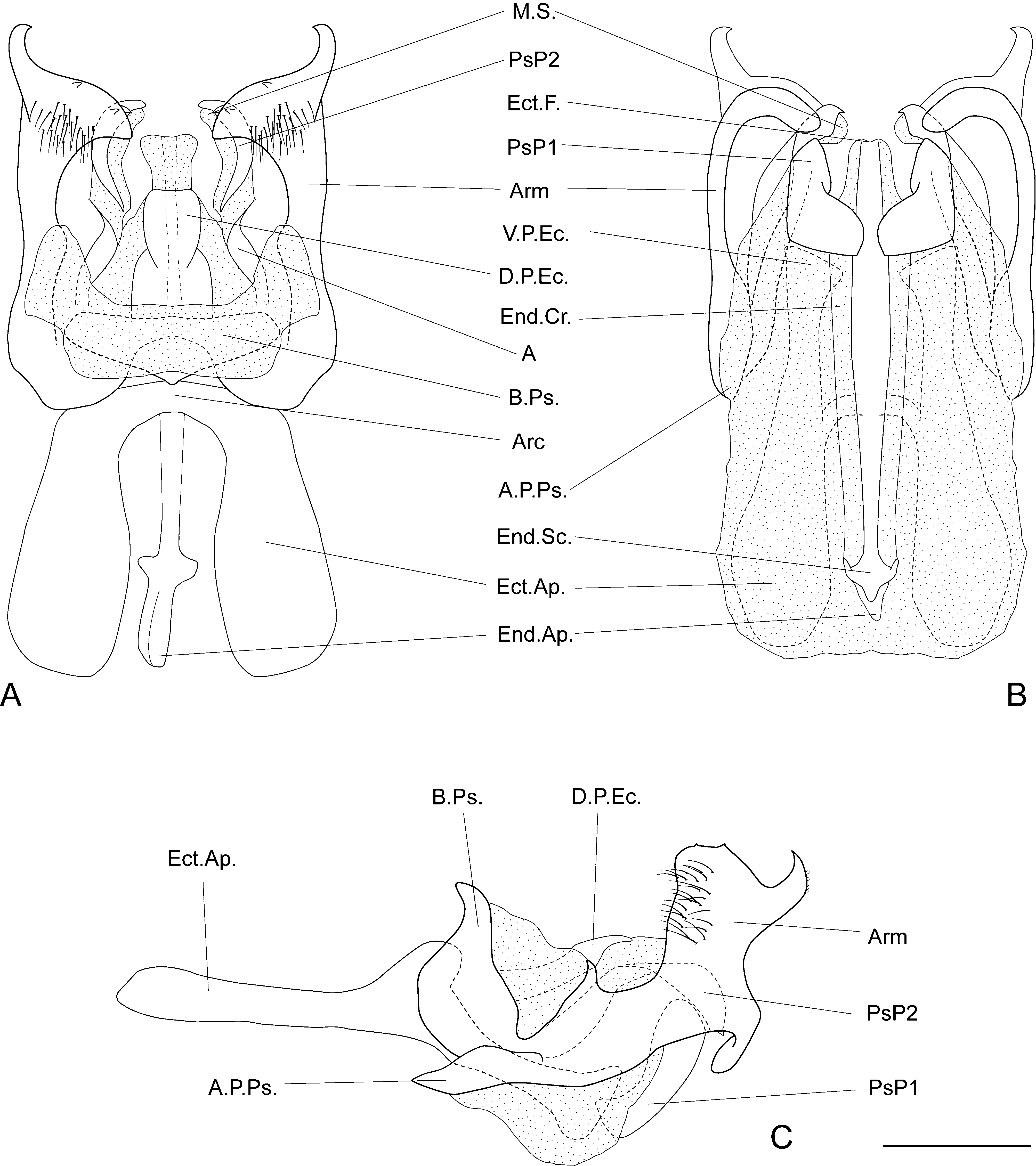
Phallic complex ( Figs. 28A–C View FIGURE 28 ; 29A–C). Pseudepiphallus: central portion of the base of pseudepiphallic sclerite thinner than lateral portions, in dorsal view; pseudepiphallic arms sclerotized, hard, upcurved forming 90° angle in lateral view; apex of pseudepiphallic arm with bristles on outer surface; superior, supero-internal and infero-internal projections reduced to spine; inferior projection hook shaped with bristles on inner surface, upcurved, ventral projection curved inwards with bristles; lateral projection present, short, apex pointed; anterior projection of pseudepiphallic sclerite short, not surpassing base of pseudepiphallic sclerite; PsP2 elongate, sclerotized, reaching posterior extremity of pseudepiphallic arms in dorsal view, apex pointed in lateral view, curved inwards in dorsal view, with membranous sphere on inner surface; sclerite A connected with base of pseudepiphallic sclerite, straight, articulation with PsP2 visible; PsP1 elongate, inner surface well sclerotized in ventral view. Ectophallic invagination: ectophallic apodeme elongate; ectophallic arc located below the base of pseudepiphallic sclerite, “v”-shaped in dorsal view; ventro-posterior projection elongate, tip slightly curved inward; dorsal projection sclerotized, fused. Endophallus: median-posterior projection of endophallic sclerite elongated, not surpassing PsP2 apex, median portion wider in ventral view; lateral-posterior lobes of endophallic sclerite elongate; endophallic apodeme elongate, anterior to ectophallic apodeme.
Female. Larger than male, general coloration similar ( Fig. 27E View FIGURE 27 ). Supra-anal plate medium brown, posterior margin light brown, anterior margin slightly concave, posterior margin rounded with long bristles ( Fig. 27L View FIGURE 27 ). Subgenital plate light brown with whitish band medially, wider than long, anterior margin sub straight, posterior margin rounded with central furrow reaching the median area of plate ( Fig. 27M View FIGURE 27 ). Ovipositor as in figs. 27N, O.
Copulatory papilla ( Figs. 29D–F View FIGURE 29 ). Sclerotized, wider than long, cylindrical, anterior margin membranous, posterior region with visible hole, anterior margin rounded.
Measurements (mm). Male (n=4): Hw, 3.37 ± 0.22 (3.22–3.72); iod, 1.65 ± 0.23 (1.42–1.86); Lpron, 3.85 ± 0.24 (3.59–4.15); awpron, 3.22 ± 0.40 (2.79–3.59); pwpron, 3.95 ± 0.13 (3.78–4.09); wpron, 5.13 ± 0.36 (4.71– 5.52); LFW, 3.36 ± 0.13 (3.16–3.47); wFW, 1.82 ± 0.15 (1.67–2.05); LFIII, 17.32 ± 0.93 (16.2–18.45); wFIII 3.52 ± 0.19 (3.3–3.75); LTIII, 17.85 ± 1.26 (16.35–18.9); Ltars 1-III, 4.8 ± 0.44 (4.35–5.4).
Female (n=5): Hw, 3.67 ± 0.17 (3.47–3.9); iod, 1.71 ± 0.22 (1.48–1.98); Lpron, 4.03 ± 0.21 (3.72–4.21); awpron, 3.72 ± 0.15 (3.53–3.9); pwpron, 4.48 ± 0.14 (4.28–4.65); wpron, 5.44 ± 0.25 (5.08–5.7); LFIII, 18.18 ± 0.72 (17.4–18.9); wFIII, 4.26 ± 0.34 (3.9–4.8); LTIII, 18.09 ± 0.77 (17.25–18.75); Ltars 1-III, 4.65 ± 0.3 (4.35– 4.95); OL, 10.92 ± 0.55 (10.5–11.85).

| MZSP |
Sao Paulo, Museu de Zoologia da Universidade de Sao Paulo |
No known copyright restrictions apply. See Agosti, D., Egloff, W., 2009. Taxonomic information exchange and copyright: the Plazi approach. BMC Research Notes 2009, 2:53 for further explanation.
|
Kingdom |
|
|
Phylum |
|
|
Class |
|
|
Order |
|
|
Family |
|
|
Genus |
Eidmanacris corumbatai Garcia, 1998
| Nihei, Silvio S. 2017 |
Eidmanacris corumbatai
| Prado 2006: 452 |
| Prado 2005: 83 |
Eidmanacris corumbatai Garcia, 1998 in Mesa, Sperber & Garcia, 1998 : 46
| Mesa 1998: 46 |