
Empoasca (Matsumurasca) thapae Dworakowska, 1994
|
publication ID |
https://doi.org/ 10.5281/zenodo.207054 |
|
DOI |
https://doi.org/10.5281/zenodo.6182030 |
|
persistent identifier |
https://treatment.plazi.org/id/03821738-8C71-FFF8-B4A4-FC850AEBC453 |
|
treatment provided by |
Plazi |
|
scientific name |
Empoasca (Matsumurasca) thapae Dworakowska, 1994 |
| status |
|
Empoasca (Matsumurasca) thapae Dworakowska, 1994 View in CoL
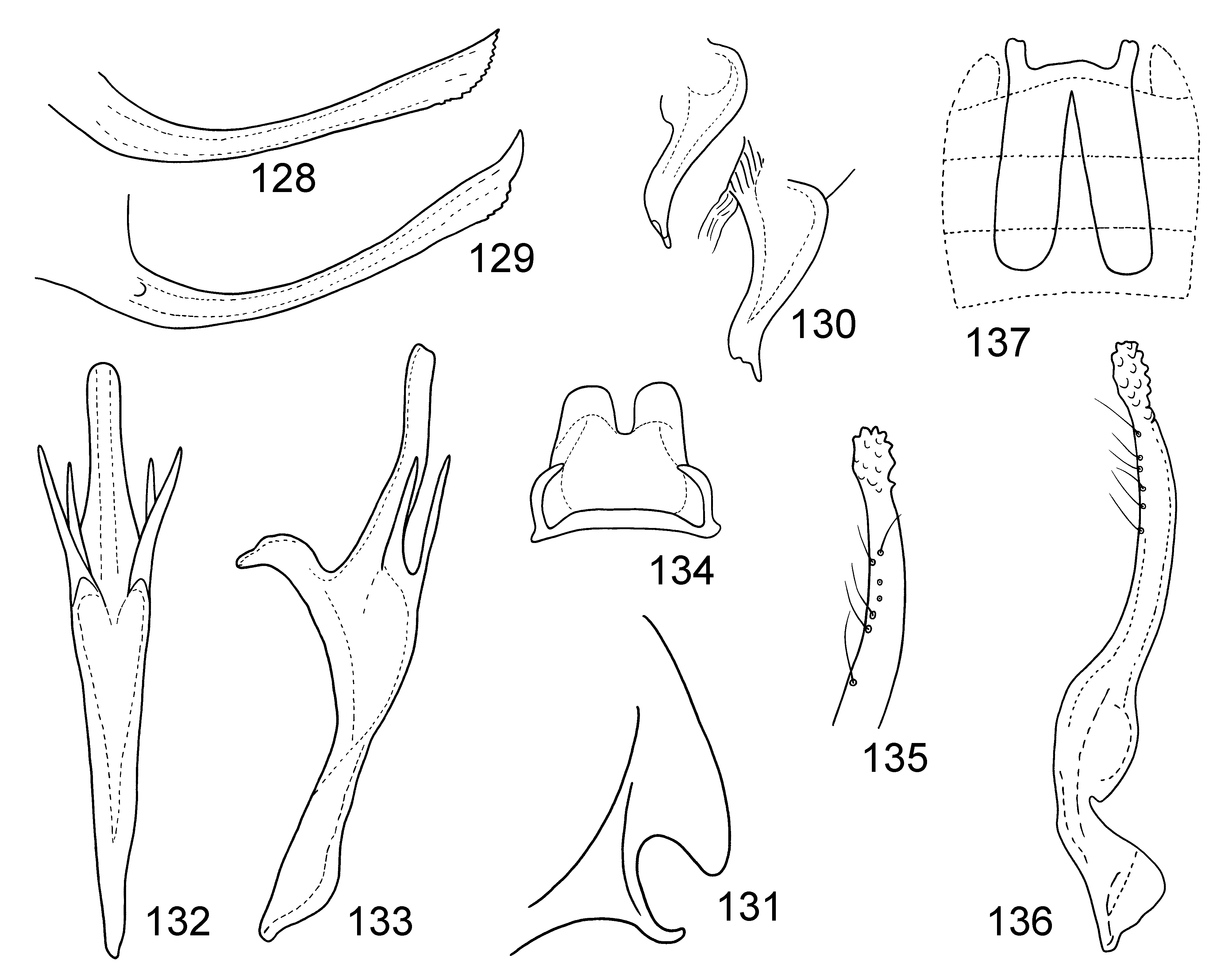
( Figs 128–137 View FIGURES 128 – 137 )
Empoasca (Matsumurasca) thapae Dworakowska, 1994: 104 View in CoL
Type locality. Rumtek, Sikkim, India. ( SMTD). Distribution. India (Sikkim).
Key to species of the subgenus Empoasca (Matsumurasca Anufriev) View in CoL (3)
1. Aedeagal shaft very short, less than half length of preatrium................................................... 2
- Aedeagal shaft long, nearly as long as, or even longer than, preatrium............................................ 3
2. Ventral pygofer appendage bent caudodorsad in basal third and adorned with tufted hairs in apical third, apex smooth on ven- tral side ( Fig. 27 View FIGURES 26 – 34 )........................................................................ E. (M.) clypealata View in CoL
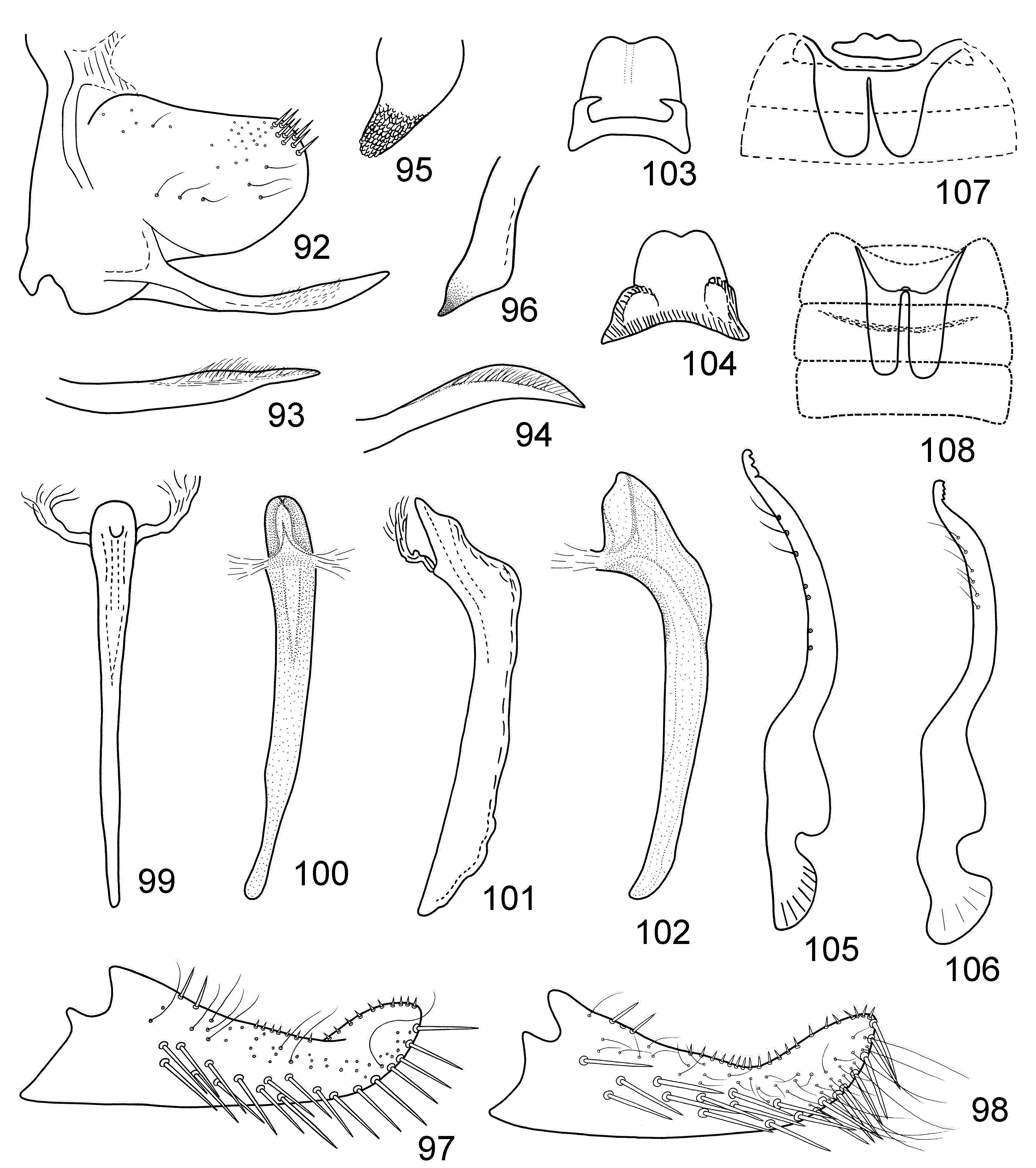
- Ventral pygofer appendage slightly curved dorsad in middle portion and adorned with sparse minute hairs subterminally, apex delicately serrated on ventral side ( Figs 93, 94 View FIGURES 92 – 108 )................................................ E. (M.) parvifacia View in CoL
3. Aedeagal shaft with ventral processes..................................................................... 4
- Aedeagal shaft without ventral processes................................................................... 9
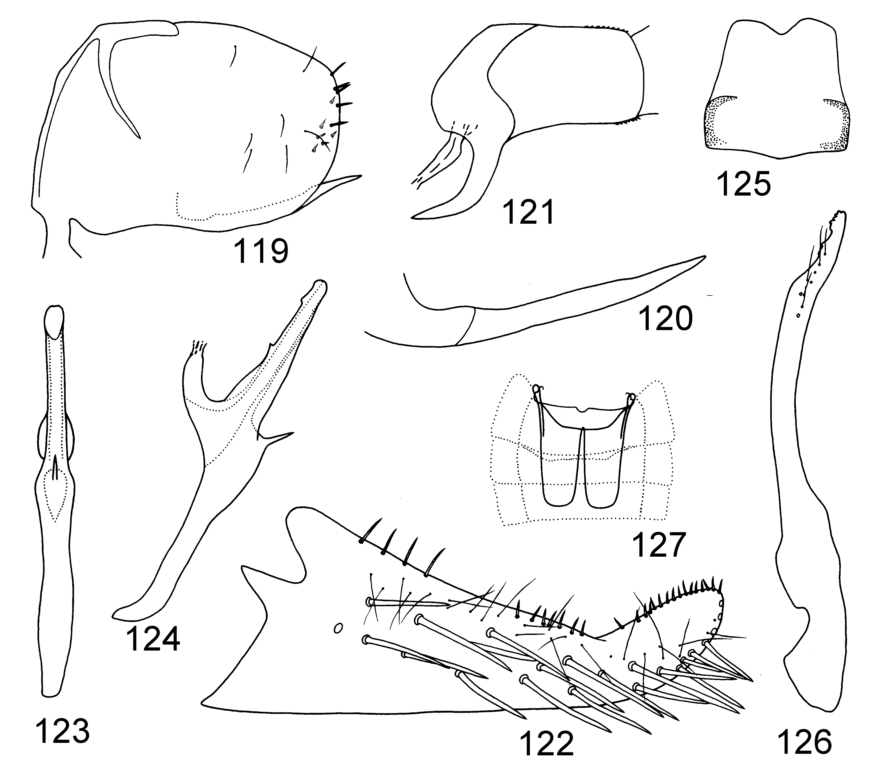
4. Aedeagal shaft with teeth on dorsal side ( Fig. 123 View FIGURES 119 – 127 )........................................ E. (M.) spinalis View in CoL , sp. nov.
- Aedeagal shaft without teeth on dorsal side................................................................. 5
5. Aedeagal shaft with two pairs of ventral processes........................................................... 6
- Aedeagal shaft with one pair of ventral processes............................................................ 7
6. Aedeagal shaft with both pairs of ventral processes at base ( Fig. 132 View FIGURES 128 – 137 )................................. E. (M.) thapae View in CoL
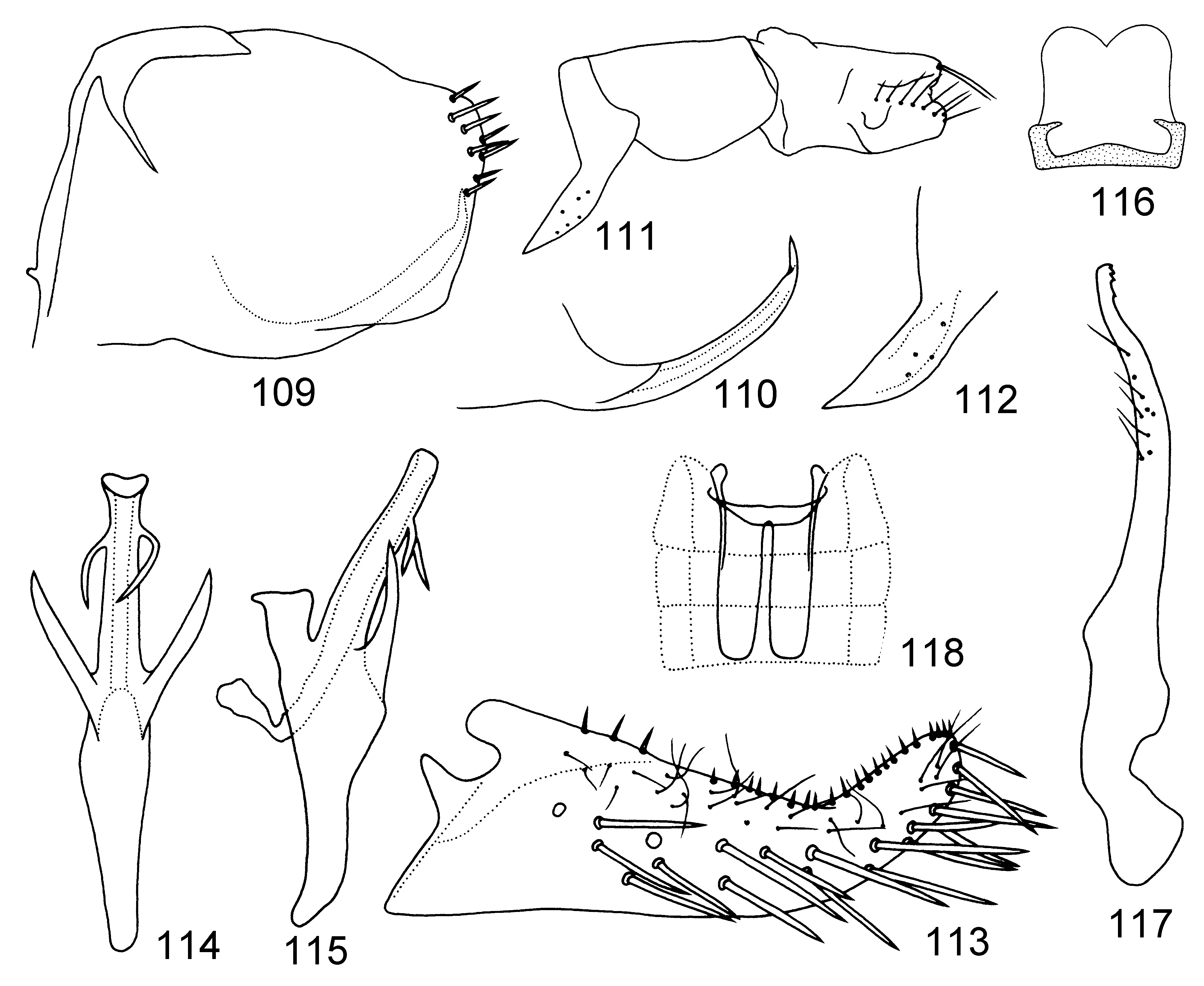
- Aedeagal shaft with one pair of ventral processes at base and second pair subapically ( Fig. 114 View FIGURES 109 – 118 )........ E. (M.) quadrialata View in CoL
7. Aedeagal shaft curved in lateral view ( Fig. 71 View FIGURES 68 – 71 ), gonopore subterminally on ventral side ( Fig. 70 View FIGURES 68 – 71 )............ E. (M.) dolichi View in CoL
- Aedeagal shaft nearly straight in lateral view, gonopore terminal................................................ 8
8. Ventral pygofer appendage slightly S-shaped, directed caudad ( Fig. 53 View FIGURES 51 – 67 )............................... E. (M.) diversa View in CoL
- Ventral pygofer appendage nearly straight, bent and directed dorsad at its base ( Fig. 2 View FIGURES 1 – 6 )...................... E. (M.) aino View in CoL
9. Aedeagal shaft with pair of processes at apex............................................................... 10
- Aedeagal shaft without pair of processes at apex............................................................ 11
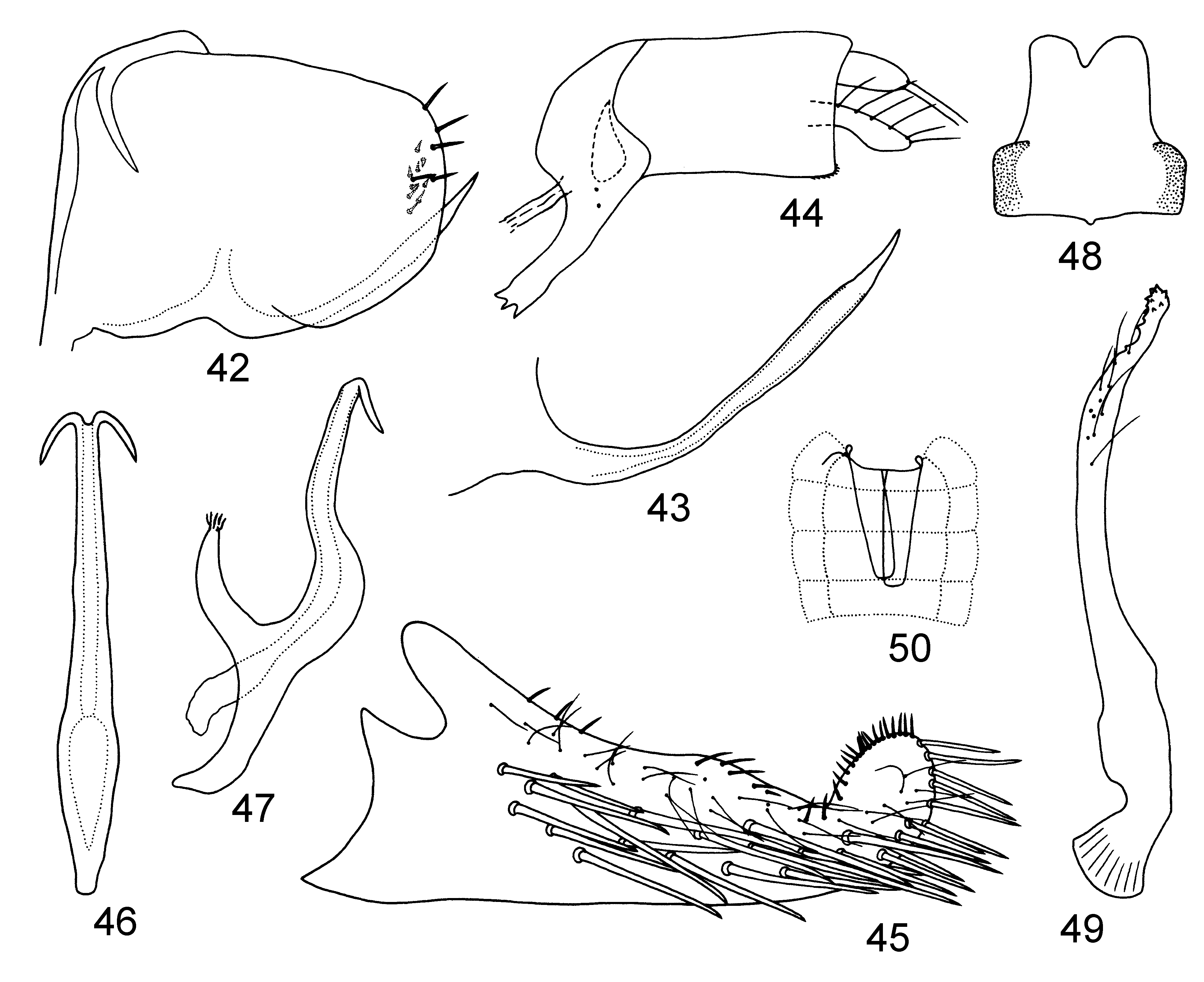
10. Anal tube process of almost the same width throughout and distinctly toothed at apex ( Fig. 44 View FIGURES 42 – 50 ).... E. (M.) dentalis View in CoL , sp. nov.
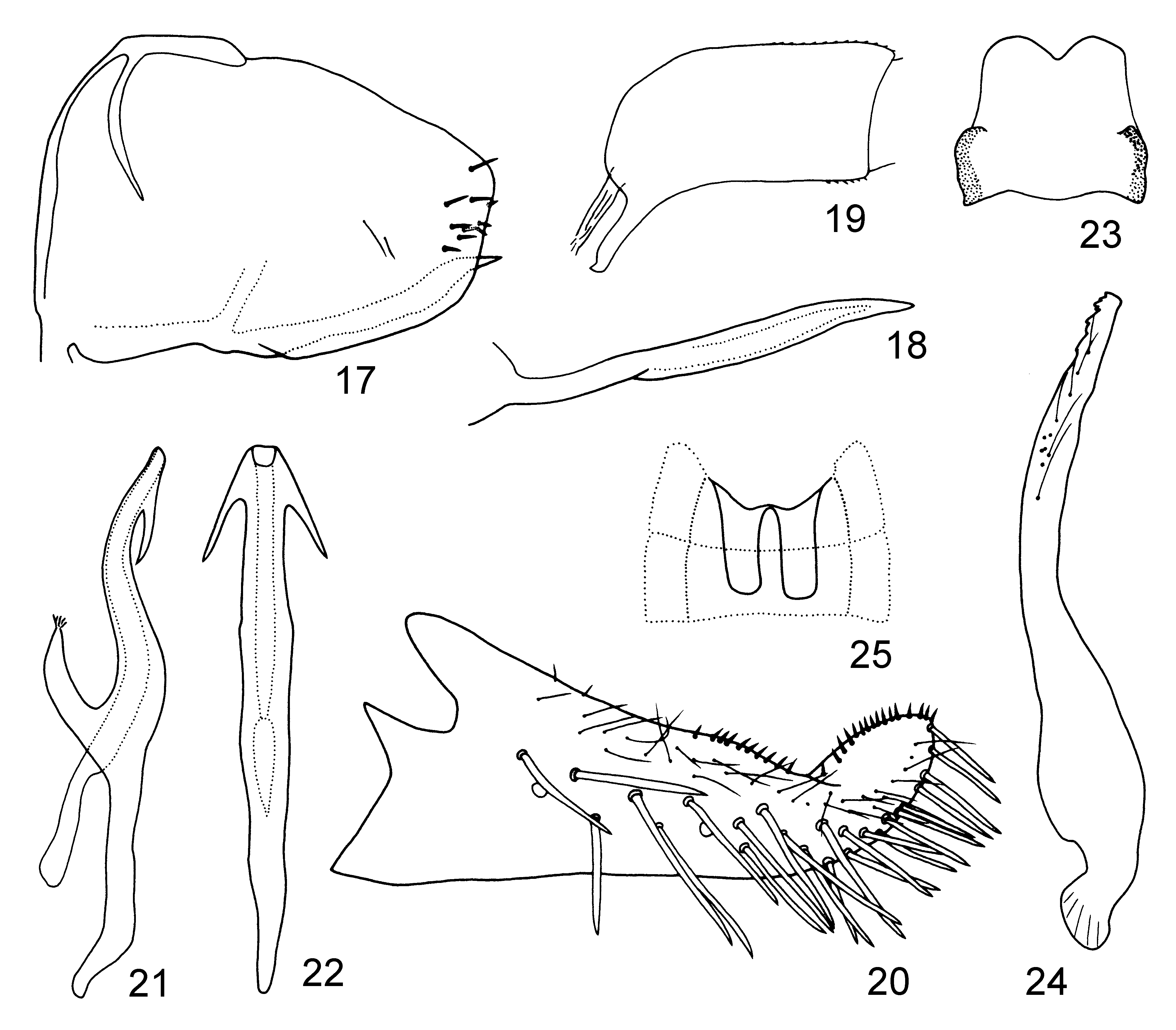
- Anal tube process strongly narrowed apically and not toothed at apex ( Fig. 19 View FIGURES 17 – 25 )................ E. (M.) biprocessa View in CoL , sp. nov.
11. Ventral pygofer appendage smooth, not serrated or coarsely sculptured.......................................... 12
- Ventral pygofer appendage serrated terminally or subterminally, or coarsely sculptured subterminally.................. 13
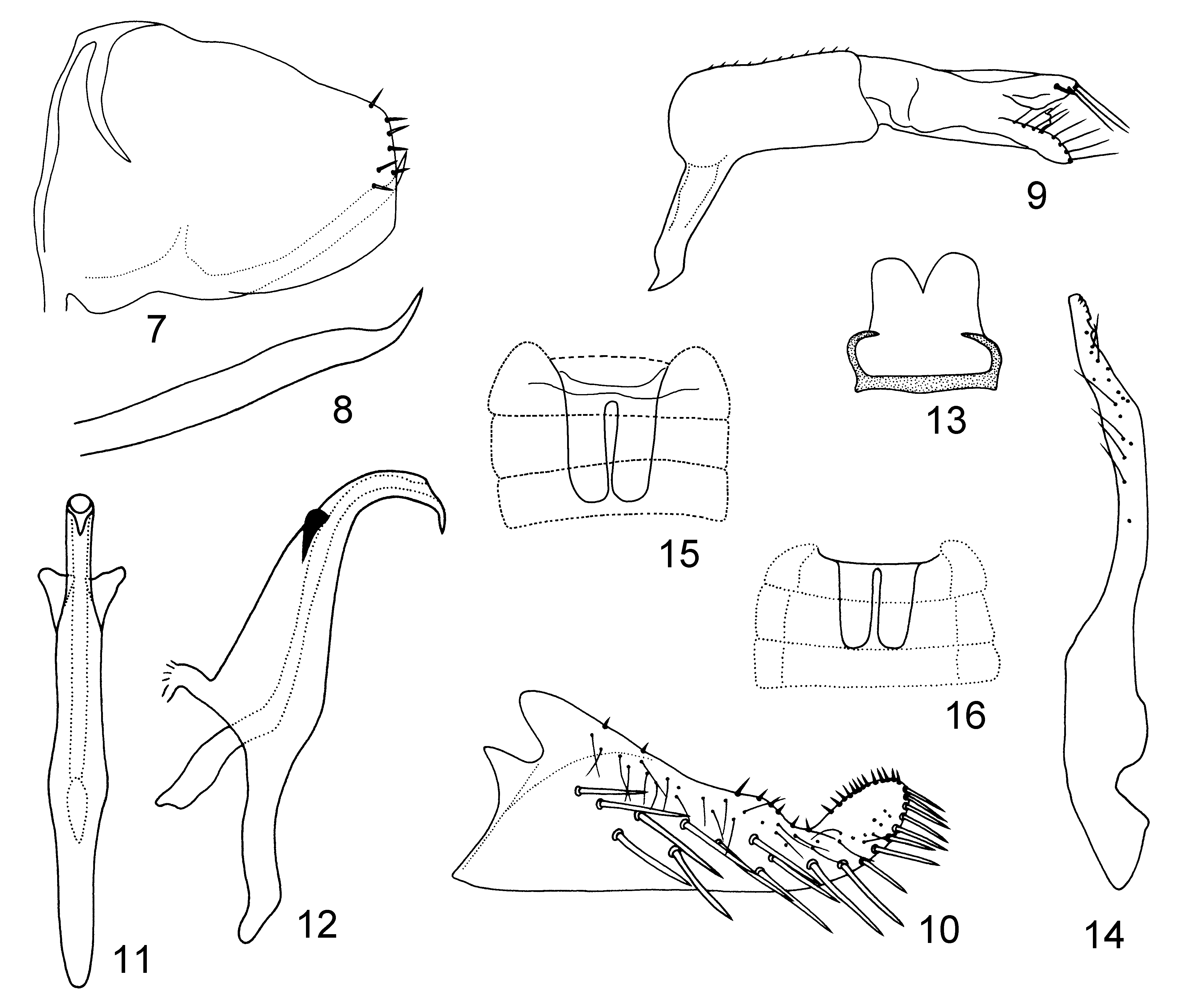
12. Aedeagal shaft arcuate in lateral view, with acute ventroapical projection ( Figs 11, 12 View FIGURES 7 – 16 ); anal tube process straight through most of length ( Fig. 9 View FIGURES 7 – 16 )............................................................................ E. (M.) biloba View in CoL
- Aedeagal shaft straight in lateral view, without acute ventroapical projection ( Figs 83–86 View FIGURES 78 – 91 ); anal tube process evenly curved ( Fig. 81 View FIGURES 78 – 91 )................................................................................... E. (M.) onukii
13. Ventral pygofer appendage serrated terminally or subterminally................................................ 14
- Ventral pygofer appendage coarsely sculptured subterminally, not serrated............................. E. (M.) schima View in CoL
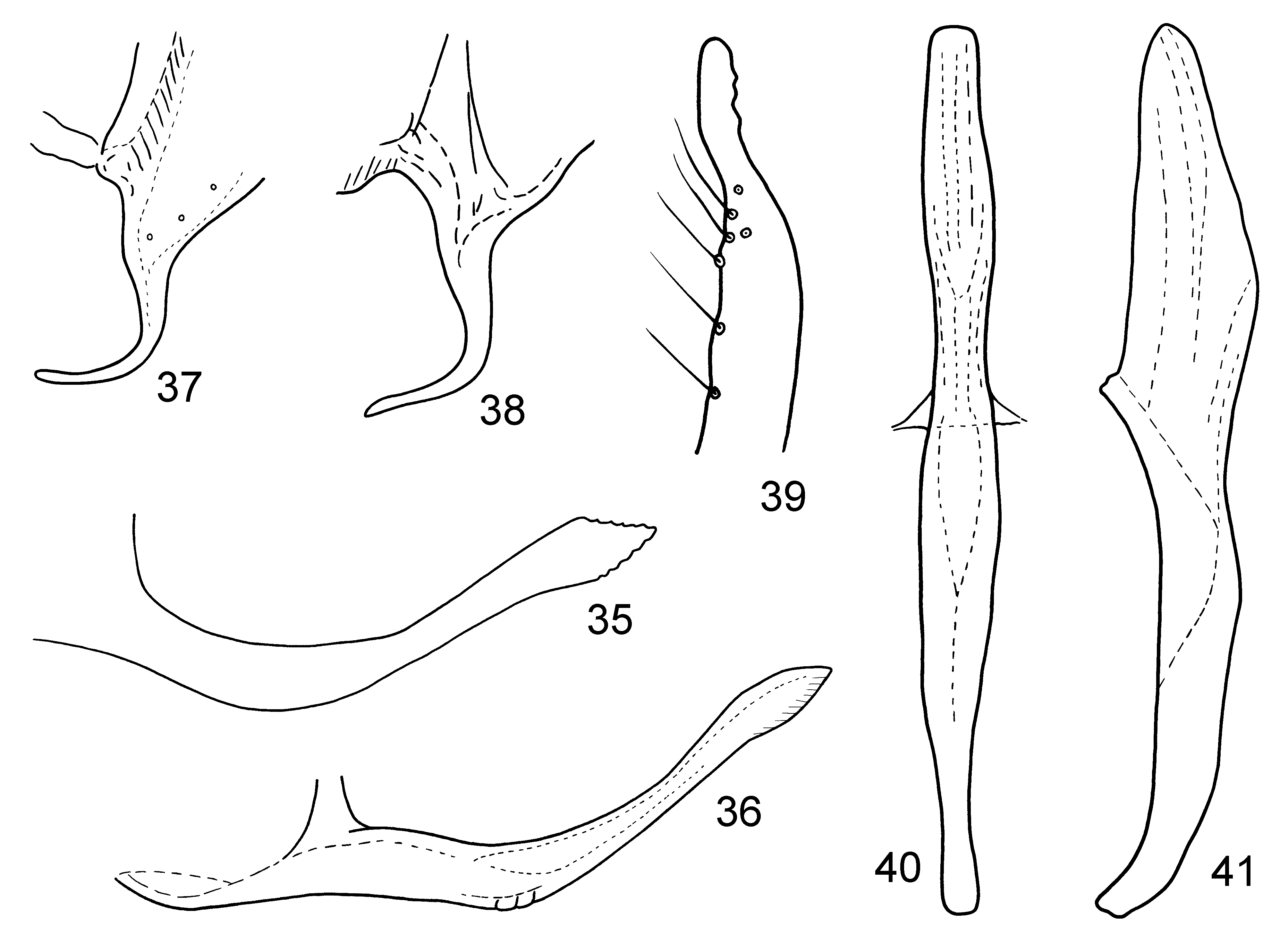
14. Ventral pygofer appendage serrated terminally, exceeding caudal margin of pygofer lobe ( Figs 35, 36 View FIGURES 35 – 41 )...... E. (M.) conifera View in CoL
- Ventral pygofer appendage serrated subterminally at ventral side, not exceeding caudal margin of pygofer lobe ( Figs 72, 73 View FIGURES 72 – 77 )........................................................................................ E. (M.) latissima View in CoL


No known copyright restrictions apply. See Agosti, D., Egloff, W., 2009. Taxonomic information exchange and copyright: the Plazi approach. BMC Research Notes 2009, 2:53 for further explanation.
|
Kingdom |
|
|
Phylum |
|
|
Class |
|
|
Order |
|
|
Family |
|
|
Genus |
Empoasca (Matsumurasca) thapae Dworakowska, 1994
| Liu, Yang, Qin, Dao-Zheng, Fletcher, Murray J. & Zhang, Ya-Lin 2011 |
Empoasca (Matsumurasca) thapae
| Dworakowska 1994: 104 |