
Eurypylus hapax Kornicker & Iliffe 2000
|
publication ID |
https://doi.org/ 10.11646/zootaxa.1565.1.1 |
|
publication LSID |
lsid:zoobank.org:pub:A2CDD9CB-CA5E-418B-A471-9EEFDC5CCF16 |
|
persistent identifier |
https://treatment.plazi.org/id/2A5087FF-3EB6-FC89-3A91-FD0EFD656959 |
|
treatment provided by |
Felipe |
|
scientific name |
Eurypylus hapax Kornicker & Iliffe 2000 |
| status |
|
Eurypylus hapax Kornicker & Iliffe 2000 View in CoL
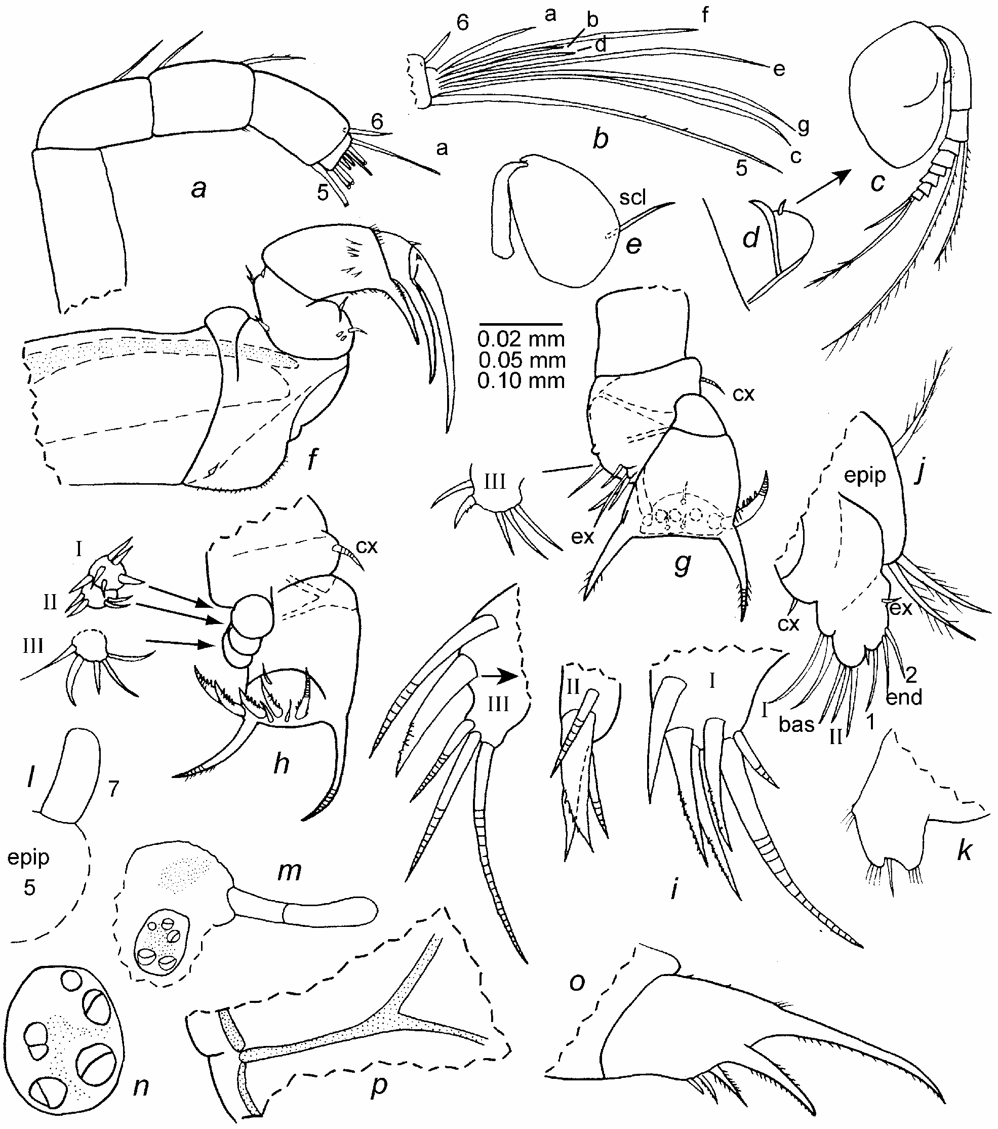
Figs. 73–75 View FIGURE 73 View FIGURE 74 View FIGURE 75
Eurypylus hapax Kornicker & Iliffe 2000: 45 View in CoL , figs. 27–29.—Kornicker et al. 2002: 64, figs. 46, 47.
Holotype. USNM 194494 View Materials , instar IV female in alcohol.
Type locality. Exuma Sound, Great Bahama Bank , depth 142 m.
Material. Sta 00-019, Mystery Cave, Stocking Island, Great Exuma, Great Bahama Bank: USNM 1021484, one instar II female on slide and in alcohol; USNM 1021485, one instar III female on slide and in alcohol; USNM 1021486, two instar III females in alcohol; USNM 1021487, one instar IV female in alcohol.
Distribution. Great Bahama Bank: Crab Cay Crevasse, Mystery Cave, and Exuma Sound; depth 35– 140 m.
Description of instar II female ( Figs. 73 View FIGURE 73 , 74 View FIGURE 74 ). Carapace similar in shape to that of instar IV male illustrated by Kornicker et al. (2002: fig. 46 a) (fig. 73 a). Anterior rostrum indicated by minute overlap at midheight of anterior margin of carapace ( Fig. 73 a,e,f View FIGURE 73 ). Posterior edge of caudal process linear ( Fig. 73 a–c View FIGURE 73 ). Anterior and anteroventral margin of valves slightly rugose ( Fig. 73 f View FIGURE 73 ). Extent of attached dorsal margin indicated in Fig. 73 b View FIGURE 73 .
Ornamentation ( Fig. 73 a,c View FIGURE 73 ): Single bristles numerous along valve margins and scattered over valve surface. Row of about 6 short straight bristles present along outer edge of caudal process.
Infold: Infold of caudal process with about 10 bristles forming row along inner margin and 2 small bristles near midwidth ( Fig. 73 d View FIGURE 73 ). Two setal bristles present just dorsal to caudal process. Usual bristle on anterior infold near rostrum not observed with certainty.
Selvage: Lamellar prolongation with smooth outer edge present along free margins and extending past posterior end of caudal process.
Central adductor muscle attachments ( Fig. 73 a,b View FIGURE 73 ): Consisting of about 17 small oval attachments.
Carapace size (length, height in mm): USNM 1021484, 0.66, 0.50.
First antenna ( Fig. 74 a,b View FIGURE 74 ): 1st segment bare. 2nd segment with distal dorsal bristle. 3rd and 4th segments fused; 3rd segment with dorsal bristle; 4th segment with terminal dorsal bristle and few terminal ventral spines. Sensory bristle of 5th segment long with 2 minute distal spines and terminal spine. 6th segment fused to 5th segment, with short terminal dorsal bristle with base on medial side. 7th segment: a-bristle almost twice length of bristle of sixth segment; b-bristle medial, slightly longer than a-bristle; c-bristle about same length as sensory bristle of 5th segment, narrower in distal half. 8th segment: d-bristle obscured but appearing to be only slightly longer than b-bristle, filament-like, bare; e-bristle almost same length as c-bristle, filament-like distally, bare; f-bristle about two-thirds length of c-bristle, bare; g-bristle same length as c-bristle, bare. Tips of c-, f-, and g-bristles with terminal spine.
Second antenna: Protopod bare ( Fig. 74 e View FIGURE 74 ). Endopod with single segment with 1 short proximal anterior bristle ( Fig. 74 d View FIGURE 74 ). Exopod ( Fig. 74 c View FIGURE 74 ): 1st segment with small terminal medial bristle; bristles of segments 2–7 with proximal ventral spines and distal natatory hairs; bristle of 8th segment with few ventral spines and distal natatory hairs; 9th segment with 2 bristles (ventral bristle with distal natatory hairs, dorsal bristle short, bare). Sclerite attached to posterior edge of protopod slender and slightly curved proximally ( Fig. 74 e View FIGURE 74 ).
Mandible ( Fig. 74 f View FIGURE 74 ): Coxa endite consisting of short stout medial spine in proximal ventral corner; ventral margin of coxa with hairs and short spines. Basis: ventral margin with 4 small medial bristles (1 longer than others); dorsal margin with 4 bristles (1 near midlength, 3 distal). Exopod absent. Endopod: 1st segment with distal medial spines, distal ventral spines, spines forming terminal medial row near dorsal margin, and stout ventral claw with short proximal spines along ventral and dorsal edge; 2nd segment with stout ventral claw and 1 or 2 minute terminal dorsal bristles; 3rd segment with stout terminal claw and 2 small bristles (1 ventral, 1 dorsal).
Maxilla ( Fig. 74 g –i View FIGURE 74 ): Endite I with 6 bristles (4 claw-like); endite II with 4 bristles (2 claw-like); endite III with 5 bristles (2 posterior (1 claw-like pectinate), 3 anterior) ( Fig. 74 i View FIGURE 74 ). Coxa with short dorsal bristle ( Fig. 74 g,h View FIGURE 74 ). Exopod with 3 bristles (1 long, 2 short). Basis with bristle near exopod. Endopod: 1st segment with distal spines on anterior margin and stout pectinate alpha- and beta-bristles; 2nd segment with 2 a-bristles, 1 c- bristles, and 5 pectinate end bristles (middle bristle shortest).
Fifth limb ( Fig. 74 j View FIGURE 74 ): Epipod with about 27 plumose bristles. Coxa endite with 1 short bristle. Basis with 2 endites: endite I lobate with 2 terminal bristles; endite II with 3 terminal bristles. Endopod with 2 fused segments: 1st segment with 2 bristles (1 short); 2nd segment with 2 terminal bristles. Exopod represented by 1 short bristle.
Sixth limb ( Fig. 74 k View FIGURE 74 ): Small with 2 spinous lobes and between them 1 bristle.
Seventh limb ( Fig. 74 l View FIGURE 74 ): Short bare lobe.
Furca ( Fig. 74 o View FIGURE 74 ): Each lamella with 5 or 6 claws; right lamella with 5 claws, left lamella with 6 claws; left lamella with spines following claw 6; claws 1 and 2 stout, nonarticulated; remaining claws articulated, and decreasing in size posteriorly along lamella; claws 1–4 with teeth along posterior edge (those on claw 4 minute); claws 2 and 3 with fine spines along anterior edge. Anterior edge of right lamella anterior to left lamella by about two-thirds of width of claw 1, with long spines near base of claw 1 and few minute anterior proximal spines.
Bellonci Organ ( Fig. 74m View FIGURE 74 ): Elongate, broadening to rounded tip, and with indistinct suture near midlength.
Eyes ( Figs. 73 a,b View FIGURE 73 , 74 m,n View FIGURE 74 ): Medial eye bare with amber colored pigment in vicinity of midlength. Lateral eye smaller than medial eye, with 5 ommatidia (4 divided; undivided ommatidium medial to others) and brownish pigment.
Upper lip: Projecting anteriorly, bare.
Y-Sclerite ( Fig. 74 p View FIGURE 74 ): With ventral branch typical for family.
Heart ( Fig. 73 b View FIGURE 73 ): Oval in lateral view.
Gut content: Amber colored unrecognizable particulate matter.
Description of instar III female ( Fig. 75 View FIGURE 75 ). Carapace similar in shape to that of instar II female except posterodorsal corner more rounded ( Fig. 75 a View FIGURE 75 ).
Ornamentation ( Fig. 75 a,c View FIGURE 75 ): Similar to that of instar II female.
Infold: Anterior infold with minute bristle ventral to rostrum ( Fig. 75 c View FIGURE 75 ). Infold of caudal process similar to that of instar II female.
Selvage: Similar to that of instar II female.
Central adductor muscle attachments ( Fig. 75 a,b View FIGURE 75 ): Consisting of 19 ovoid attachments.
Carapace size (length, height in mm): USNM 1021485 View Materials , 0.75, 0.58. USNM 1021486 View Materials , 2 specimens : 0.73, 0.56; 0.79, 0.68.
First antenna: Differs from that of instar II female in having 2 ventral spines on fused 3rd and 4th segments (1 at midlength, 1 subterminal), and 1 terminal ventral bristle on 4th segment.
Second antenna: Similar to that of instar II female except tip of endopod slightly tapered ( Fig. 75 d View FIGURE 75 ).
Mandible: Similar to that of instar II female.
Fifth limb: Epipodite with 30 plumose bristles. Remainder of limb similar to that of instar II female.
Sixth limb ( Fig. 75 e View FIGURE 75 ): Single endite with 1 bristle; end segment with 8 bristles followed by space and 2 stout posterior bristles.
Seventh limb ( Fig. 75 f View FIGURE 75 ): Elongate, bare, with indistinct narrowly separated sutures and rounded tip.
Furca ( Fig. 75 g View FIGURE 75 ): With 5 claws on right lamella and 6 on left, similar to those on instar II female; few spines present on lamellae following claws. Right lamella with few proximal anterior spines and longer spines near base of claw 1. Right lamella anterior to left by width of base of claw 1.
Bellonci Organ: Similar to that of instar II female.
Eyes ( Fig. 75 h View FIGURE 75 ): Medial eye and lateral eyes similar to those of instar II female.
Upper lip: With bare anterior projection (upper lip stippled in Fig. 75 g View FIGURE 75 ).
Posterior of body ( Fig. 75 f View FIGURE 75 ): With two clusters of spines at posterodorsal corner just anterior to dorsal end of girdle.
Y-Sclerite ( Fig. 75 i View FIGURE 75 ): Similar to that of instar II female except posterior end with small flare.
Gut content: Unidentified amber-colored particulate matter.
Supplementary description of instar IV female. Carapace size (length, height in mm): USNM 1021487, 0.90, 0.71.
Comparisons. E. hapax and E. concentricostatus ( Hartmann 1974) are the only species known in the genus with a furca without secondary claws and with five claws on each lamella ( Kornicker & Iliffe 2000). The carapace of E. hapax is without the concentric rib present on E. concentricostatus .
Discussion. The instar IV male and female of this species have been described ( Kornicker & Iliffe 2000:45; Kornicker et al. 2002:64). Instars II (female) and III (female) are described herein, and the carapace size is given for an instar IV female. All specimens are identified as females, mainly because the seventh limbs of all are fairly well developed, and also the endopods of the second antennae consist of single segments. Adults of the species are unknown.


| USNM |
Smithsonian Institution, National Museum of Natural History |
No known copyright restrictions apply. See Agosti, D., Egloff, W., 2009. Taxonomic information exchange and copyright: the Plazi approach. BMC Research Notes 2009, 2:53 for further explanation.
|
Kingdom |
|
|
Phylum |
|
|
Class |
|
|
Order |
|
|
Family |
|
|
Genus |
Eurypylus hapax Kornicker & Iliffe 2000
| Kornicker, Louis S., Iliffe, Thomas M. & Harrison-Nelson, Elizabeth 2007 |
Eurypylus hapax
| Kornicker, L. S. & Iliffe, T. M. 2000: 45 |