
Euschistus (Mitripus) convergens
|
publication ID |
https://doi.org/ 10.11646/zootaxa.1984.1.1 |
|
persistent identifier |
https://treatment.plazi.org/id/03C31150-FFF4-1019-FF1B-FCCFE53FF96C |
|
treatment provided by |
Plazi |
|
scientific name |
Euschistus (Mitripus) convergens |
| status |
|
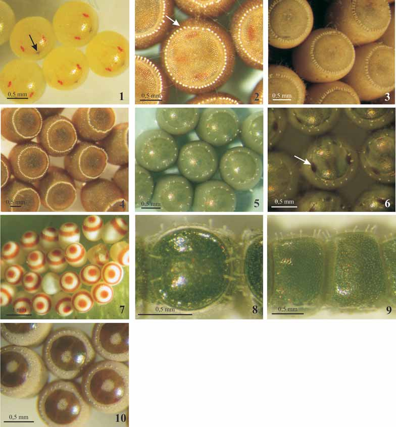
Euschistus (Mitripus) convergens , Euschistus (Mitripus) hansi , and Euschistus (Lycipta) picticornis
( Figs. 1 View FIGURES 1–10 , 17–36 View FIGURES 11–25 View FIGURES 26–40 ; Tab. 2 View TABLE 2 )
Eggs barrel-shaped to spherical ( Figs. 17, 18, 24, 25 View FIGURES 11–25 , 33 View FIGURES 26–40 ); operculum circular, with variable convexity; usually convex ( Figs. 17, 24 View FIGURES 11–25 ), flat in C. picticornis ( Fig. 33 View FIGURES 26–40 ). Chorion surface spinose and translucent after hatching of nymphs. Before hatching, eggs milky-white in E. hansi and E. picticornis to yellow in E. convergens ( Fig. 1 View FIGURES 1–10 ); red eyes and dark brown ruptor ovis become visible with the development of embryo. Aero-micropylar processes white, slightly clavated at apex ( Figs. 22 View FIGURES 11–25 , 28 View FIGURES 26–40 ), entirely tubular in E. picticornis ( Fig. 35 View FIGURES 26–40 ).
In the SEM, eggs of Euschistus spp. studied show chorion surface spinose ( Figs. 17, 18, 24, 25 View FIGURES 11–25 , 33 View FIGURES 26–40 ), although it has been described as finely reticulated in E. hansi ( Martins & Campos 2006) . In areas where the egg is fixed to another in the egg mass or to the substratum, chorion is covered by an adhesive substance that prevents visualization of sculpture pattern ( Figs. 17, 24 View FIGURES 11–25 , 33 View FIGURES 26–40 ). In all areas of the egg, a great number of spines projects from the surface, uniformly distributed in the chorion of E. picticornis ( Figs. 34, 35 View FIGURES 26–40 ), or laminate expansions form polygonal figures, with long and acute spines along those projections, of E. hansi and E. convergens ( Figs. 19, 20 View FIGURES 11–25 , 26 View FIGURES 26–40 ). In the E. hansi egg, spine-shaped projections with different lengths can emerge from the smooth chorion, composing nearly polygonal figures in some areas of chorion ( Fig. 27 View FIGURES 26–40 ). Lateral walls near the aero-micropylar processes, and operculum with longer and numerous spines, almost always tied by fine sheets ( Figs. 21, 22 View FIGURES 11–25 , 28, 35 View FIGURES 26–40 ), or sometimes inserted by smaller spines as in E. hansi ( Figs. 29, 30 View FIGURES 26–40 ). Limits of the operculum are not easily visualized in noneclosioned eggs ( Figs. 21 View FIGURES 11–25 , 29 View FIGURES 26–40 ). Aero-micropylar processes can be distinguished from chorionic spines for being longer and larger in diameter; the processes are tubular in E. picticornis ( Fig. 35 View FIGURES 26–40 ) and slightly clavated at apex in the other two species ( Figs. 22 View FIGURES 11–25 , 28, 29 View FIGURES 26–40 ). Its apical portion bears a central hole; at the magnification used, aero-micropylar processes walls seem smooth, instead of spongy ( Figs. 23 View FIGURES 11–25 , 31, 32, 36 View FIGURES 26–40 ).


No known copyright restrictions apply. See Agosti, D., Egloff, W., 2009. Taxonomic information exchange and copyright: the Plazi approach. BMC Research Notes 2009, 2:53 for further explanation.