
Frontonia lynni, Long & Song & Gong & Hu & Ma & Zhu & Wang, 2005
|
publication ID |
https://doi.org/ 10.11646/zootaxa.1003.1.4 |
|
DOI |
https://doi.org/10.5281/zenodo.10532416 |
|
persistent identifier |
https://treatment.plazi.org/id/03BBE87D-FF97-FFDF-FEC6-3A90561EC374 |
|
treatment provided by |
Felipe |
|
scientific name |
Frontonia lynni |
| status |
sp. nov. |
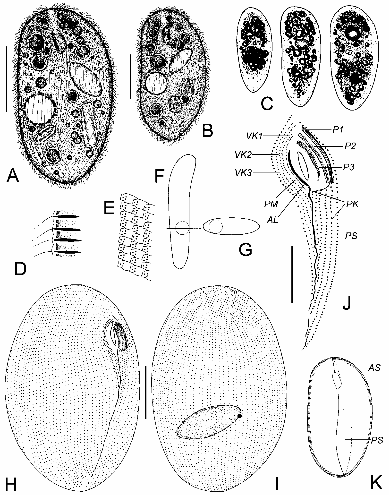
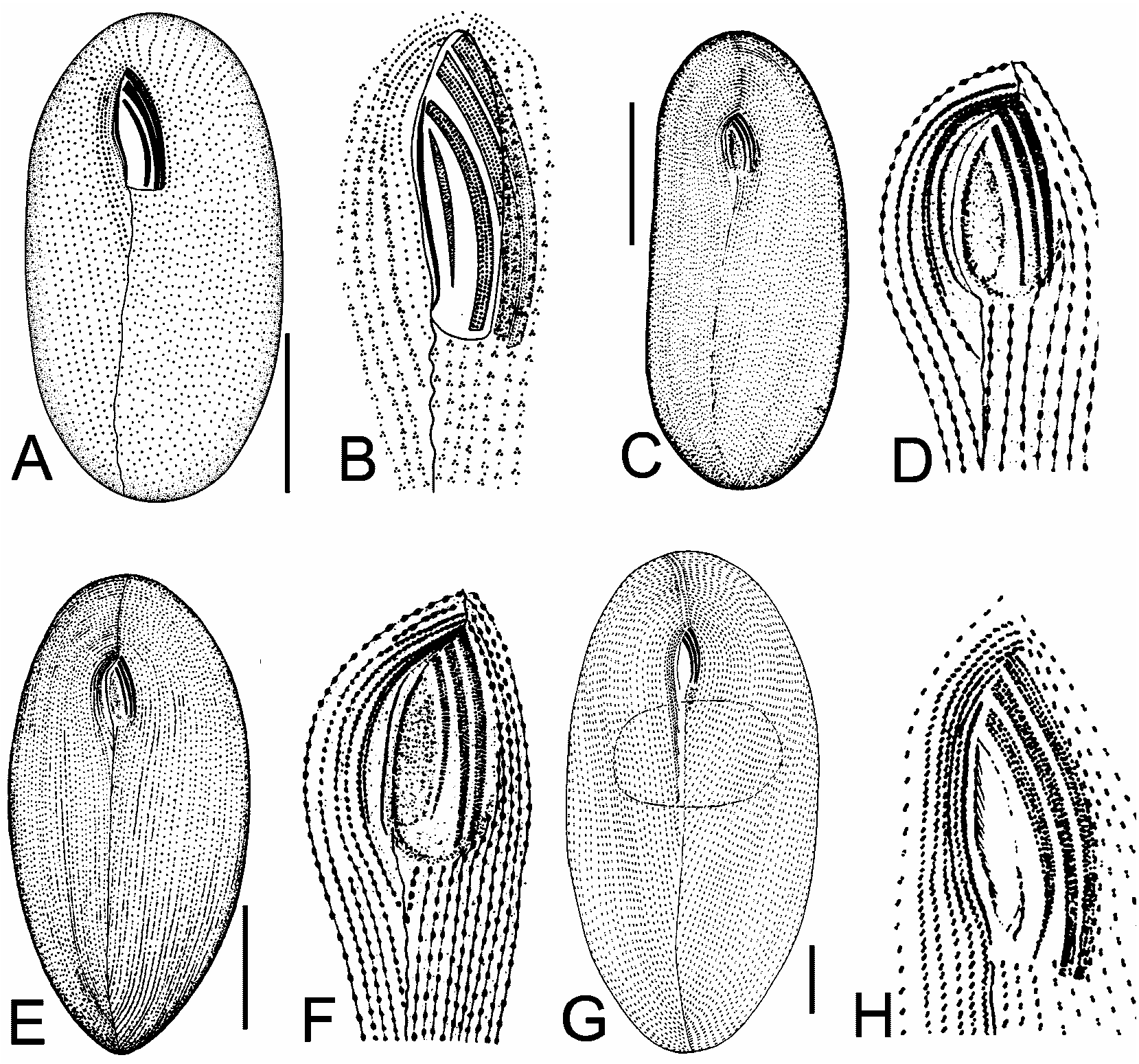
Frontonia lynni n. sp. ( Figs. 13 View FIGURE 1 View FIGURE 2 View FIGURE 3 ; Table 1)
Diagnosis: Marine Frontonia about 100–210 x 70–150 µm in vivo, body shape ellipsoidal, dorsoventrally strongly flattened of about 3:1 with conspicuously small buccal cavity. 71– 83 somatic, three vestibular and five postoral kineties. Peniculi 1 and 2 each with four rows, whereas peniculus 3 has five ciliary rows. Macronucleus ellipsoidal. Single contractile vacuole, equatorially located on the right margin. Extrusomes spindleshaped.
Type location: A mesotrophic sand beach (salinity 26 ‰) in Qingdao (36°08 N, 120°43), China .
Type specimens: One holotype with silver nitrate impregnated specimens is deposited in the Natural History Museum, London, UK with registration number 2005:24:12. Two paratypes with silver nitrate impregnated specimens are deposited in the Laboratory of Protozoology , OUC (slides number: 20041115011, 20041115012) .
Dedication: We dedicate this species to Prof. Dr. Denis H. Lynn, University of Guelph, Canada, a worldfamous protozoologist, who has greatly contributed to the ciliate taxonomy and systematics.
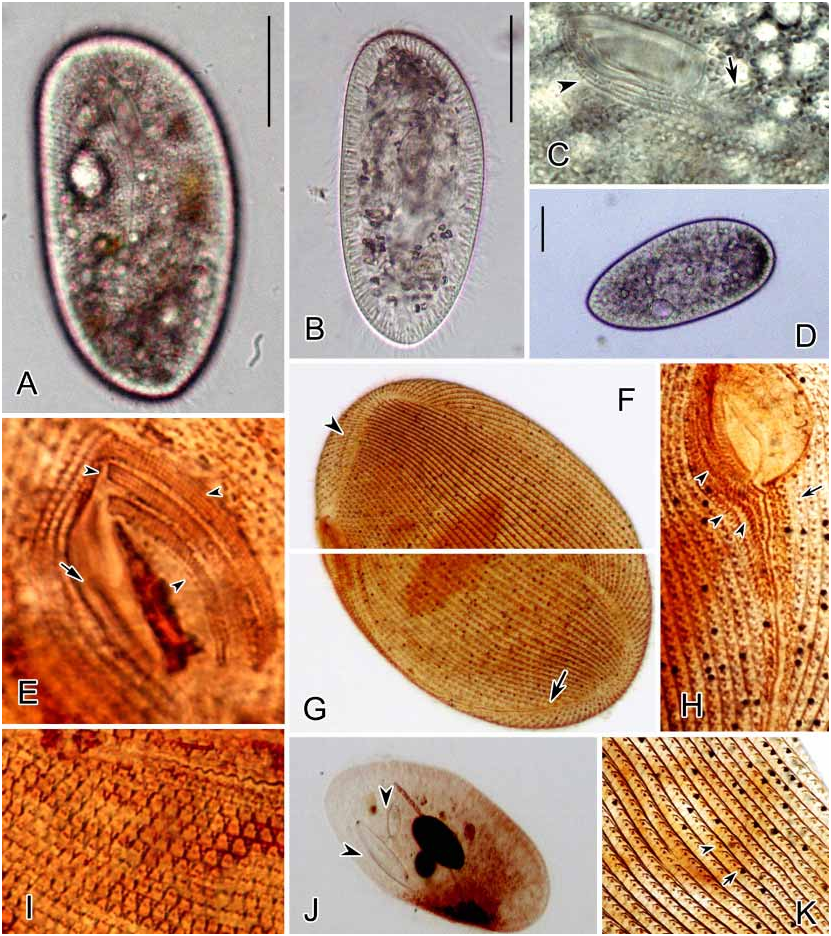
Description: Size highly variable, in vivo about 100–210 × 70–150 µm, but mostly 130–170 µm in length; ratio of length: width about 3:2. Body shape constant, ellipsoidal in outline when viewed from ventral or dorsal; right margin straight while left margin slightly convex ( Figs. 1 View FIGURE 1 AC; 3A, 3B). Both ends broadly rounded. Dorsoventrally conspicuously flattened about 3:1 ( Fig. 1G View FIGURE 1 ). Macronucleus ellipsoidal, positioned at body center; micronucleus spherical, adjacent to macronucleus ( Fig. 1I View FIGURE 1 ). One contractile vacuole, about 20 µm in diameter, positioned equatorially and conspicuously right of median. Extrusomes spindleshaped, about 6 µm long, densely arranged in cortex ( Figs. 1D View FIGURE 1 ; 3B, 3C, 3K View FIGURE 3 ). Cytoplasm transparent, colorless to slightly greyyellowish, usually filled with many food vacuoles (> 10 µm in diameter), as well as dark granules and crystals (35 µm across) ( Figs. 1C View FIGURE 1 ; 3B View FIGURE 3 ), containing bacteria, diatoms and other small ciliates ( Figs. 1A View FIGURE 1 ; 3J View FIGURE 3 ). Somatic cilia about 8 µm long. Movement mostly by gliding back and forth on substrate; when swimming, moderately rapid with rotation around the long axis of the cell.
Buccal cavity small and shallow, triangular in outline, occupying about 1/7 of body length ( Figs. 1A, 1B, 1J, 1K View FIGURE 1 ; 3C View FIGURE 3 ). Three short vestibular kineties (VK1–3) run from anterior vertex of cavity to postoral suture, with densely arranged kinetosomes ( Figs. 1H, 1J View FIGURE 1 ; 3C, 3H View FIGURE 3 ). Three inconspicuous peniculi deeply located on the left wall of cavity: peniculi 1 and 2 (P1, 2) about equally long, positioned close to each other, parallel to the edge of the left vestibular wall and each composed of four rows of kinetosomes; peniculus 3 (P3) slightly curved to right, composed of five relatively shorter kineties, of which two are complete, another two are slightly shortened and the leftmost one is about half the length of the two on the right ( Figs. 1J View FIGURE 1 ; 3E View FIGURE 3 ).
Singlerowed paroral membrane (PM) on the right edge of the buccal cavity runs from the anterior of the buccal overture to the posterior edge. Anterior portion of argentophilic line (AL) positioned parallel to paroral membrane ( Figs. 1J View FIGURE 1 ; 3E View FIGURE 3 ).
As with its congeners, silverline system as quadrangular cortical meshes that can be observed after silver nitrate impregnation ( Figs. 1E View FIGURE 1 ; 3I View FIGURE 3 ).
Abbreviations: CV = coefficiency of variation in %, Max = maximum, Mean = arithmetic mean, Min = minimum, n = number of individuals examined, SD = standard deviation, SE = standard error of arithmetic mean.
No known copyright restrictions apply. See Agosti, D., Egloff, W., 2009. Taxonomic information exchange and copyright: the Plazi approach. BMC Research Notes 2009, 2:53 for further explanation.
|
Kingdom |
|
|
Phylum |
|
|
Class |
|
|
Order |
|
|
Family |
|
|
Genus |