
Galethalea davidi Dognin, 1889
|
publication ID |
https://doi.org/ 10.11646/zootaxa.4078.1.30 |
|
publication LSID |
lsid:zoobank.org:pub:98DA2F2D-03D9-4283-AAF6-E21EF7F5EDFE |
|
DOI |
https://doi.org/10.5281/zenodo.6090745 |
|
persistent identifier |
https://treatment.plazi.org/id/03FFB604-EB29-FFB4-71DC-F9F770981BF9 |
|
treatment provided by |
Plazi |
|
scientific name |
Galethalea davidi Dognin, 1889 |
| status |
|
Galethalea davidi Dognin, 1889 , resur. stat.
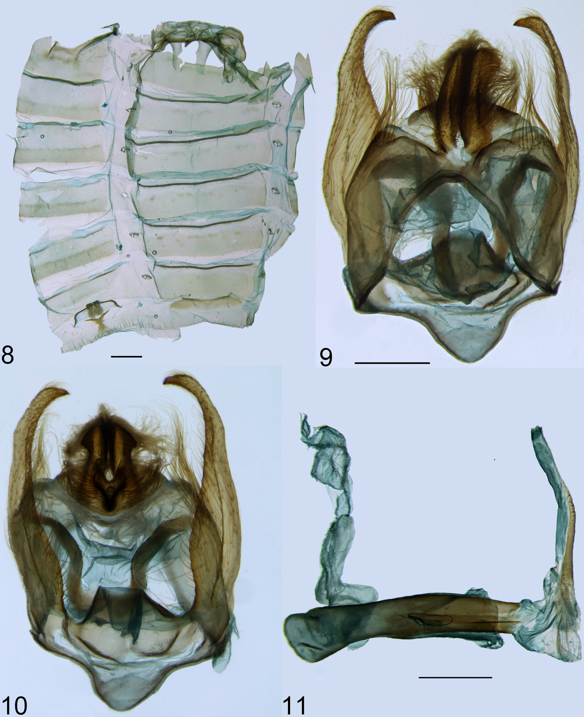
( Figures 1 View FIGURES 1 – 4 , 8–11 View FIGURES 8 – 11 )
Galethalea davidi Dognin, 1889: 14 . Syntype male: ECUADOR, Loja, August (NMNH) [photograph examined]. Nelphe davidi ( Dognin, 1889) ; Kirby, 1892: 172; Kirby, 1892: 904.
Eucereon davidi ( Dognin, 1889) ; Hampson, 1898: 490.
Eucereum davidi ( Dognin, 1889) ; Zerny, 1912: 139; Draudt, 1915: 179, pl. 25, row e. Eucereum davidi clarius Draudt, 1915: 179 .
Diagnosis ♂. Antennae black, except for the lateral surfaces of the scape, white. Frontoclypeus black ventrally and white dorsally. Mesoscutellum with three white markings. Metascutellum black. FW predominantly black, with various white spots. HW partially hyaline. T8 black.
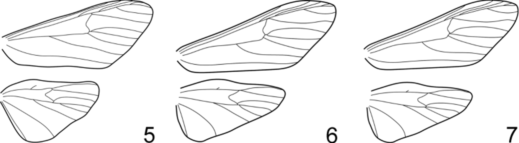
Redescription ♂. Head. Proboscis light brown. Palpi three segmented, reaching vertex. First and second segments black. Third segment approximately three times longer than wide, black, except for the dorsal apical surface, white. Antennae black, except for the scape, with few white scales anteriorly and posteriorly. Pectination starting in the second flagellomere. Frontoclypeus as wide as long, black ventrally and white dorsally; ventral half smooth-scaled, dorsal half rough-scaled. Vertex mainly black, white scales laterally. Occiput and ocular ring black. Cervical scales orange. Thorax. Mesothorax predominantly black, with three white areas, one anterior and two posterior, parallel. Metascutellum black. Patagia black, except for the external margin, white. Tegulae predominantly black, white scales at the anterior margin and near the external margin of the posterior portion. Epimera and episterna with long black scales. Ventral scales on the first pre-episternum black, like the surrounding scales. Anterior surface of the forecoxae predominantly black proximally, distal margin white. Lateral surface black. Forefemora black with a white proximal end, and two white mid-ventral spots. Foretibiae black with two white spots at the distal end. Foretarsi black. Midcoxae white anteriorly and laterally. Midfemora as forefemora, but with the white ventral spots more conspicuous. Midtibiae predominantly black, with a white dorsal subproximal area, and white distal end; spurs white. Midtarsi black, except for the distal end of the first segment, white. Hindcoxae as midcoxae. Hindfemora as the others, but with the distal end white. Hindtibiae black with white distal end and white spurs. Hindtarsi predominantly black, first segment white at both ends, and second segment white at the posterior end. FW. Entirely scaled. Axillary scales white. Dorsal surface predominantly covered by black scales. Pattern of the dorsal surface of the forewings consisting of various white spots. Fringe of the external margin with black scales, except for the area corresponding to cells CuA1-CuA2 to CuA2-CuP and for the apex, with white scales. Pattern of the ventral surface simpler than that of the dorsal surface, composed of black scales and the following white areas: the distal portion of the discal cell, subproximal portion of cells R5-M1 and M1-M2, apex, subdistal portion of cell M3-CuA1, and distal margin of cell CuA1-CuA2. Venation as in Fig. 7 View FIGURES 5 – 7 : R1 branching before the transversal vein, and R2 after it. M1 branching either beside the transversal vein, or together with it, with a short branch connecting both to the R stalk. M2 and M3 branching very near each other, without a common stalk. HW. Margins and veins with black scales, part of the central portion completely hyaline. Discal cell predominantly scaled, distal portion of the posterior half hyaline. Subproximal portion of cell M1-M2 partially hyaline, as well the proximal portions of cells M2-M3 and M3-CuA1. Cells CuA2-CuP and CuP-1A entirely hyaline, except for the external margin. Cell 1A-2A covered only by long brown scales. Venation as in Fig. 7 View FIGURES 5 – 7 : Sc rudimentary, but distinguishable. M3 and CuA1 not stalked. Abdomen. T1–2 black. T3–7 predominantly black, with two latero-posterior orange spots. T8 black. Hair-like scales present on T1–4. S2–7 whitish ventrally and black laterally. S8 black. Coremata present on ventral intersegmental membrane 7–8, but reduced in size. Anterior margin of T8 with two small sacular projections. Male genitalia. Ejaculatory duct almost as long as the aedoeagus, inserted dorsally. Coecum rounded. Aedoeagus straight, approximately the same width throughout. Vesica as long as the aedoeagus when fully everted, mostly membranous, with a longitudinal sclerotization on the inner dorsal surface. Saccus developed, slightly asymmetrical; posterior margin somewhat pointed. Tegumen composed of two parallel plates connected by the posterior margin, which bears long setae laterally. Two dorsal projections arising from the intersegmental membrane 9–10, with apex somewhat pointed, hiding the uncus in dorsal view. Dorsal surface of the projections densely setose, ventral surface glabrous. Base of the uncus much wider than its lobe, with setae dorsally and laterally. Lobe of the uncus not compressed laterally or dorsally, with dorsal setae, longer than the dorsal projections of the tegumen in lateral view; apex pointed. Valvae subequal, exceeding the dorsal projections of the tegumen, with a single lobe. Most part of the inner and ventral surface of the valvae covered by setae, external side covered by shorter setae at the distal end of the valvae; apex sharp. Transtilla and juxta heavily sclerotized, the former bearing setae laterally, and the latter connected with the valvae.
Material examined (12 ♂). BOLIVIA, Rio Songo, 750 m, Coll. Fassl ( BMNH), 1 male; Chaco, Garl[epp] ( BMNH), 1 male; ECUADOR, Napo Prov., km 23, via Santa Barbara-La Bonita, elev. 2400 m, 7–9 April 1986, Stuart McKamey ( MZSP), 1 male; PERU, Oconeque, Carabaya, SE Peru, 7000 ft., G. Ockenden, J. Joicey bequest Brit. Mus. 1934-120 ( BMNH), 3 males; idem, dry s., vii.1904, G. Ockenden, Rothschild bequest B. M. 1939-1 ( BMNH), 5 males; Agualani, Carabaya, 9000 ft., viii.[19]05, dry s., G. Ockendeni , Rothschild bequest B. M. 1939- 1 ( BMNH), 1 male; Huancabamba, Cerro de Pasco, 6–10000 ft., Böttger, Rothschild bequest B. M. 1939-1 ( BMNH), 1 male.
Remarks. Galethalea davidi was described from an undetermined number of specimens, which is why the type specimen kindly photographed by Donald Harvey is here treated as a syntype. In his description, Dognin (1889) mentioned a similarity between this species and Nelphe confinis , especially because of the abdomen. In fact, this is true of all species currently placed in Nelphe , and many of the species placed in Eucereon , some of which were discussed in the introduction of this paper.
It is unclear whether the poorly developed coremata are characteristic of the species, or if this is due to a developmental restraint caused by the low pyrrolizidine alkaloid content of the foodplant available to the larva, similarly to what Davenport & Conner (2003) described for another arctiid.

| MZSP |
Sao Paulo, Museu de Zoologia da Universidade de Sao Paulo |
No known copyright restrictions apply. See Agosti, D., Egloff, W., 2009. Taxonomic information exchange and copyright: the Plazi approach. BMC Research Notes 2009, 2:53 for further explanation.
|
Kingdom |
|
|
Phylum |
|
|
Class |
|
|
Order |
|
|
Family |
|
|
Genus |
Galethalea davidi Dognin, 1889
| Pinheiro, Lívia R. 2016 |
Eucereum davidi (
| Zerny 1912: 139 |
Eucereon davidi (
| Hampson 1898: 490 |
Galethalea davidi
| Kirby 1892: 172 |
| Kirby 1892: 904 |
| Dognin 1889: 14 |