
Gnathorhiza Cope, 1883
|
publication ID |
https://doi.org/ 10.5194/fr-19-17-2016 |
|
DOI |
https://doi.org/10.5281/zenodo.11045704 |
|
persistent identifier |
https://treatment.plazi.org/id/C47687C2-FF82-2803-A373-5ABEFD45FA4F |
|
treatment provided by |
Felipe |
|
scientific name |
Gnathorhiza Cope, 1883 |
| status |
|
Genus Gnathorhiza Cope, 1883
Type species: Gnathorhiza pusilla ( Cope, 1877) Gnathorhiza otschevi Minikh, 1977
( Fig. 1 View Figure 1 )
2003 Gnathorhiza sp. – Borsuk-Białynicka et al. (2003) 2010 Gnathorhiza – Brinkmann et al. (2010)
Type locality and horizon: Bolshoye Bogdo Mountain , southern Russia, Early Triassic , late Olenekian , Yarenskian Supergorizont, Fedorovskian Gorizont .
Referred material: ZPAL P. VII/5, P. VII/6 upper tooth plates, ZPAL P. VII/7, P. VII/9 lower tooth plates. The material comes from Czatkowice 1 locality (southern Poland), Early Triassic, early late Olenekian, Yarenskian Supergorizont, Fedorovskian Gorizont.
Emended diagnosis (modified after Minikh, 1977): Medium-sized tooth plates with an obtuse inner angle (lower about 125 ◦, upper about 140 ◦); radiating ridges straight, narrow and acute, originate from one point at the mediolingual junction; four ridges in upper tooth plates, three in lower ones; lingual edge slightly concave; big, cone-shaped cusps present on ridges crests; in upper tooth plates the first ridge about 2 times longer than the second and fourth ridges; in lower tooth plates the first ridge about 2 times longer than the second one, and about 1.5 times longer than the third one; pterygopalatine ascending process originates above the second ridge of the upper tooth plates, and is short and slightly curved posteriad; prearticular sulcus single and shallow; prearticular symphysis half-oval; furrows between ridges are throughout; inter-ridge furrows very deep; occlusal pits absent; enamel to bone junction straight; tooth plates do not contact in the midline.
5.1 General description
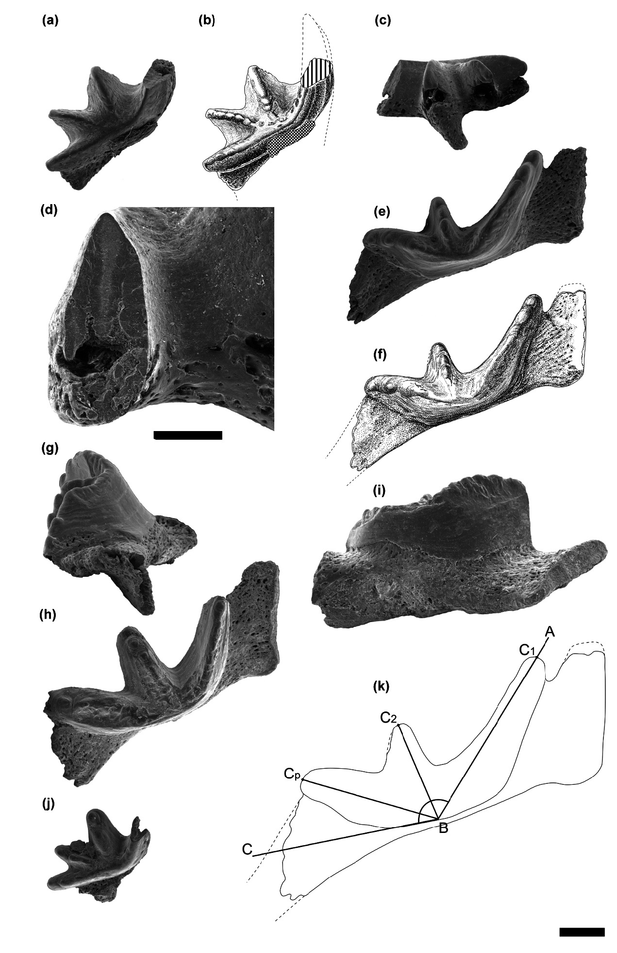
There are two upper tooth plates and two lower ones known from the Czatkowice 1 locality ( Fig. 1 View Figure 1 ). All the tooth plates of Gnathorhiza otschevi from Czatkowice 1 are small, with the last ridge not exceeding 4 mm. They are high-crowned, triangular in outline, with an obtuse inner angle and an almost indistinguishable occlusal surface. All ridges are straight, narrow and acute, and originate medially from one point at the mediolingual junction. However, the first ridge is slightly curved posteriorly near its origin. Cusps are present on the labial part of all the ridges ( Fig. 1 View Figure 1 ). Inter-ridge furrows are wide, very deep and reach the mediolingual junction. There is no wear facet on the medial faces of both upper and lower tooth plates, which indicates that they do not contact each other along the midline.
5.2 Upper tooth plates
The upper tooth plates ( Fig. 1a, b, c and j View Figure 1 ) bear four ridges. The first ridge is broken in both specimens, but it seems to be the longest or as long as the last one, as similar to all other representatives of Gnathorhiza . The first ridge is also the highest in ZPAL P. VII/5, although in ZPAL P. VII/6 the first broken ridge is slightly lower than the second ridge. The pterygopalatine bone is thin and short. Sulci of the pterygopalatine bone are almost indistinguishable, and the ascending process originates from the bone above the second ridge ( Fig. 1c View Figure 1 ). The process is short, oval in section and slightly curved posteriorly. ZPAL P. VII/5 has two big cusps on both the second and third ridge, as well as three cusps on the fourth ridge. On ZPAL P. VII/6 the cusps are nearly worn out, but few can be still observed on the second and fourth ridge. The inner angle of the upper tooth plates equals about 135 ◦ in ZPAL P. VII/5 and about 141 ◦ in ZPAL P. VII/6 ( Table 1 View Table 1 ). Specimen ZPAL P. VII/5 is the smallest tooth plate from Czatkowice 1. This specimen could have belonged to a juvenile fish as can be assumed from relatively big cusps ( Fig. 1j View Figure 1 ) in comparison to other specimens from the collection.
5.3 Lower tooth plates
Three ridges separated by deep furrows can be observed on each of the lower tooth plates ( Fig. 1e–i View Figure 1 ). The first ridge is the longest and highest one. Only in one specimen ( ZPAL P. VII/7) the first ridge is complete, and it is almost 2 times longer than the second one and 1.2 times longer than the third one ( Table 1 View Table 1 ). The sulcus on the prearticular bone is single and very shallow. The symphyseal shaft of the prearticular bone is long and slightly curved medially. Despite the fact that the posterior shaft of this bone is incomplete, it is evident that it was not much longer before it broke. The prearticular symphysis is long, half-oval in section and devoid of grooves on the surface ( Fig. 1i View Figure 1 ). ZPAL P. VII/9 bears five to seven cusps on each of the ridges, while ZPAL P. VII/7 has only three cusps on each ridge. On the last ridge of the latter specimen a newly formed cusp can be seen, as well as horizontal growth lines of enamel ( Fig. 1g View Figure 1 ). The inner angle reaches almost 130 ◦ in both lower tooth plates ( Table 1 View Table 1 ) .
5.4 Microstructure
A natural section of the ridge is visible on a broken first ridge of ZPAL P. VII/6 upper tooth plate ( Fig. 1d View Figure 1 ). The ridge section is roughly oval in shape. In the place where the pterygopalatine bone fuses to the tooth plate, in one-third of the section length, a small pulp cavity can be seen. The bone has a spongy appearance inside the pulp cavity. A moderately narrow core of petrodentine stretches from the pulp cavity up to the occlusal surface of the ridge. It has a lighter colour than the surrounding dentine ( Fig. 1d View Figure 1 ). The core of petrodentine has the shape of a narrow, strongly elongated triangle, broadened toward the occlusal surface of the ridge.
Petrodentine (or pleromic hard tissue of Oervig, 1967) is a specific hypermineralized dentine present in adult dipnoan tooth plates (e.g. Smith, 1984; Kemp, 2001; Reisz et al., 2004). This tissue “is formed by the continued growth of the core dentine of the cusps in the hatchling tooth plate in those species that have this tissue” ( Kemp, 2001, p. 424). Due to its continuous growth, growth lines can be seen in petrodentine ( Smith, 1984). This tissue is free of denteons, almost free of collagen, but rich in calcium hydroxyapatite ( Lund et al., 1992). Petrodentine is clearly distinguishable from the osteodentine (trabecular dentine), because its secretion begins in the larval stages of tooth plate development ( Smith, 1984). Circumdenteonal dentine does not surround petrodentine; it is enclosed in interdenteonal dentine ( Kemp, 2001). Smith (1984) defined 12 characters (which were later reduced to 9 in Reisz et al., 2004) enabling distinction of petrodentine from other tissues, both in extant and extinct dipnoans. However, Lund et al. (1992) did not agree with Smith (1984) and stated that petrodentine is present only in gnathorhizid and lepidosirenid tooth plates. But Kemp (2001) showed petrodentine also in some other derived taxa (e.g. Mioceratodus ).
Originally the term was used by Lison (1941) to describe such tissue in tooth plates of extant Protopterus and Lepidosiren , but petrodentine is present also in some extinct taxa, e.g. Gnathorhiza ( Smith, 1984; Lund et al., 1992; Kemp, 2001). On the occlusal surface of gnathorhizid tooth plates petrodentine is visible as a hard raised dentine that encloses regions of circumdenteonal and interdenteonal dentine ( Lund et al., 1992). Gnathorhiza has extensive masses of petrodentine similar in structure to petrodentine of Mioceratodus ( Kemp, 2001) . The petrodentine in G. otschevi upper tooth plate from Early Triassic Czatkowice 1 locality ( Fig. 1d View Figure 1 ) presented here shows fairly comparable structure with those described earlier for Gnathorhizidae by Lund et al. (1992).
| ZPAL |
Zoological Institute of Paleobiology, Polish Academy of Sciences |
No known copyright restrictions apply. See Agosti, D., Egloff, W., 2009. Taxonomic information exchange and copyright: the Plazi approach. BMC Research Notes 2009, 2:53 for further explanation.
|
Kingdom |
|
|
Phylum |
|
|
Class |
|
|
Order |
|
|
Family |
Gnathorhiza Cope, 1883
| Skrzycki, P. 2016 |
Gnathorhiza
| Cope 1883 |