
Gonioctena (Spartoxena) pseudogobanzi Kippenberg
|
publication ID |
https://doi.org/ 10.5281/zenodo.181638 |
|
DOI |
https://doi.org/10.5281/zenodo.6233336 |
|
persistent identifier |
https://treatment.plazi.org/id/0399D92F-FFF2-3B77-C79E-FAF5FC7DF82D |
|
treatment provided by |
Plazi |
|
scientific name |
Gonioctena (Spartoxena) pseudogobanzi Kippenberg |
| status |
|
Gonioctena (Spartoxena) pseudogobanzi Kippenberg , mature larva
( Figs. 1–5 View FIGURE 1 View FIGURE 2 View FIGURES 3 – 9 , 10 View FIGURE 10 )
Material examined. SPAIN. Málaga, Frigiliana (UTM 30SVF1972), 350 m asl, 12 May 2007, 17 mature larvae; Málaga, Villanueva de Cauche (UTM 30 SUF 7095), 750 m asl, 11 May 2007, 15 mature larvae.
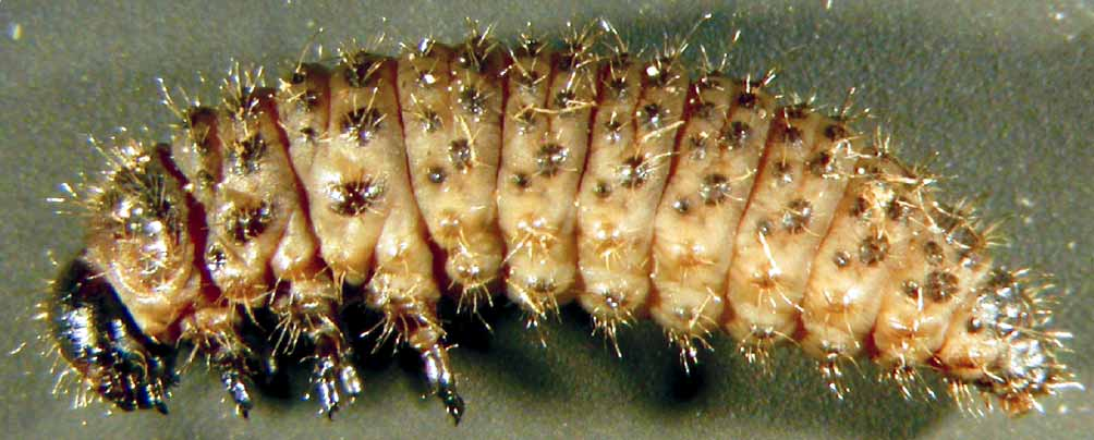
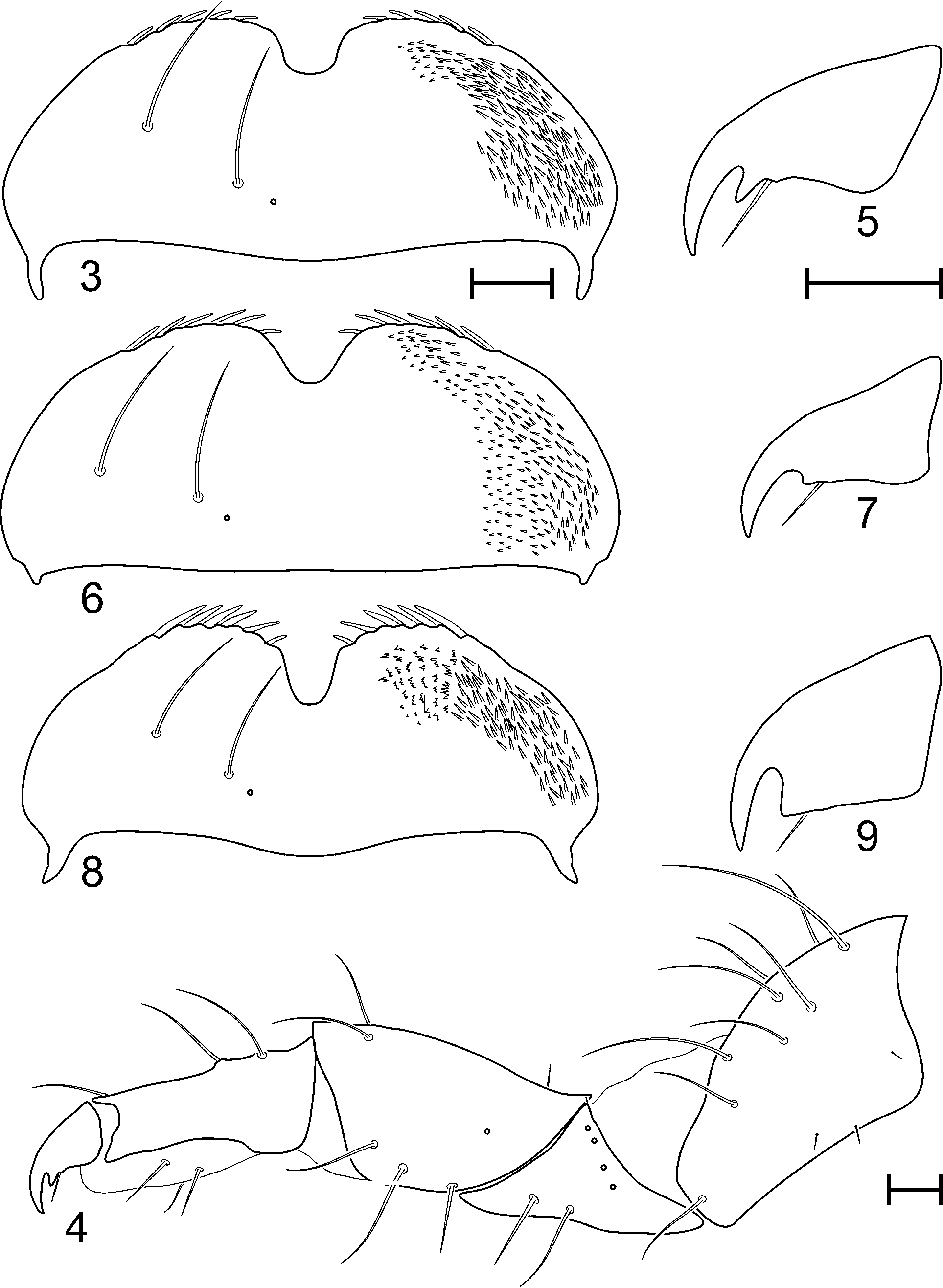
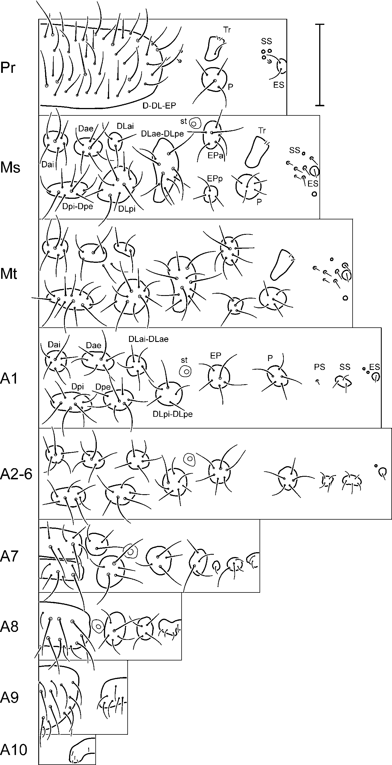
Description. Length: 8.5–12.5 mm. Body eruciform ( Fig. 1 View FIGURE 1 ), convex and slightly arched in preserved specimens. Inter-tubercular plates pale yellow-brown, tubercles pale brown in ventral region, dark brown in dorsal and dorsolateral regions, as well as pronotum. Head. Hypognathous, well sclerotized. Great part of vertex and frons dark brown coloured, the anterior part of frons, clypeus and mouth parts, paler. Epicranial suture well developed and long, frontal arms distinct, V-shaped and almost straight ( Fig. 2 View FIGURE 2 ). Endocarina present, extending almost to clypeus. Vertex bearing 5 large primary setae (v1, v3, v4, v5, v6) along with many shorter ones on each side. Frons with 5 primary setae (f1, f2, f3, f4, f6) and 7–10 slightly shorter ones on each side. Antennae very short and well sclerotized, three-segmented: first joint highly transverse; second joint almost as long as wide, bearing a conical membranous sensillum, 3 minute setae and 1 placoid sensillum; distal joint narrow, subconical, with membranous apex bearing 5 highly minute setae and 1 placoid sensillum. Stemmata arranged in two groups, 1 pair located below the base of antenna and 2 pairs behind the antenna. Clypeus with 3 pairs of setae. Mouthparts. Labrum ( Fig. 3 View FIGURES 3 – 9 ) bearing 2 pairs of setae on upper surface and 1 pair of placoid sensilla, anterior border with a wide U-shaped notch and 5–6 stout setae on each side. Epipharynx with 2 bands of microtrichia situated laterally to anterior notch ( Fig. 3 View FIGURES 3 – 9 ), microtrichia mostly isolated (but sometimes paired). Mandibles symmetrical, 5-toothed, bearing 2 setae on external face and 1 placoid sensillum on dorsal side. Maxillae: cardo transverse, with 1 seta in external border; stipes elongate, with 2 large setae near base of maxillary palp; mala bearing 12–15 setae on internal margin and apex, basal setae longer than apical ones, maxillary palpi 4-segmented, first joint slightly wider than long, bearing 2 long setae basally and another minute seta apically on external margin, second joint highly transverse, third joint longer than wide with 2 setae on internal face and 1 on external side, and fourth joint conical with 1 minute seta on internal face and membranous apex bearing 11–12 highly minute setae. Labium with postmentum membranous, bearing 3 pairs of setae, anterolateral one very short; prementum with 4 pairs of minute setae, 1 pair posterior and 3 pairs anterior to labial palpi along with 1 pair of placoid sensilla; palpi two-segmented, first joint transverse, distal joint conical with membranous apex bearing 10 highly minute setae. Thorax. All tubercles multisetose ( Fig. 10 View FIGURE 10 ). Prothorax with tubercles D (dorsal), DL (dorsolateral) and EP (epipleural) fused together in a pronotal sclerite, pronotum (D-DL-EP) bearing 10 pairs of primary setae along with many other slightly shorter ones; tubercle P (pleural) with 4–6 setae; ventral region with slightly sclerotized tubercles, tubercle SS (sternellar) reduced to 3–4 sclerotized spots bearing 1 seta, midventral tubercle ES (eusternal) bearing 4 setae. Meso- and metathorax with 6 tubercles on each side of dorsal region: Dai (dorsal anterior interior, with 5–7 setae), Dae (dorsal anterior exterior, 6–7 setae), Dpi-Dpe (dorsal posterior interior and dorsal posterior exterior fused together, 7–13 setae), DLai (dorsolateral anterior interior, 2–5 setae), DLpi (dorsolateral posterior interior, 10– 15 setae), DLae-DLpe (dorsolateral anterior exterior and dorsolateral posterior exterior fused together, 12–15 setae); epipleural region with 2 tubercles, EPa (epipleural anterior, 8–11 setae) and EPp (epipleural posterior, 5–6 setae); mesothoracic spiracle isolated from EPa tubercle, located in front of DLae-DLpe one; P tubercle bearing 4–6 setae; SS and ES tubercles reduced to numerous sclerotized dots bearing isolated setae. Legs. All pairs similar in size; trochantin located in front of P tubercle ( Fig. 10 View FIGURE 10 ), bearing 2 minute setae in anterior half; prothoracic trochantin also with a larger seta in postero-ventral angle; coxa almost twice longer than wide in lateral view ( Fig. 4 View FIGURES 3 – 9 ), with 10–11 large setae on dorsal face and 3–5 shorter ones in each lateral declivity; trochanter triangular in lateral view, with 2 large setae on each side, 1 minute seta and 4 placoid sensilla near coxal articulation on anterior side, and 2 placoid sensilla on posterior side; femur wider apically than basally in lateral view, with 2 large and 1 small setae dorsally, 3 large setae and 1 placoid sensillum on anterior side, and 2 large setae on posterior side; tibio-tarsus twice longer than wide, bearing 3 large and 1 minute setae dorsally and 4 ventrally; unguis wide basally, curved apically, with marked acute tooth and seta on lower side ( Fig. 5 View FIGURES 3 – 9 ). Abdomen. All tubercles multisetose ( Fig. 10 View FIGURE 10 ). Segments 1–6 with 6 tubercles on each side of dorsal region: Dai (3–6 setae), Dae (6–8 setae), Dpi (6–9 setae), Dpe (6–8 setae), DLai-DLae (4–7 setae) and DLpi- DLpe (6–8 setae); epipleural region with tubercle EP bearing 7–10 setae; spiracle isolated from EP tubercle, located in front of DLpi-DLpe one; P tubercle with 6–11 setae; sternal region presents the following tubercles: PS (parasternal, 2–5 setae, but it is extremely reduced in segment 1, substituted by 1 isolated setae), SS (4–6 setae), ES separated in two halves, each one with 1–3 setae. Segment 7 with tubercle Dai fused to Dae, and Dpi to Dpe; segment 8 and 9 with all dorsal and dorsolateral tubercles fused together, ventral ones also fused in segment 9; segment 10 forming anal pseudopod, without dorsal tubercles, ventral ones fused together.
Distribution and ecology. G. pseudogobanzi is distributed in the southeast region of Spain, from the Penibetic mountains to the South and ranging from eastern Malaga to Murcia ( Kippenberg, 2001; Baselga, 2007). Its host plant is known after the original description ( Kippenberg, 2001): Genista umbellata (L'Hér.) Dum. Cours. [ Fabaceae ]. The larvae described in this paper were collected in the same plant [field identification]. Genista umbellata is present in the Iberian Peninsula but also in Morocco and Algeria. The Iberian range is restricted to the Southeast of Spain, but its distribution is wider than that of G. pseudogobanzi , reaching the provinces of Badajoz, Sevilla, Albacete and Valencia to the North ( Talavera, 1999). Although phytophagous insects usually have narrower distribution ranges than their hosts ( Gaston, 2003), further research is needed to clarify to which extent both distributions are different.

| SUF |
Shimonoseki University of Fisheries |
No known copyright restrictions apply. See Agosti, D., Egloff, W., 2009. Taxonomic information exchange and copyright: the Plazi approach. BMC Research Notes 2009, 2:53 for further explanation.