
Hybusa occidentalis ( Westwood, 1843 )
|
publication ID |
https://doi.org/ 10.11646/zootaxa.5380.4.4 |
|
publication LSID |
lsid:zoobank.org:pub:EE5E50F4-3A8B-4270-8412-BEA89FAB87C5 |
|
DOI |
https://doi.org/10.5281/zenodo.10250410 |
|
persistent identifier |
https://treatment.plazi.org/id/03AB87BF-8319-2D18-FF78-FA81FEECFE66 |
|
treatment provided by |
Plazi |
|
scientific name |
Hybusa occidentalis ( Westwood, 1843 ) |
| status |
|
Hybusa occidentalis ( Westwood, 1843) View in CoL
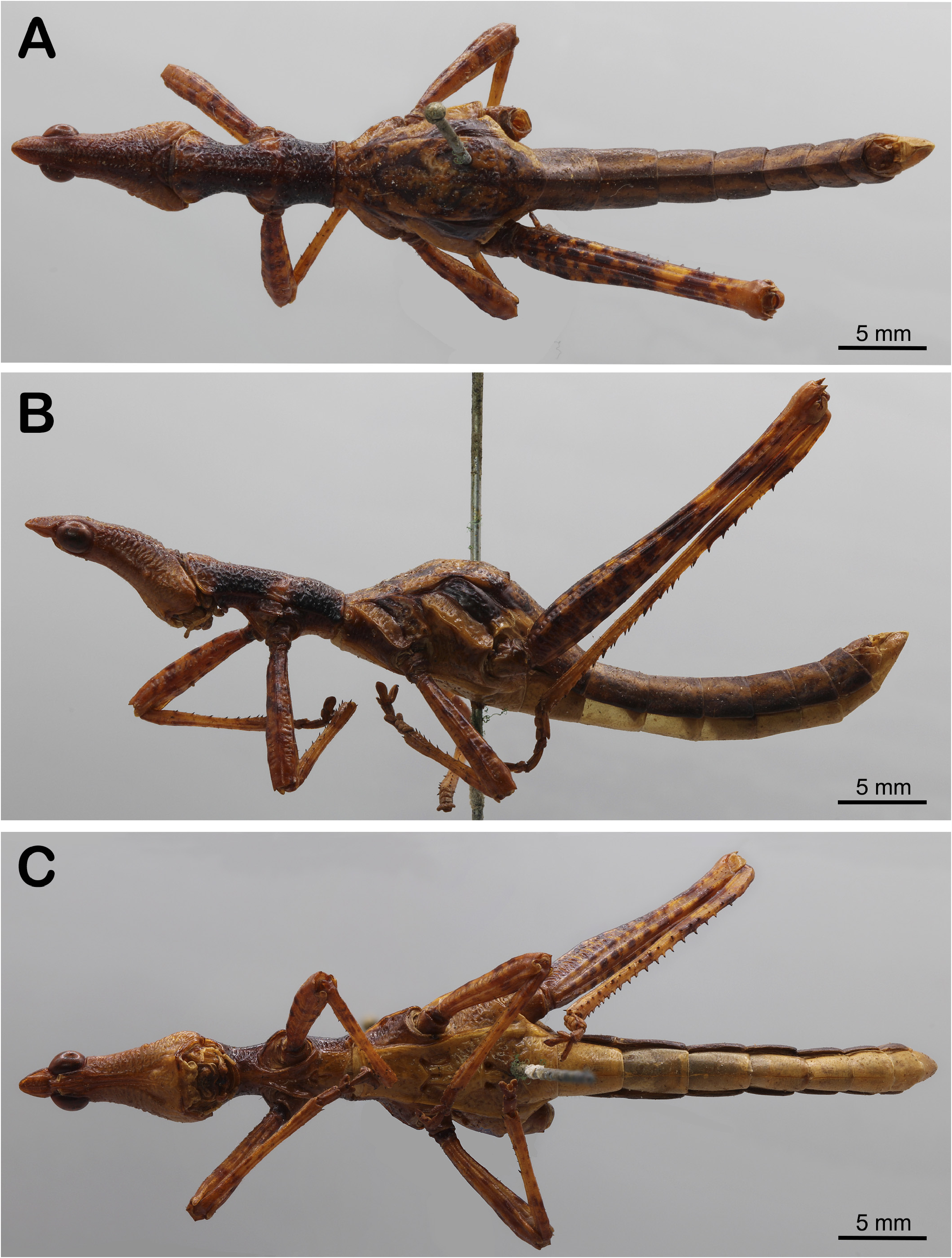
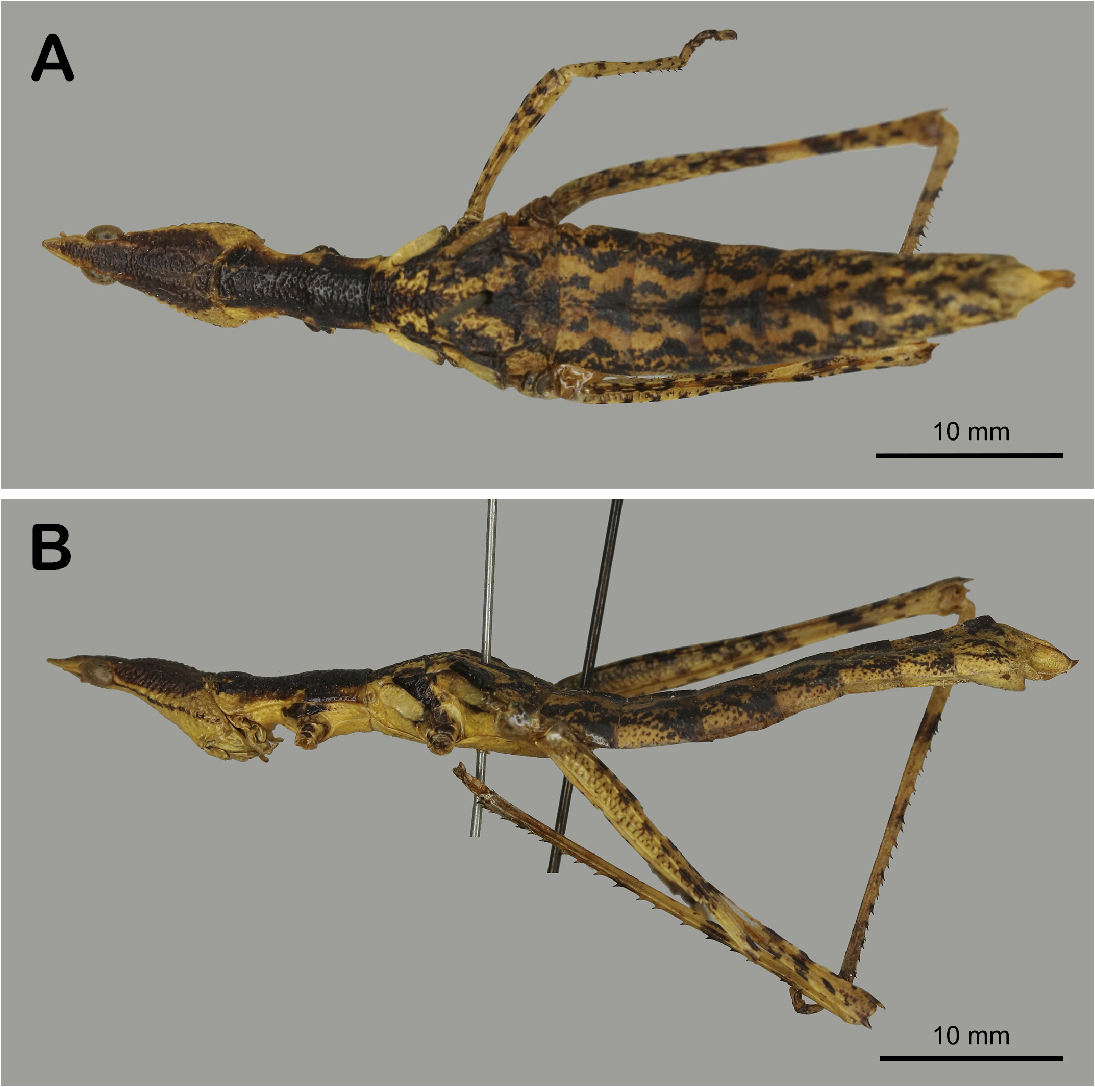
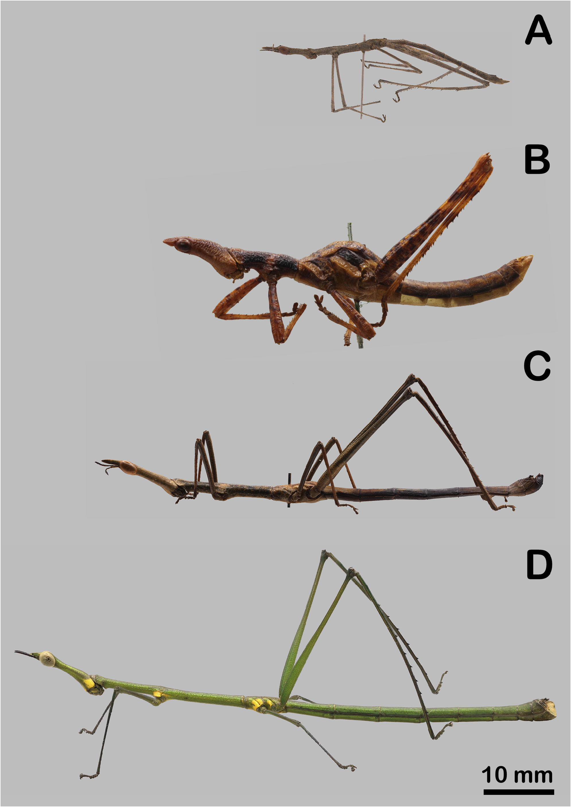
Figs. 2A–C View FIGURE 2 , 3A View FIGURE 3 , 7A–B View FIGURE 7 , 10B View FIGURE 10
Westwood, 1843; Erichson, 1844: transferred from Proscopia to Hybusa ; Jago, 1989: male genitalia description; Alfaro et al. 2013: updated geographic distribution.
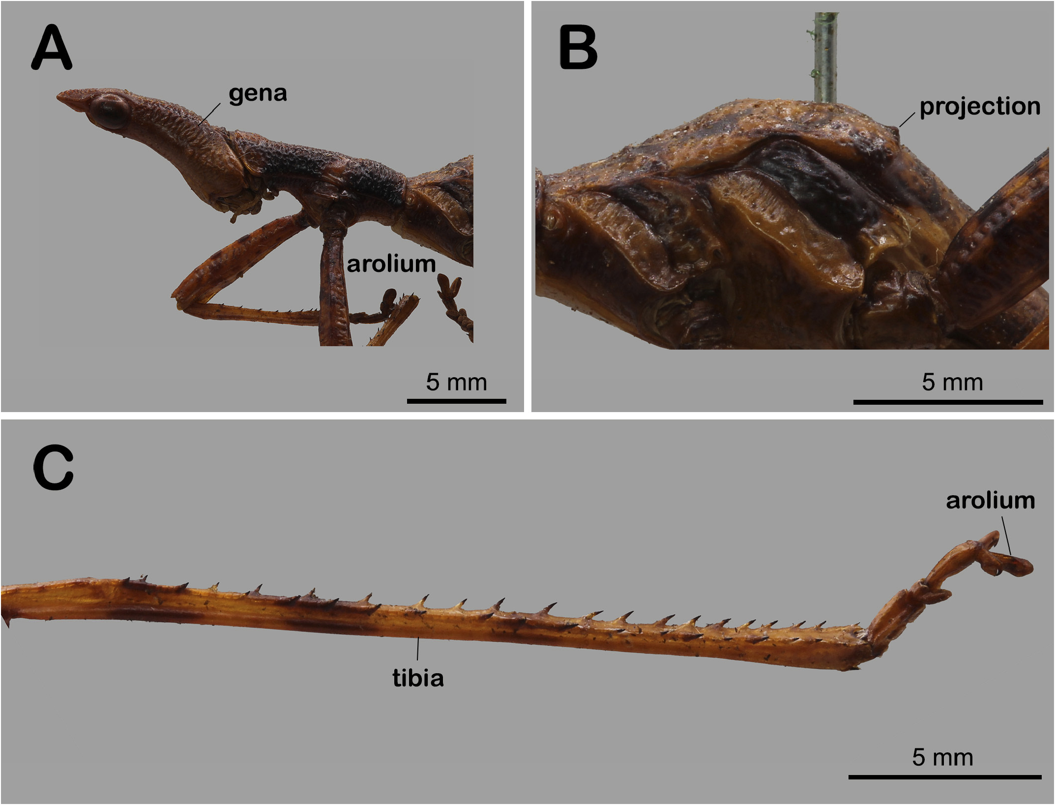
Diagnosis. Frons light-brown, vertex blackened ( Figs. 2A–B View FIGURE 2 ; 3A View FIGURE 3 ; 7A–B View FIGURE 7 ; 11A View FIGURE 11 ). Female head in dorsal view with triangular black spot; gena with a conspicuous black line from base to compound eye ( Figs. 7A–B View FIGURE 7 ). Pronotum dark-brown to black, with light-brown spots at proepisternum ( Figs. 2A–B View FIGURE 2 ; 3A View FIGURE 3 ; 11A View FIGURE 11 ). Meso- and metanotum with arched black spots ( Figs. 2A View FIGURE 2 ; 7A View FIGURE 7 ; 12 View FIGURE 12 ). Metepimeron black ( Figs. 2A–B View FIGURE 2 ; 3B View FIGURE 3 ; 12 View FIGURE 12 ). All femora with black dots ( Figs. 2A–C View FIGURE 2 ; 7A–B View FIGURE 7 ). Hind tibia with three black spots, two at basal half and one at distal half ( Fig. 3C View FIGURE 3 ). Tergite 1–8 with black medial longitudinal thick line ( Fig. 2A View FIGURE 2 ). Epiproct widened ( Figs. 4A–B View FIGURE 4 ). Male subgenital plate undivided; 1.5 times longer than tergite 8; dorsal margin nearly straight ( Figs. 4B–C View FIGURE 4 ; 13A View FIGURE 13 ). Female abdomen with conspicuously large black dots ( Figs. 7A–B View FIGURE 7 ). Female tergites 1–8 with black Y-shaped spots ( Fig. 7A View FIGURE 7 ).
Redescription. Male. Body light-brown with several black spots, shiny ( Figs. 3A–C View FIGURE 3 ). Head and thorax are conspicuously rugose ( Figs. 2A–B View FIGURE 2 ; 3A View FIGURE 3 ; 11A View FIGURE 11 ).
Head. Vertex light-brown, with blackened spot ( Fig. 2A View FIGURE 2 ). Fastigium as high as long, 1.3 times shorter than compound eye ( Figs. 3A View FIGURE 3 ; 11A View FIGURE 11 ); in dorsal view subtriangular, with apex nearly truncated, with base 3.4 times wider than apex ( Fig. 2A View FIGURE 2 ); in lateral view with longitudinal sulcus, apex rounded ( Figs. 3A View FIGURE 3 ; 11A View FIGURE 11 ); in ventral view conspicuously concave, with medial longitudinal ridge ( Fig. 2C View FIGURE 2 ). Coronal suture inconspicuous. Transversal ridge of frons slightly smoother than the remaining parts of head. Clypeus with dorsal margin straight, lateral margin sinuous with lateral sulcus; ventral margin with conspicuous emargination. Labrum asymmetrical ( Fig. 2C View FIGURE 2 ).
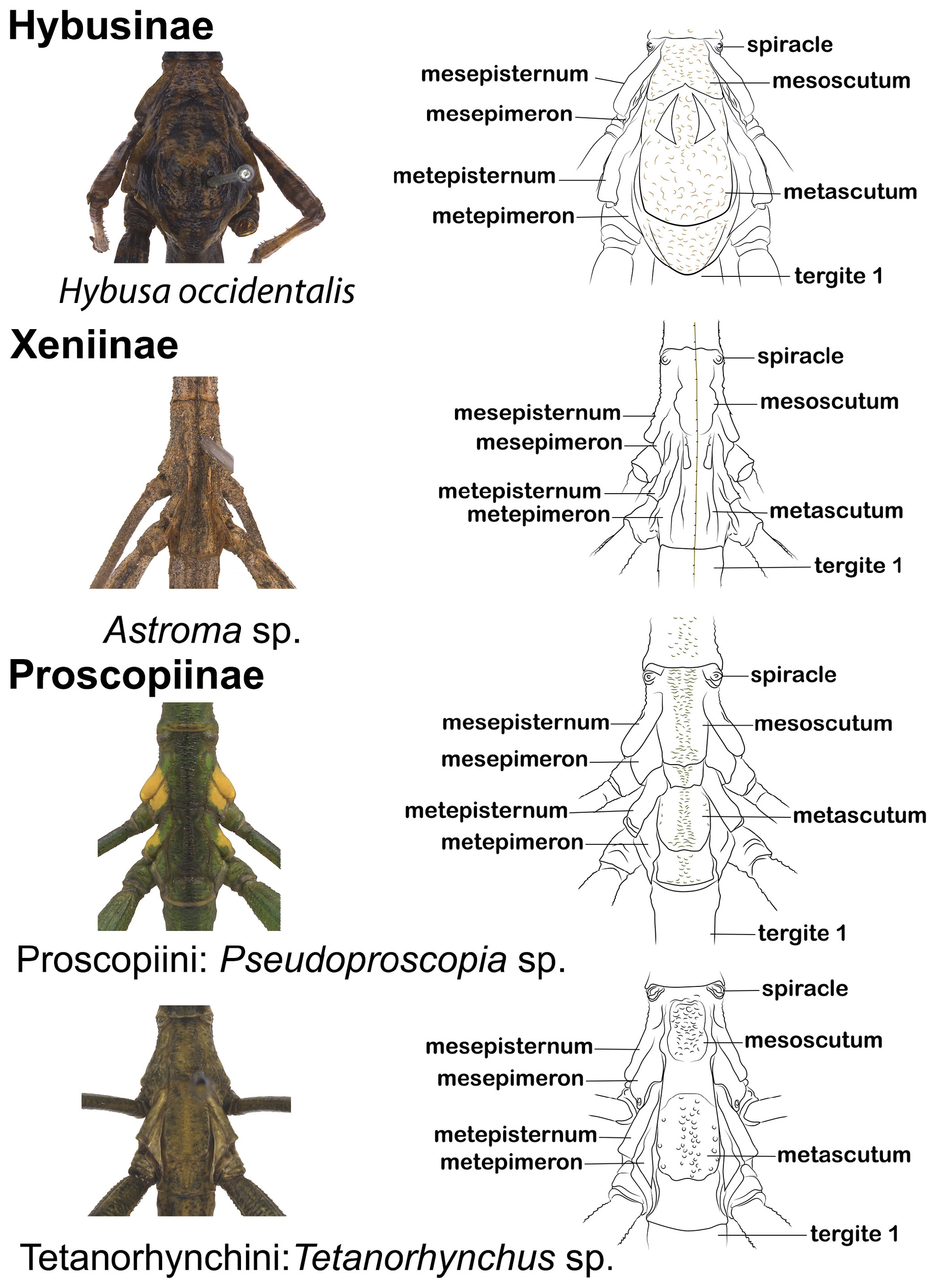
Thorax. Pronotum dark-brown to black, with light-brown spots at proepisternum ( Figs. 2A–B View FIGURE 2 ; 3A View FIGURE 3 ; 11A View FIGURE 11 ). Pro-, meso- and metaepisternum laterally projected, gradually increasing in length ( Fig. 2A–B View FIGURE 2 ). Meso- and metanotum with arched black spots ( Figs. 2A View FIGURE 2 ; 12 View FIGURE 12 ). Mesoscutum trapezoidal ( Figs. 2A View FIGURE 2 ; 12 View FIGURE 12 ). Metepimeron black ( Figs. 2B View FIGURE 2 ; 12 View FIGURE 12 ). Prosternum dark-brown, shiny, with a conspicuous ridge at coxa insertion ( Fig. 2C View FIGURE 2 ). Meso- and metasternum fused, light-yellow, smooth, gradually increasing in width from base to apex ( Fig. 2C View FIGURE 2 ). Pro-, meso- and metasternal processes conspicuous; mesosternal process resembling a “M” ( Fig. 2C View FIGURE 2 ).
Legs. All femora with black dots and medial longitudinal carina ( Figs. 2A–B View FIGURE 2 ); apex with pair of large parallel spines ( Fig. 2B View FIGURE 2 ). Fore and mid legs with nearly the same length ( Figs. 2A–B View FIGURE 2 ). Fore and mid tibiae with small black spines at ventral surface. Anterior and mid tarsi with almost half-length of all respective tibiae ( Figs. 2A–B View FIGURE 2 ). Hind tibia with three black spots, two at basal half and one at distal half; dorsally with several black spines ( Fig. 3C View FIGURE 3 ). Hind tarsus with approximately ¼ of hind tibia length.All tarsal claws and arolium long, wide, robust, with ellipsoid aspect ( Figs. 2B View FIGURE 2 ; 3C View FIGURE 3 ).
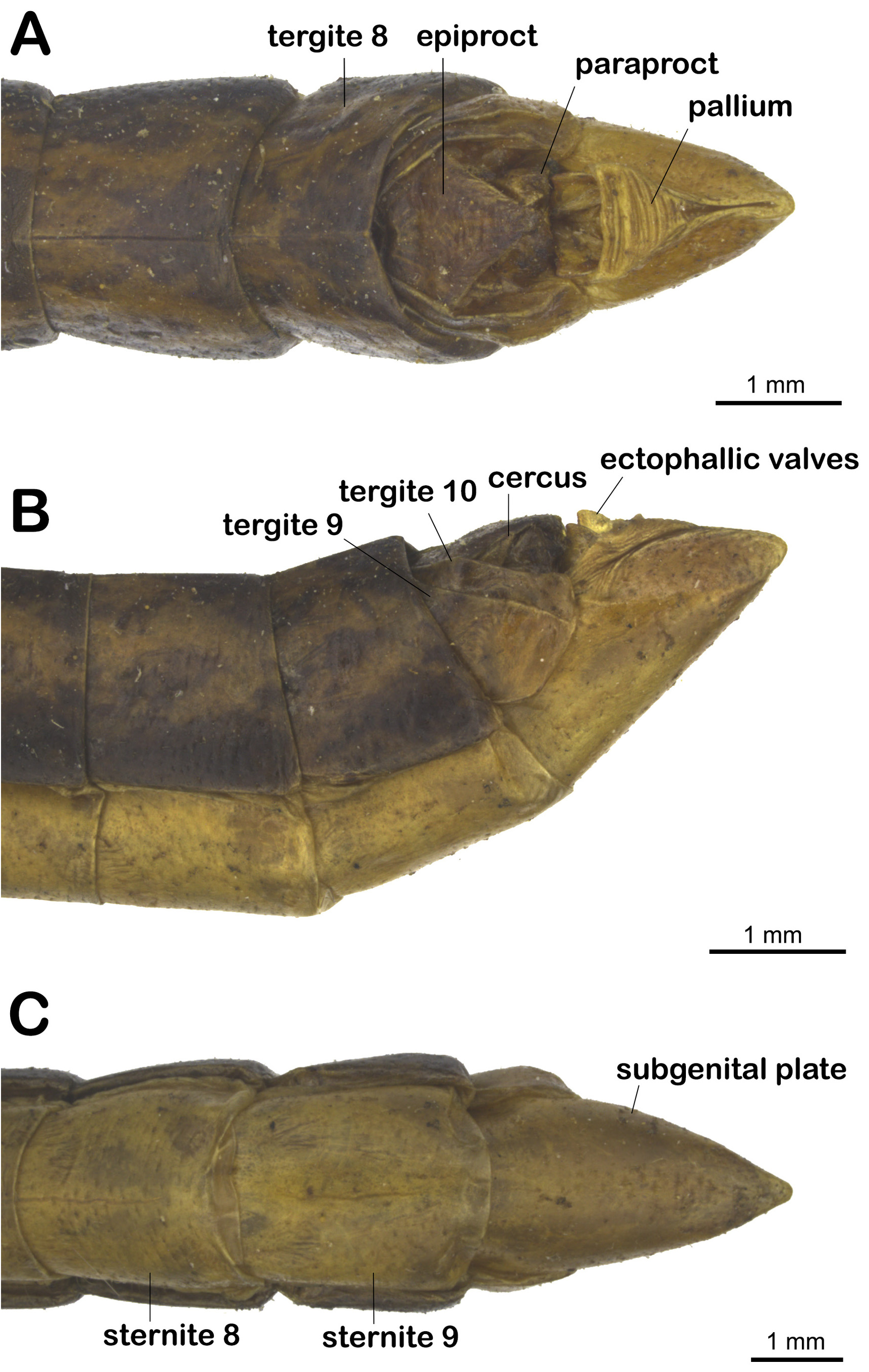
Abdomen. Tergite 1–8 with black medial longitudinal thick line ( Fig. 2A View FIGURE 2 ). Tergite 1 D-shaped, half-length of tergite 2. Tergites 2–8 rectangular, longer than wide, gradually decreasing in length, with the same length as respective sternites ( Fig. 2A View FIGURE 2 ). Tergite 9 trapezoidal, approximately two times higher than tergite 10 ( Figs. 4A–B View FIGURE 4 ; 13A–B View FIGURE 13 ). Tergite 10 with dorsal margin sinuous ( Figs. 4A–B View FIGURE 4 ; 13A–B View FIGURE 13 ). Epiproct rugose, pentagonal, conspicuously widened at anterior half; posterior half narrowing; apex curving ventrally ( Figs. 4A–B View FIGURE 4 ; 13A–B View FIGURE 13 ). Pallium striated, subtriangular, membranous ( Figs. 4A View FIGURE 4 ; 13B View FIGURE 13 ). Cercus simple, inconspicuous, short ( Figs. 4A–B View FIGURE 4 ; 13A–B View FIGURE 13 ). Subgenital plate undivided; 1.2 times longer than tergite 8; dorsally with an apical cleft; ventrally conical, with narrow, rounded apex ( Figs. 4A–C View FIGURE 4 ; 13A–B View FIGURE 13 ).
Genitalia. Male genitalia. Epiphallus rim wide, nearly covering lophi ( Fig. 5C View FIGURE 5 ). Transverse sclerite mostly membranous, laterally with slender sclerotized portion, ending in hook-like apex; apex connected to epiphallic rim ( Figs. 5A–B View FIGURE 5 ; 14A View FIGURE 14 ). Lophi ventral-most portion connected to transverse sclerite at sclerite’s mid-length; ventral portion U-shaped, projecting towards dorsal portion; dorsal portion bifurcating in two fused elongated projections, these pointed antero-dorsally; projections sinuous, asymmetrical ( Figs. 5A–D View FIGURE 5 ; 6A View FIGURE 6 ; 14A View FIGURE 14 ). Ectophallic valves not connected, parallel, with nearly half-length of transverse sclerite; in dorsal view sub-triangular, widened ( Figs. 5A–B View FIGURE 5 ; 14A View FIGURE 14 ; posterior portion projecting dorsally and outwards body, better observed in lateral view ( Figs. 5C–D View FIGURE 5 ). Phallotreme conspicuous, sclerotized, laterally pigmented; medially striated; posterior portion projected into sac-like membrane ( Figs. 5A–B View FIGURE 5 ; 14A View FIGURE 14 ). Endophallus with conspicuous proximal and distal sacs; proximal sac membranous, pigmented; distal sac sclerotized, sinuous, projecting inwards, conspicuously pigmented ( Figs. 6B–D View FIGURE 6 ). Ejaculatory duct simple, widened at the base, slender at medial portion, ending in a slightly widened apex; apex with two rounded sclerotizations, with a small membranous portion projecting from it ( Figs. 6B–D View FIGURE 6 ).
Female. As in male, except for the body more robust ( Fig. 7A View FIGURE 7 ). Head broader, with acute fastigium ( Figs. 7A–B View FIGURE 7 ) Gena with a conspicuous line from compound eye towards apex ( Fig. 7B View FIGURE 7 ). Abdomen with several black spots ( Figs. 7A–B View FIGURE 7 ).


No known copyright restrictions apply. See Agosti, D., Egloff, W., 2009. Taxonomic information exchange and copyright: the Plazi approach. BMC Research Notes 2009, 2:53 for further explanation.
|
Kingdom |
|
|
Phylum |
|
|
Class |
|
|
Order |
|
|
Family |
|
|
Genus |
Hybusa occidentalis ( Westwood, 1843 )
| Queiroz, Larissa Lima De, Rafael, José Albertino, Pádua, Diego Galvão De, Araujo, Rodrigo De Oliveira & Heleodoro, Raphael Aquino 2023 |
Hybusa
| Erichson 1844 |
Proscopia
| Klug 1820 |