
Kawingasaurus, Cox, 1972
|
publication ID |
https://doi.org/ 10.1093/zoolinnean/zlac033 |
|
DOI |
https://doi.org/10.5281/zenodo.7922177 |
|
persistent identifier |
https://treatment.plazi.org/id/3F6A87AB-FFB1-FFE7-71E5-FE79FC25FA3E |
|
treatment provided by |
Plazi |
|
scientific name |
Kawingasaurus |
| status |
|
KAWINGASAURUS FOSSILIS COX, 1972
Specimen: GPIT-PV-117032 (formerly GPIT/RE/9272).
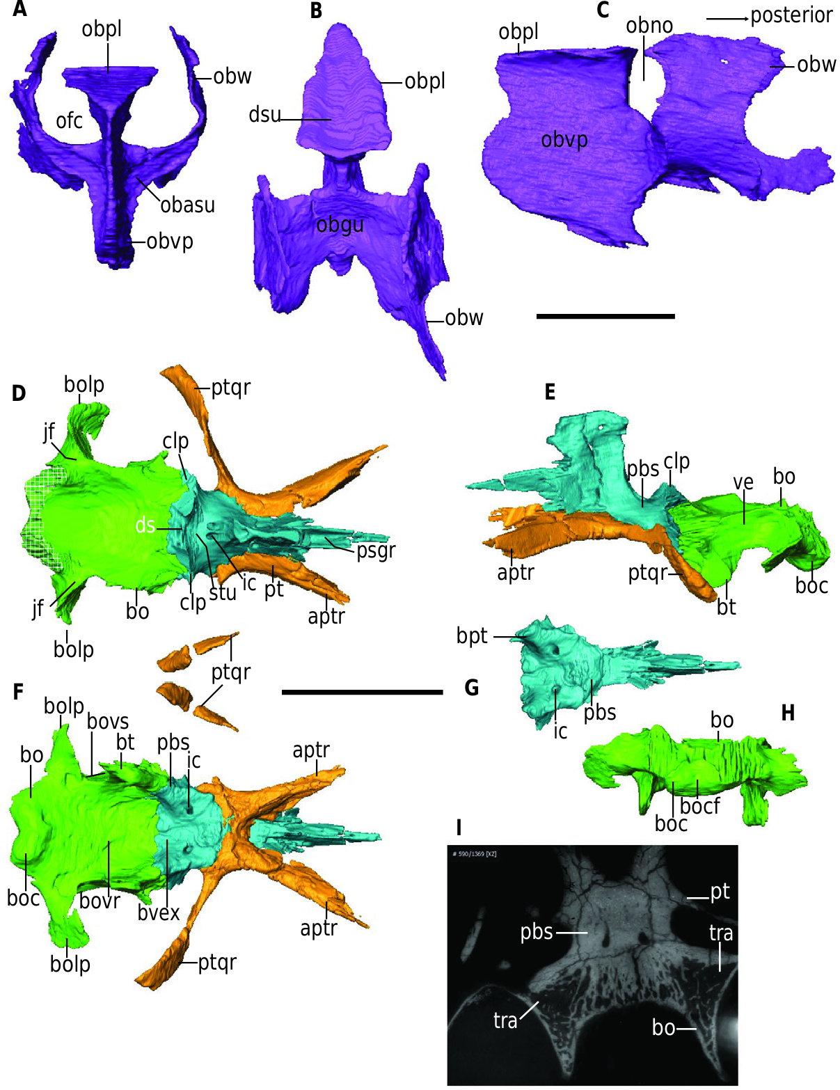
Orbitosphenoid: The orbitosphenoid ( Fig. 9 View Figure 9 ) is a complex composed of two dorsally directed wings posteriorly and the vertical median process anteriorly often called the mesethmoid in cistecephalids ( Keyser, 1973; Laass & Kaestner, 2017; Angielczyk et al., 2019). It contacts the frontal dorsally and the parasphenoid ventrally. The orbitosphenoid is nearly rectangular in lateral view and hosts the olfactory bulbs on its dorsal portion.
The wings (obw) in GPIT-PV-117032 have the usual Y-shaped cross-section. The lateral wings are oriented almost vertically in anterior view and connect to the body of the bone at a right angle ( Fig. 9A View Figure 9 ). The dorsal vertical portion of the wings is thin, whereas the horizontal portion is thicker. In anterior view, the base of the wing is excavated by a shallow sulcus (obasu, Fig. 9A View Figure 9 ) that descends ventromedially for a short distance. The anteriormost half of the orbitosphenoid comprises the median vertical process (obvp), which forms a horizontal dorsal plate (obpl) dorsally and descends to form a thick vertical plate ventrally that articulates with the parasphenoid rostrum ( Fig. 9A View Figure 9 ). The dorsal horizontal plate articulates with the frontal dorsally. The median process separates the olfactory cavities (ofc, Fig. 9A View Figure 9 ), which are relatively expanded in Kaaeingasaurus compared to Myosaurus and Pristerodon (height-to-width ratio 2.0 in Kaaeingasaurus vs. 1.7 in Myosaurus vs. 1.8 in Pristerodon ). In dorsal view, the horizontal plate of the vertical process is triangular and is excavated by a long anteroposterior sulcus (dsu, Fig. 9B View Figure 9 ).
In lateral view, the orbitosphenoid in Kaaeingasaurus is relatively longer anteroposteriorly than in Myosaurus ( Fig. 9C View Figure 9 ). The wings are well separated from the mesethmoid and their lateral surfaces are slightly convex. Their dorsal margins are horizontal and flat to articulate with the frontal, as in Pristerodon . The ventral margin of the wing expands posteriorly and horizontally, giving it a C-shaped posterior margin. The anterior margin of the wings appears S-shaped in lateral view. In dorsal view, the gutter (obgu) formed by the two wings is anteroposteriorly short and inclined posteriorly. There is a notch between the median vertical process and the orbitosphenoid wings in lateral view (obno). In lateral view, the vertical plate of the median vertical process has a horizontally oval outline topped by the rectangular shape of the dorsal plate dorsally.
Pterygoid: The pterygoid (pt, Fig. 9 View Figure 9 D-F) displays the typical X-shape dicynodont morphology in ventral view. In GPIT-PV-117032, the pterygoid is almost completely preserved except for the right quadrate ramus. The quadrate ramus in Kaaeingasaurus diverges from the median sagittal plane at an angle of about 80°. The anterior palatal ramus of the pterygoid has a concave lateral surface observed in dorsal view, and it forms an angle of 30° with the median sagittal axis of the skull. The angle between the palatal and quadrate rami of the pterygoid is 83°. As observed in lateral view, the palatal ramus is taller than the quadrate ramus ( Fig. 9E View Figure 9 ). The posterior end of the quadrate ramus deepens ventromedially in lateral view. Medially, the posteriormost region of the quadrate ramus has a convex surface that articulates with the quadrate.
The palatal ramus of the pterygoid is thicker ventrally than the quadrate ramus and terminates anteriorly in an irregular suture with the ectopterygoid ( Fig. 9F View Figure 9 ). In ventral view, the two pterygoids meet at the midline of the median horizontal plate in Kaaeingasaurus, as in most cistecephalids. The ventral exposure of the pterygoid median plate in Kaaeingasaurus is short compared to the parabasisphenoid at the median sagittal axis ( Fig. 9F View Figure 9 ), as noted in Angielczyk et al. (2019). Kaaeingasaurus lacks the crista oesophagea, with the median pterygoid plate being flat ( Fig. 9F View Figure 9 ), and also lacks an interpterygoid vacuity, as noted by Cox (1972). An interpterygoid vacuity is also essentially absent in Cistecephalus ( Keyser, 1973; Angielczyk et al., 2019), contrasting with the condition in cistecephalids like Kembaaeacela and Cistecephaloides ( Cluver, 1974a; Kammerer et al., 2016; Angielczyk et al., 2019), which have small openings for this structure. The quadrate ramus is broad posteriorly but mediolaterally compressed towards the median plate in ventral view.
No known copyright restrictions apply. See Agosti, D., Egloff, W., 2009. Taxonomic information exchange and copyright: the Plazi approach. BMC Research Notes 2009, 2:53 for further explanation.