
Laophontodes sarsi, George, 2018
|
publication ID |
https://doi.org/ 10.5852/ejt.2018.439 |
|
DOI |
https://doi.org/10.5281/zenodo.5989060 |
|
persistent identifier |
https://treatment.plazi.org/id/03E55261-2277-FFEA-6E7E-FAE068245603 |
|
treatment provided by |
Plazi |
|
scientific name |
Laophontodes sarsi |
| status |
sp. nov. |
Laophontodes sarsi sp. nov.
urn:lsid:zoobank.org:act:D78FE397-C13D-4AEF-96A8-A2AFE61829EE
Figs 9–11 View Fig. 9 View Fig. 10 View Fig. 11
Etymology
The epitheton ‘sarsi’ is given in respectful memory of Georg Ossian Sars, esteemed Norwegian scientist. who provided valuable contributions to our knowledge of Copepoda Harpacticoida , including the description of numerous species.
Type material
Two females collected and identified as Laophontodes typicus by G.O. Sars. Due to the limited number of (partially damaged) individuals, specimens were not dissected and, therefore, some appendages could not be observed or described.
Holotype
NORWAY: adult ♀, northern Norwegian coast, Skjerstad fjord , G.O. Sars leg., preserved in 75% ethanol ( NHM F20298-1 ).
Paratype
NORWAY: adult ♀, same data as for holotype ( NHM F20298-2).
Description
Female
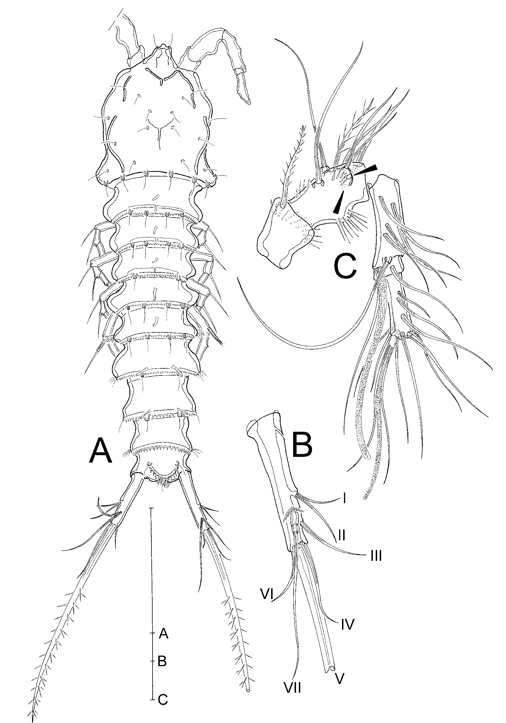
HABITUS ( Fig. 9A View Fig. 9 ). Cylindrical, tapering distally, body length (R to end of FR) 387 µm (female 1) and 350 µm (female 2). Cphth slightly as long as broad, 25% of total body length, posteriorly with bulge on each side. R small, fused to cphth, with 2 sensilla on apical margin. Body somites distinct. Posterior margins of thoracic somites serrated dorsally, with sensilla arising from small socles and 1 tube pore centrally. Genital double somite formed by fusion of last thoracic and first abdominal somites, juncture detectable by dorsal serration and constricted outer margins. Abdominal part of GDS with finely serrated posterior margin and pair of tube pores; second and third abdominal somites dorsally strongly serrated at posterior margins; second somite with, third somite without sensilla-bearing socles and pair of tube pores. Telson slightly smaller than preceding somite, FR widely separated proximally. Anal operculum with pair of sensilla and moderately long spinules at apical margin.
FURCAL RAMI ( Fig. 9A–B View Fig. 9 ). Slender, about 3.7 times longer than its broadest part, proximally with tube pore (tube not discernible in Fig. 9B View Fig. 9 ), and with 7 setae: I and II arising centrally from outer margin, II slightly longer than I; III subapical on dorsal side; IV and V apical, IV very narrow and less than half the length of V, which is tripinnate; VI apical on inner margin, as long as II; VII dorsal, tri-articulated at base, arising from pedestal, base surrounded by row of spinules.
ANTENNULE ( Fig. 9C View Fig. 9 ). 5-segmented. First segment with 1 bipinnate seta and apical row of spinules; second segment with 9 setae (2 setae broken in Fig. 9C View Fig. 9 ), one bipinnate; third segment with 7 bare setae and 1 aes; fourth segment partially overlapped by preceding one, with 1 bare seta; fifth segment with 9 bare setae, and 2 additional apical setae and small aes forming trithek.
SETAL FORMULA. 1-1/2-9/3-7+aes/4-1/5-11+aes.
ANTENNA ( Fig. 10A View Fig. 10 ). With enp slightly shorter than allobasis, exp represented by minute bare seta. Allobasis with 1 abexopodal seta and scant spinules. Enp with 2 rows of spinules, 2 bare spines and 1 minute seta on distal edge, 3 strong geniculate setae and 2 spines apically, and denticulate frill subapically.
MD, MXL AND MX. Not drawn due to unfavourable position. However, unilobate md palp with 6 setae and mx with minute 1-segmented enp carrying 2 setae observed.
MAXILLIPED ( Fig. 10B View Fig. 10 ). With small syncoxa bearing 1 biplumose seta surrounded by small spinules; basis twice as long as syncoxa, with 1 row of spinules; enp (turned around in Fig. 10B View Fig. 10 ) forming long claw with minute bare seta.
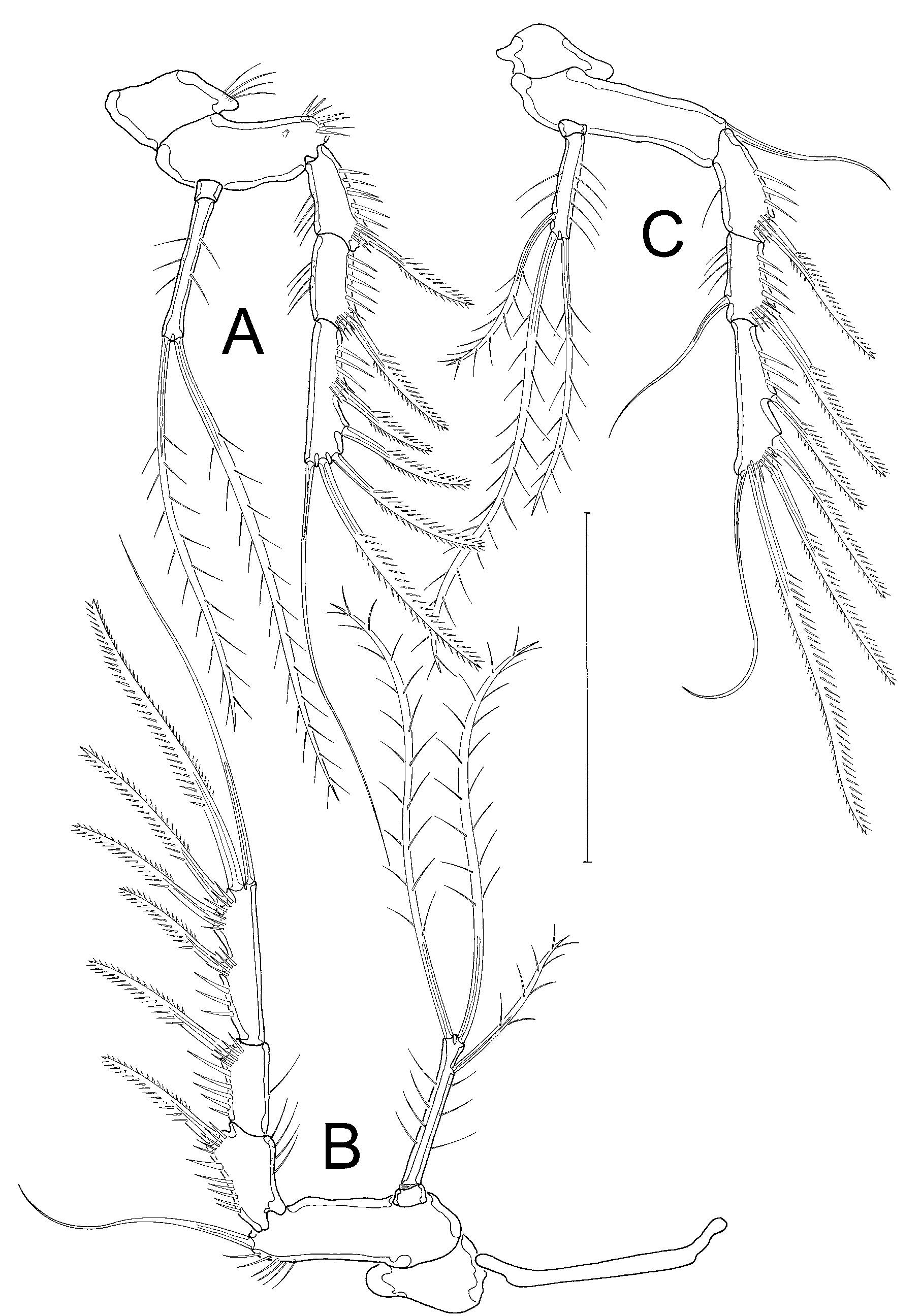
SWIMMING LEGS. P1 ( Fig. 10C View Fig. 10 ) with squarish coxa; basis as long as coxa, with 1 bipinnate outer seta and 1 uniplumose inner seta displaced to anterior surface. Enp 2-segmented, enp-1 elongated, with row of spinules on outer and inner margins; enp-2 small, ⅓ length of enp-1, apically with large claw, 1 long slender bare seta and 1 small seta. Exp 3-segmented, half the length of enp. All exopodal segments with spinules on outer margins; exp-1 with 1 uniplumose outer spine; exp-2 with 1 long bare geniculated outer seta; exp-3 with 4 long bare geniculated setae (innermost broken in Fig. 10C View Fig. 10 ). P2–P4 ( Fig. 11A– C View Fig. 11 ) with transversely elongated bases, 3-segmented exopods and 2-segmented endopods. Exp-3 longest, all segments with rows of spinules on outer margin; exp-1 and exp-2 with fine spinules sparsely on inner margin; P2 ( Fig. 11A View Fig. 11 ) and P3 ( Fig. 11B View Fig. 11 ) exps without inner seta; P4 ( Fig. 11C View Fig. 11 ) exp-1 and exp-3 without, exp-2 with 1 bare inner seta; P2–P4 exp-3 with 3 outer bipinnate spines, and apically with 1 bipinnate outer seta and 1 slender bare inner seta. P2–P4 enp-1 small, without setae. P2 enp-2 elongated, with spinules on outer and inner margins, and 2 biplumose apical setae; P3 and P4 enp-2 as in P2 (shorter in P4), but additionally with 1 biplumose inner seta. P5 ( Fig. 10D View Fig. 10 ) small, benp and exp fused. Benp with outer basal seta arising from setophore. Endopodal lobe represented by 2 setae, one biplumose and one fish-bone. Exp short, with 2 outer, 1 subapical, and 2 apical setae, all bipinnate; no tube pores evident.
GENITAL FIELD ( Fig. 10E View Fig. 10 ). Small, with single gonopore; P6 strongly reduced, limbs fused forming a single small clasp, with pair of minute bare setae.
Male
Unknown.

| NHM |
University of Nottingham |
No known copyright restrictions apply. See Agosti, D., Egloff, W., 2009. Taxonomic information exchange and copyright: the Plazi approach. BMC Research Notes 2009, 2:53 for further explanation.