
Laophontodes scottorum, George, 2018
|
publication ID |
https://doi.org/ 10.5852/ejt.2018.439 |
|
DOI |
https://doi.org/10.5281/zenodo.5989058 |
|
persistent identifier |
https://treatment.plazi.org/id/03E55261-2272-FFF7-6E5E-FD726A23513F |
|
treatment provided by |
Plazi |
|
scientific name |
Laophontodes scottorum |
| status |
sp. nov. |
Laophontodes scottorum sp. nov.
urn:lsid:zoobank.org:act:9A5CB956-4F5E-4928-BA06- A29001 View Materials DC3E97
Figs 7–8 View Fig. 7 View Fig. 8
Etymology
The epitheton ‘scottorum’ is given in respectful remembrance of Thomas Scott, who firstly described L. typicus , and of his son Andrew Scott, who provided the illustrations.
Type material
Holotype
UNITED KINGDOM: ♀, the Scottish Firth of Forth , labelled as [co-type, collection Norman, collection number BMNH 1911.11.8.44995 ].
Dissection not permitted owing to single individual: description based on intact specimen resulting in certain perspective-related bias and preventing examination of some appendages. Uncertainty with an observation is denoted with (?). Male partially described by T. Scott (1907) (as L. typicus ).
Description
Female
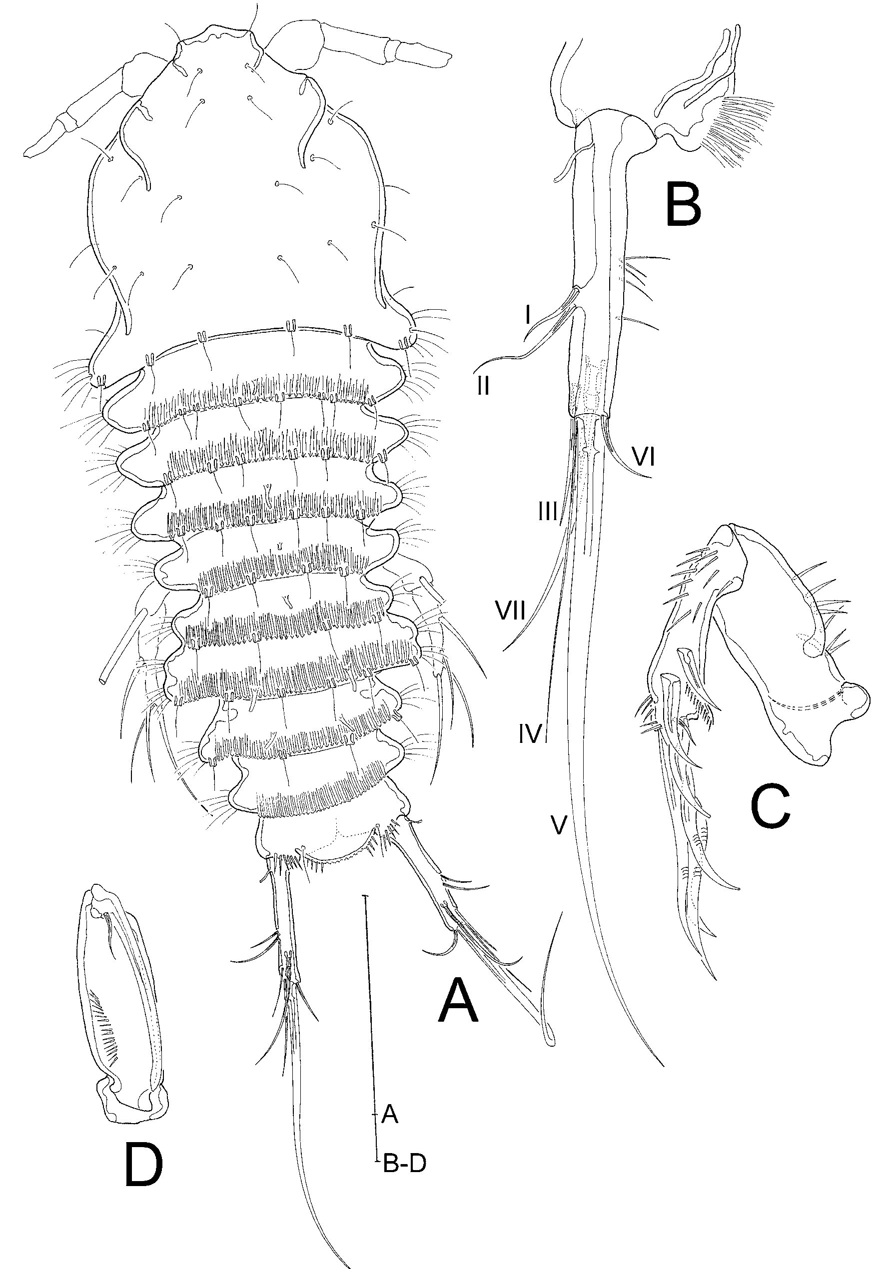
HABITUS ( Fig. 7A View Fig. 7 ). Slightly flattened dorsoventrally and longitudinally compressed, body length (R to end of FR) 438 µm. Cphth as long as broad, ⅓ of body length. R small, fused to cphth, with 2 sensilla on apical margin. Posterior margin of cphth extended laterally, rounded at outer margin and carrying fine setular tufts. Pedigerous somites 1–4 about 4.5 times as broad as long, clearly distinct, laterally extended, with setular tufts on outer lateral margins. Posterior margins serrated dorsally and with fine ripples running anteriorly; additionally with sensilla arising from small socles and 1 tube pore centrally. Genital double somite formed by fusion of last thoracic and first abdominal somites, juncture detectable by serration, sensilla-bearing socles and constricted outer margins; abdominal half with pair of tube pores on distal margin. Second and third abdominal somites remarkably smaller than thoracic and first abdominal somites, being about 3.5 times as broad as long; posterior margins similarly serrated and with fine ripples running anteriorly, second somite also with pair of tube pores on distal margin and with sensilla-bearing socles, third somite lacking sensilla and tube pores. Telson 2.9 times as broad as long, FR arising widely separated. Anal operculum broad, with moderately long spinules at apical margin (several spinules broken in Fig. 7A View Fig. 7 ).
FURCAL RAMI ( Fig. 7A–B View Fig. 7 ). Slender, about 3.8 times longer than broadest point, proximally with tube pore, and with 7 setae: I and II arising centrally from outer margin, II being twice as long as I; III slightly smaller than II, subapically at dorsal side; IV and V apical, IV very narrow and less than half the length of V; VI apical at inner margin, as long as I; VII dorsal, arising from pedestal, tri-articulated at base.
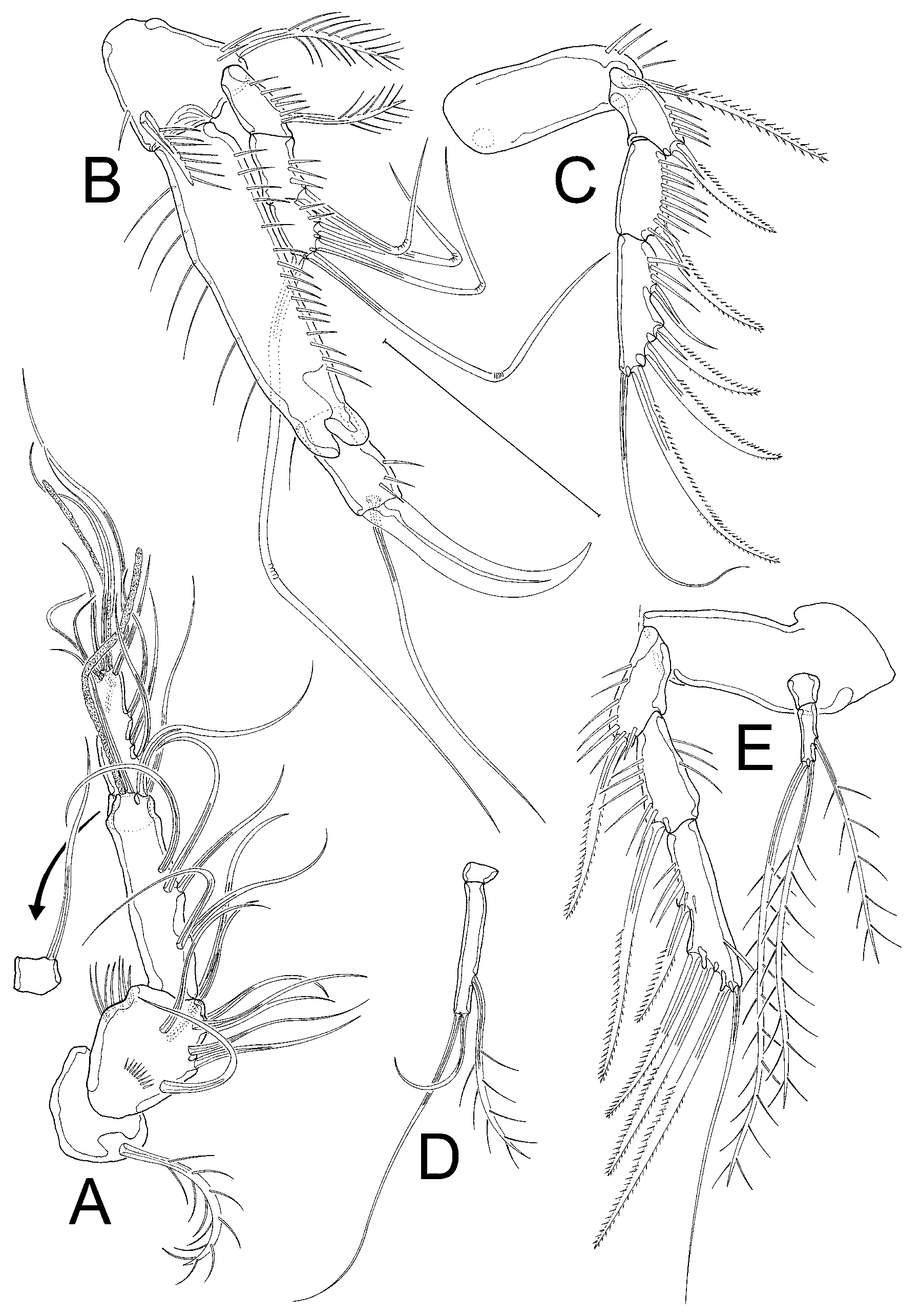
ANTENNULE ( Fig. 8A View Fig. 8 ). 5-segmented. First segment with 1 biplumose seta; second segment with 9 bare setae; third segment longest (?), equipped with 7 bare setae and 1 aes; fourth segment overlapped by preceding one, with 1 bare seta; fifth segment with 10 bare setae and small apical aes.
SETAL FORMULA. 1-1/2-9/3-7+aes/4-1/5-10+aes.
ANTENNA ( Fig. 7C View Fig. 7 ). Sturdy, enp slightly shorter than allobasis, exp represented by small bare seta. Allobasis without abexopodal seta, but with abexopodal row of spinules. Enp with several spinules, 2 bare spines plus 1 minute seta on distal edge, and 4 strong geniculate setae and 1 spine apically; subapical denticulate frill.
MD, MXL AND MX. Not examined.
MAXILLIPED ( Fig. 7D View Fig. 7 ). Syncoxa partially obscured; basis with row of spinules; enp formed into strong claw, same length as enp, with minute bare seta.
SWIMMING LEGS. P1 ( Fig. 8B View Fig. 8 ) coxa obscured; basis triangular with 1 biplumose outer seta and 1 uniplumose inner seta displaced to anterior surface. Enp 2-segmented, enp-1 strong, elongated, with row of spinules on both outer and inner margin; enp-2 small, ⅓ length of enp-1, with large claw and 1 long, slender bare seta apically, small seta not discernible. Exp 3-segmented, less than half the length of enp, each segment with spinules on outer margin, also exp-1 with 1 biplumose outer seta; exp-2 with 1 long, bare and geniculate outer seta; exp-3 with 4 long, bare geniculate setae. P2–P4 ( Fig. 8C–E View Fig. 8 ) with transversely elongated bases, 3-segmented exopods and 2-segmented endopods (not discernible in P2). Exopodal segments increasing in size from exp-1 to exp-3, each segment with row of spinules on outer margin, no inner setae. Exp-1 and exp-2 with 1 bipinnate outer spine; exp-3 with 3 outer spines, and apically with 1 outer spine and 1 slender flexible and bare inner seta. P2 enp ( Fig. 8C View Fig. 8 ) missing or broken, not drawn. P3 enp-1 very small and bare; enp-2 about 7 times longer than enp-1, with 1 inner biplumose seta and 2 apical bare setae, inner apical seta half the length of outer apical seta ( Fig. 8D View Fig. 8 ). P4 enp small, enp-1 bare, enp-2 about twice as long as enp-1, with 1 inner and 2 apical setae, all biplumose ( Fig. 8E View Fig. 8 ). P5, GF, and P6 not examined, ventral urosome covered by detritus.
No known copyright restrictions apply. See Agosti, D., Egloff, W., 2009. Taxonomic information exchange and copyright: the Plazi approach. BMC Research Notes 2009, 2:53 for further explanation.