
Leptogomphus schieli, Dow & Stokvis & Ngiam, 2017
|
publication ID |
https://doi.org/ 10.11646/zootaxa.4358.2.1 |
|
publication LSID |
lsid:zoobank.org:pub:8861BCC0-022F-4803-98E8-D28B90F666E4 |
|
DOI |
https://doi.org/10.5281/zenodo.5631254 |
|
persistent identifier |
https://treatment.plazi.org/id/03C3A90C-9844-FF90-FF6A-8DD1FDD6ED1A |
|
treatment provided by |
Plazi |
|
scientific name |
Leptogomphus schieli |
| status |
sp. nov. |
Leptogomphus schieli View in CoL sp. nov.
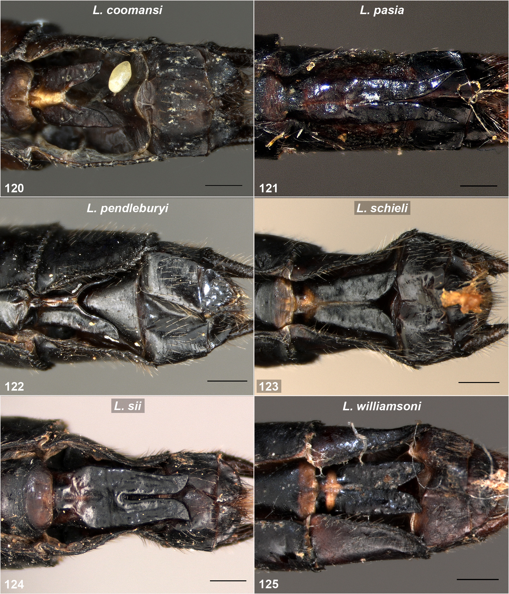
( Figs 8 View FIGURES 4–9 , 16 View FIGURES 14–17 , 25 View FIGURES 24–27 , 33 View FIGURES 32–35 , 43 View FIGURES 42–45 , 48 View FIGURES 46–51 , 58 View FIGURES 56–59 , 62 View FIGURES 60–63 , 75 View FIGURES 74–77 , 85 View FIGURES 84–87 , 91 View FIGURES 88–93 , 97 View FIGURES 94–99 , 103 View FIGURES 100–105 , 109 View FIGURES 106–111 , 115 View FIGURES 112–117 , 123 View FIGURES 120–125 , 128 View FIGURE128 )
Leptogomphus new species;—Dow et al. 2016: 10, 13 (both sexes Gunung Penrissen).
Holotype 1 ♂ ( SAR 15_GOM8), by very small, steep stream below peak of Gunung Penrissen, Borneo Highlands resort area on Gunung Penrissen, Kuching Division, 1.11905N, 110.22793E, 14 vii 2015, leg. G.T. Reels, to be deposited in BMNH.
Paratypes. 2 ♀♀ ( SAR 15_GOM6, 7; supposition), seepage below peak of Gunung Penrissen, same area as holotype, 1.11905N, 110.22793E, 15 vii 2015, leg. R.A. Dow, in collection Dow.
Etymology. Schieli, a noun in the genitive case. Named for Franz-Josef Schiel in honour of his contributions to Odonatology.
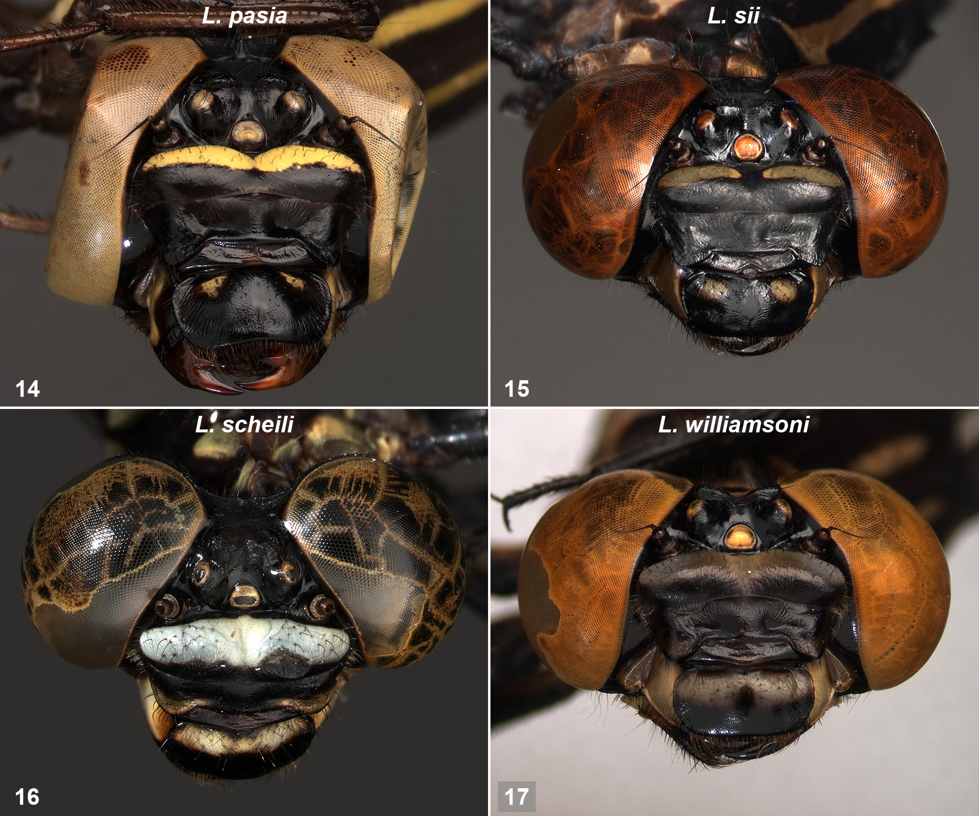
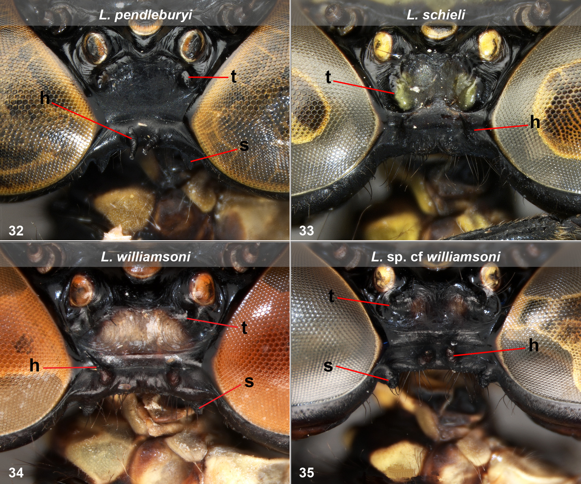
Diagnosis. The male is easily distinguished from those of all other Leptogomphus known from Borneo by the convergent branches of the epiproct. The female is distinguished from those of all species known from Borneo except L. williamsoni (and L. sp. cf williamsoni ) by the dorsal markings of the thorax, and from the female of L. williamsoni by the head lacking occipital spurs.
Description of holotype male. Head ( Figs 8 View FIGURES 4–9 , 16 View FIGURES 14–17 ). Labium largely pale, very dark brown basally centrally, apical part median lobe, narrowly on apical outer margin of palps, and hooks. Labrum black, transverse basal pale stripe. Mandible bases pale, genae pale adjacent to mandible bases, otherwise black. Clypeus black with pair of widely separated yellow marks at anterior lateral corners postclypeus. Ante- and postfrons not very sharply divided, postfrons almost entirely pale bluish ( Fig. 16 View FIGURES 14–17 ). Pair of prominent, robust tubercles behind lateral ocelli, occupying almost all of vertex between ocelli and occipital plate; ocelli pale yellowish. Vertex and occiput black.
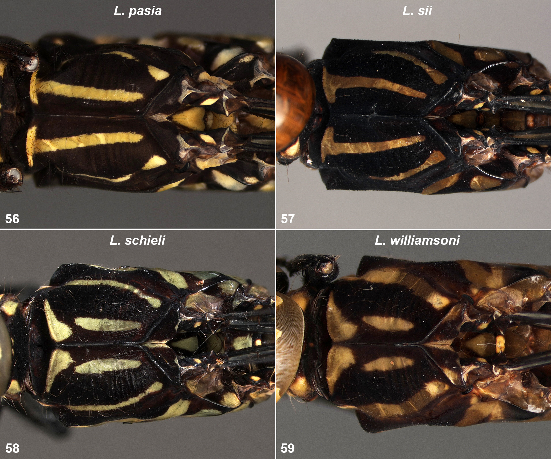
Thorax. Prothorax mostly black, pale irregular transverse stripe anteriorly on anterior pronotal lobe, greenish yellow on lateral parts, brownish orange centrally. Pair of greenish-yellow lateral marks, small central posterior mark (divided centrally) on middle pronotal lobe. Propleuron mottled dark brown and pale. Synthorax black and dark brown with pale greenish markings as follows ( Figs 58 View FIGURES 56–59 , 62 View FIGURES 60–63 ): broad mesothoracic collar, divided at middorsal carina. Narrow dorsal thoracic stripes reaching level of apex of mesothoracic collar but separated from it, extending well past apex of antealar crest. Narrow antehumeral stripes just separated from mesokatepisternum. Mesepimeron with broad stripe, irregular along its lower margin. Metepisternum with large subrectangular mark near antealar carina, metepimeron with broad stripe. Mesokatepisternum yellow in line with antehumeral stripe, black above. Metakatepisternum largely yellowish. Metaposternum greyish and pale brown. Legs robust and relatively short. Coxae mottled pale, grey and brown, trochanters largely black. Rest of legs black except inner surface of anterior femora with pale stripe. Wings: sectors of arculus separated at origin with 5 cross veins up to and at first bifurcation of superior sector in Fw, 3 in Hw. Discoidal field with 2 rows of cells from origin, transitioning to three rows before level of nodus in both wings. 14 (right) to 15 (left) Ax in Fw, 10 in Hw, 10 (left) or 12 (right) Px in Fw, 14 (right) to 12 (left) in Hw. Pt brown, covering ca 4 underlying cells.
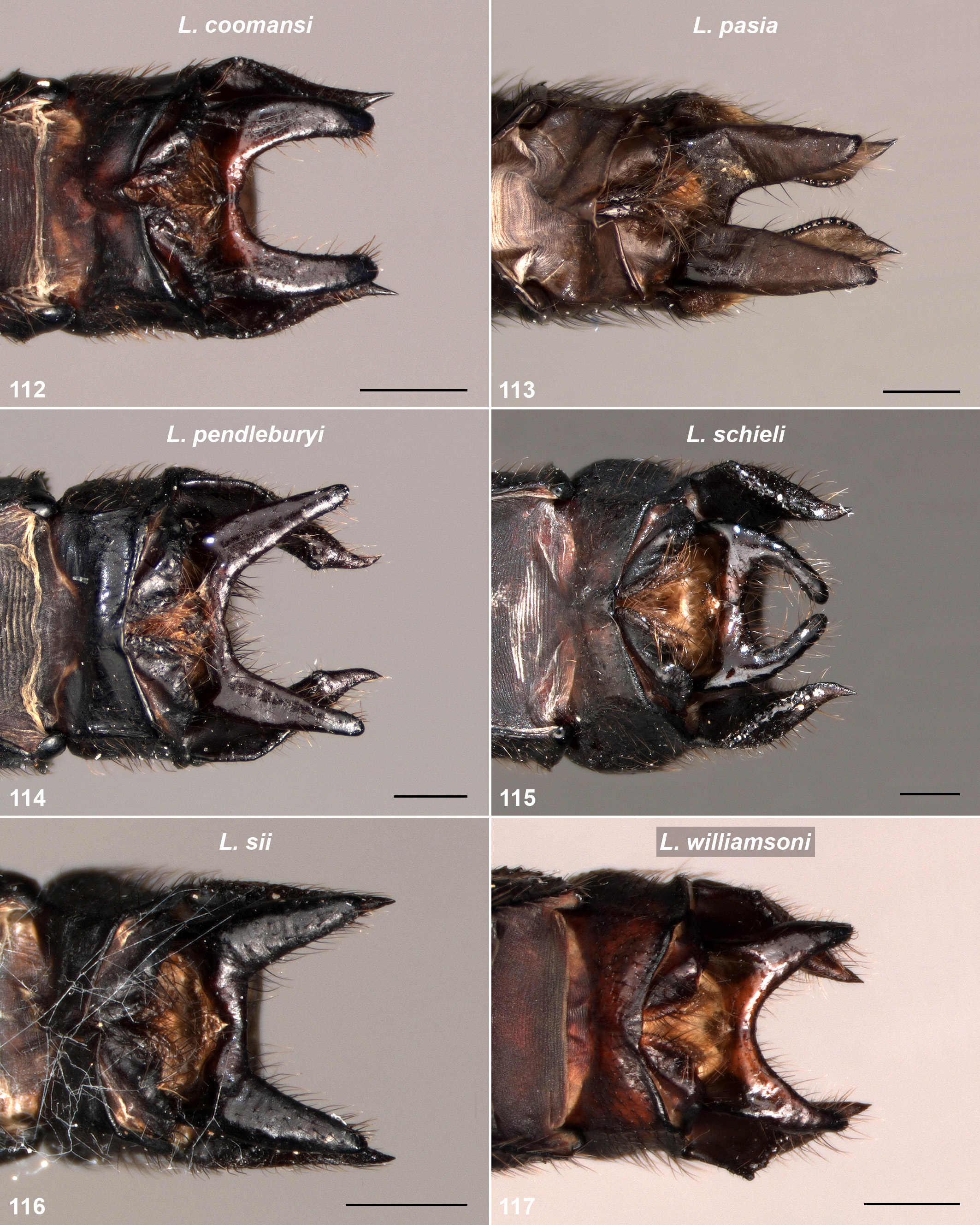
Abdomen. Slender after base of S3, expanding moderately from base of S7, maximum width reached apical part of S8, then almost constant. Black with pale markings as follows: S1 large pale lateral mark, dorsal mark. S2 central lateral pale mark from base, including part of auricle, narrowly divided from posterior yellow mark, narrow apical middorsal stripe. S3–6 with small lateral markings at base, tiny, barely visible on S6. On left S8 with tiny apical lateral brown mark. S3–7 with very narrow, irregular middorsal line, only present in apical half S3, on S7 slight expanded apically. Cerci ( Figs 103 View FIGURES 100–105 , 109 View FIGURES 106–111 ) black, broad at base in lateral view ( Fig. 109 View FIGURES 106–111 ), tapering to sharp slightly upturned tip, along upper margin sloping steeply down from base to just before mid-length, then gently curved to apex, along lower margin sloping down from base to just before half length, then abruptly up to apex, forming a prominent corner, small teeth present on up-turned section. In dorsal view ( Fig. 103 View FIGURES 100–105 ) outer margin running very slightly outward from base, then inward to tips at just after half length; prominent corner on lower margin just visible. Dorsally dark brown at apex. Epiproct ( Figs 103 View FIGURES 100–105 , 115 View FIGURES 112–117 ) mostly black, shorter than cerci, deeply divided, branches narrow, turned up and strongly inwards so rounded apices only narrowly separated. In lateral view ( Fig. 109 View FIGURES 106–111 ) strongly curved, apices before tips of cerci, turned almost to rear; upper margin obscured by cerci. Accessory genitalia as shown in Figures 91 View FIGURES 88–93 , 97 View FIGURES 94–99 , with anterior hamule moderate sized, tapering toward sharp apex. Posterior hamule large, ca twice length of anterior hamule, directed postero-ventrally, anterior margin converging on posterior margin for much of length, then whole curving gently forward so directed down, tapering to sharp tip, directed slightly outward.
Penis ( Fig. 97 View FIGURES 94–99 ). Penultimate segment extended to rear of join with terminal segment, bifurcated in this part, apices slender, curved down. Terminal segment extended beyond penultimate segment, approximately dumbbell shaped in lateral view, then shallow contracted before strongly expanding dorsally, apically with short rearward directed, curled, cornua; short, rearward directed flap just below cornua. Penis vesicle prominent, slightly more than half height of posterior hamule.
Measurements (mm). Hw 28, abdomen excluding anal appendages 32.5, cerci ca 1.4.
Description of female (SAR15_GOM6). As male except as noted. Head ( Figs 25 View FIGURES 24–27 , 33 View FIGURES 32–35 , 43 View FIGURES 42–45 , 48 View FIGURES 46–51 ). Transverse basal pale stripe of labium narrowly divided into large lateral parts and small central part. Yellow anterior lateral marks on post clypeus larger, small indistinct upper central mark. Stripe on postfrons greenish yellow. Pair of small tubercles immediately behind and inside of lateral ocelli, narrowly separated from slightly larger tubercles adjacent to occipital plate, these separated from occipital plate by narrow but deep depression. Rear tubercles green on their inner faces, area between them and onto rear of anterior pair of tubercles pale greyish green. Occipital plate black bearing pair of widely separated horns, directed up and slightly forward, pair of small inconspicuous tubercles between these at occipital ridge. No occipital spurs, but pair bulges on occiput below compound eye.
Thorax ( Figs 75 View FIGURES 74–77 , 85 View FIGURES 84–87 ). Prothorax with stripe on anterior pronotal lobe all yellow, tiny yellow marks on anterior dorsum middle pronotal lobe. Dorsal thoracic stripe just touching mesothoracic collar on left. Trochanters with some pale marks. Wings: sectors of arculus with 6 cross veins up to first bifurcation of superior sector in left Fw. 13 (right) or 14 (left) Ax in Fw, 10 in Hw, 10 (left) or 13 (right) Px in Fw, 10 (right) or 11 (left) in Hw. Pt pale brown.
Abdomen. Very gradually tapering from base to S8, S9 constricted at base then expanded again apically. Black with more extensive pale markings than male: S1 almost entirely yellowish. S2 with lateral yellow stripe running length of segment, including small, rounded auricle, stripe narrowly divided immediately after auricle. S3 with stripe running from base for most of segment, narrowly divided before its midpoint. S4–7 with basal lateral spot and small streak just after midpoint, the latter, diminishing in size on successive segments, tiny and very faint on S7. Middorsal stripe narrow but wider than in male, running S2–7. Cerci black, tapering to sharp slightly upturned tip, ca same length as S10. Vulvar scale ( Fig. 123 View FIGURES 120–125 ) as long as sternum of S9, divided at ca three-quarters length.
Measurements (mm). Hw 27.5, abdomen excluding anal appendages 29.5.
Variation. The second female paratype is very similar to the one described, but has no central pale mark on the postclypeus, the dorsal thoracic stripe is connected to mesothoracic collar on the right rather than left, the non-basal lateral streak on S7 is larger and brighter. 14–16 Ax in Fw, 12–13 Px in Fw, 11–12 in Hw.
Measurements (mm). Hw 28, abdomen excluding anal appendages 30.
Remarks. The females are associated with the male by reasonable supposition; the seepage in which they were caught is very close to the location where the male was taken, and they resemble the male in the length of the dorsal thoracic stripes, longer than those in the most similar species, L. williamsoni , or the females treated as L. sp. sp. cf williamsoni below, also from Gunung Penrissen. They also differ from both L. williamsoni and L. sp. cf wiliamsoni in the structures on the occiput and vertex; lacking occipital spurs and with markedly different tubercles on the vertex, and with much wider spacing between the occipital horns than in L. sp. cf wiliamsoni. The bulges on the rear occiput below the compound eyes are also not present on the taxa with similar markings.
Leptogomphus schieli was collected too late to include in the molecular analysis, but can be expected to cluster with L. williamsoni and L. sp. cf wiliamsoni.
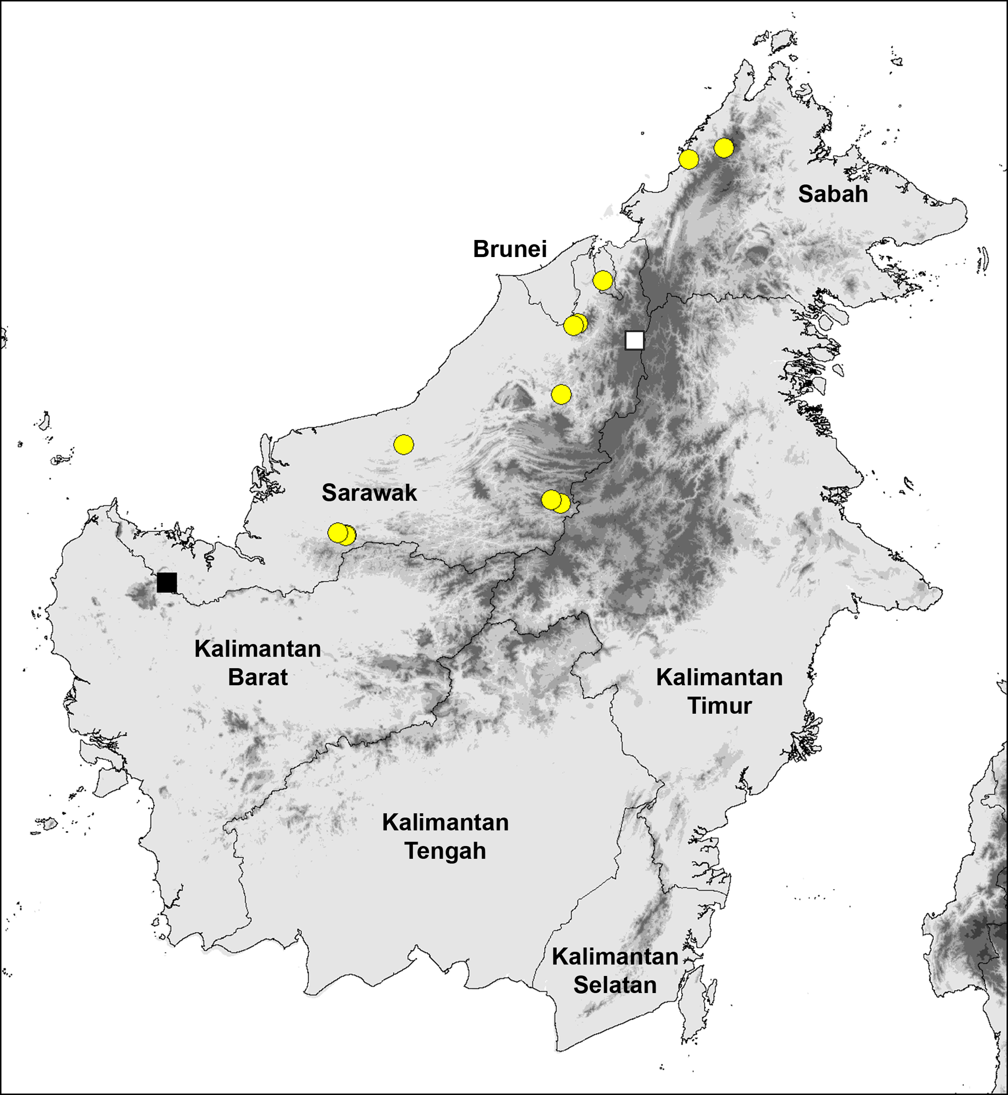
At present L. schieli is only known from near the peak of Gunung Penrissen in south-west Sarawak ( Fig. 128 View FIGURE128 ), at an altitude of approximately 1,100 m a.s.l. It should be looked for in the nearby Bungo Range, and over the border in Kalimantan on Gunung Niut.


| SAR |
Department of Forestry |
No known copyright restrictions apply. See Agosti, D., Egloff, W., 2009. Taxonomic information exchange and copyright: the Plazi approach. BMC Research Notes 2009, 2:53 for further explanation.