
Macrothrix cf. flagellata ( Smirnov and Timms, 1983 )
|
publication ID |
https://doi.org/ 10.1080/00222930701689937 |
|
persistent identifier |
https://treatment.plazi.org/id/03E32C46-B16D-FFBB-FE80-FE80DAE715EF |
|
treatment provided by |
Felipe |
|
scientific name |
Macrothrix cf. flagellata ( Smirnov and Timms, 1983 ) |
| status |
|
Macrothrix cf. flagellata ( Smirnov and Timms, 1983) View in CoL
( Figures 19 View Figure 19 , 20 View Figure 20 )
? Echinisca flagellata Smirnov and Timms 1983, p 80 –81, Figure 93.
? Macrothrix flagellata (Smirnov and Timms) View in CoL in Smirnov 1992, p 62, Figures 252–259.
Material examined here
Macquarie Island: Langdon Point, collected by H. J. G. Dartnall, AAK 1999-012; unknown locality ‘‘Ros 204’’, coll. by B. A. N. Z. A. R. Expedition, NHM 1966.7.20.3. and NHM 1966.7.20.4.
Diagnosis (based exclusively on population from Macquarie)
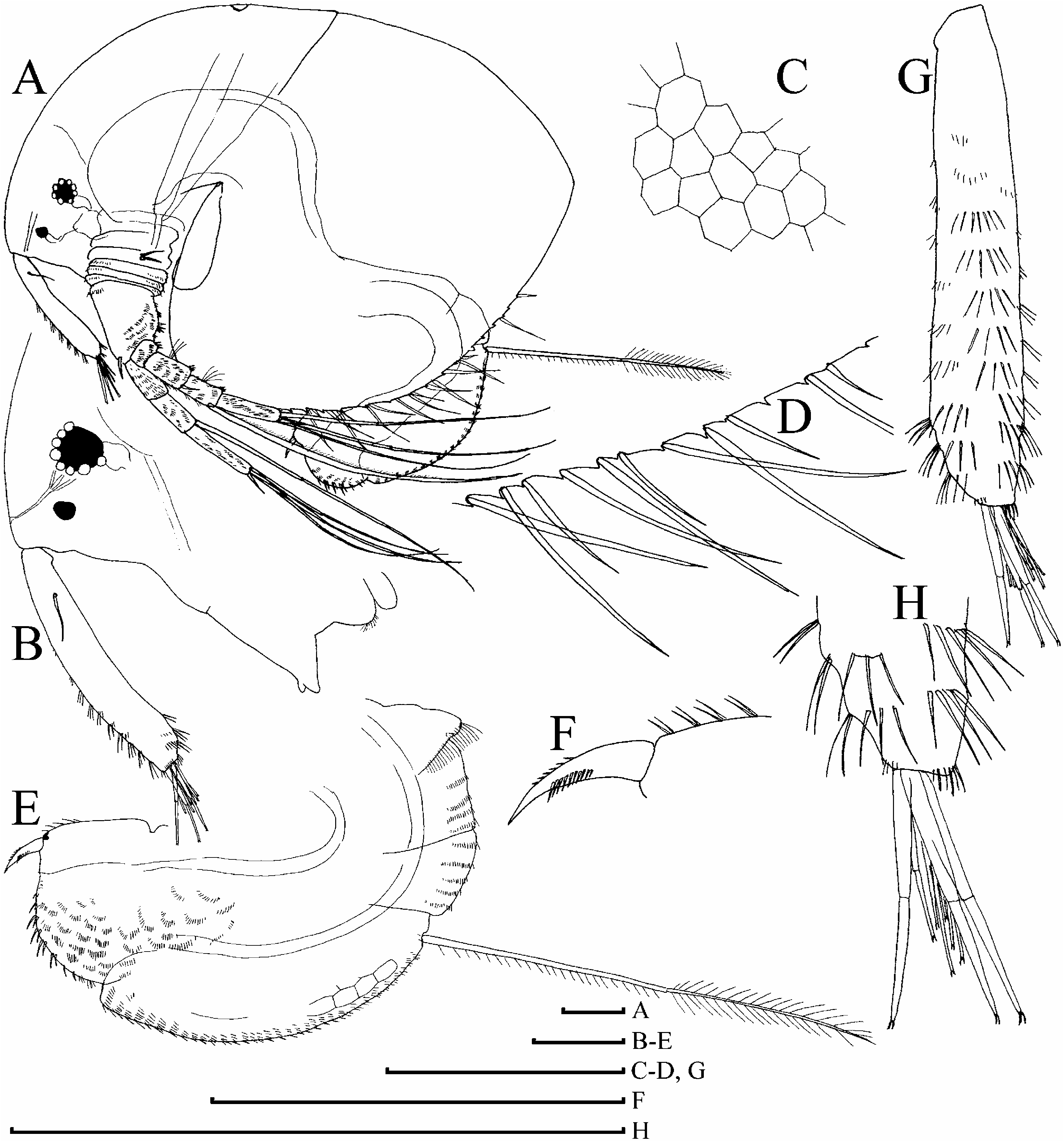
Parthenogenetic female. In lateral view body subovoid, relatively high (height/length50.65– 0.70), cervical depression absent, dorsal margin not interrupted by a ‘‘step’’ in posterior boundary of head, dorsal margin of valves without any serration, postero-dorsal as rounded angle, lies in level of middle of body height ( Figure 19A View Figure 19 ). No dome above eye. Ocellus small, less than half of eye diameter. Dorsal organ ovoid, small. Labrum with a strongly projected triangular apex bearing few tubercles ( Figure 19B View Figure 19 ). Valves with fine reticulation ( Figure 19C View Figure 19 ) and marginal setae as in previously described congeners ( Figure 19D View Figure 19 ).
Postabdomen subovoid, with rounded distal extremity, without ‘‘heel’’ basally, with a reticulation on sides ( Figure 19E View Figure 19 ). Ventral margin straight, with four to five series of moderately long, thin spinules. Dorsal margin distinctly bilobed; preanal margin with transversal series of minute setules, anal margin with groups of thicker setules. Postabdominal seta distal segment slightly shorter than basal one, densely armed with relatively short setules; basal segment with numerous, short setules. On external side of postabdominal claw, a series of 10–12 fine spinules; medial row of about six to eight denticles ( Figure 19F View Figure 19 ).
Antenna I very slightly widened distally, straight to slightly curved, without any traces of subapical external angulation; sensory seta at distance of about 1.5–2 antennular diameters (at base) from antenna I joint; on anterior face about seven to ten transverse rows of spinules, but no reticulation ( Figure 19G View Figure 19 ). Nine short aesthetascs, three of them significantly larger than the rest ( Figure 19H View Figure 19 ). Antenna II with distal burrowing spine on basal segment slightly shorter than proximal segment of exopod ( Figure 20A View Figure 20 ), distal sensory seta ( Figure 20B View Figure 20 ) somewhat longer than distal burrowing spine. Length of all apical swimming setae subequal. Lateral seta on proximal endopod segment larger than other setae, lacking robust denticles in middle ( Figure 20C–G View Figure 20 ). A spine on second segment of exopod longer than half of next segment. On posterior side of segments 1–3 of exopod there are a series of small, thin additional denticles.
Limb I outer distal lobe with longest apical seta having distal segment unilaterally armed with robust setules, inner-distal lobe with three bisegmented setae of different size, unilaterally setulated in distal part, smallest one with whole distal segment and distal portion of basal segment setulated; two ejector hooks of different size; maxillar process with two setae ( Figure 20H View Figure 20 ), anterior setae as represented in Figure 20I–K View Figure 20 . On limb II, scrapers 1–2 with delicate feathering, scrapers 3–7 with robust denticles of size characteristic for the genus; a solitary posterior seta near gnathobase; filter plate II with four setae, without a small hillock in position of rudiment of fifth seta ( Figure 20L View Figure 20 ). On limb III epipodite with a distal group of three long setae, seta 1 shortest, armed with robust denticles; setulated projections proximally to seta 3 and between setae 2 and 3 ( Figure 20M View Figure 20 ); on inner-distal limb portion, seta 1 with short and robust denticles; seta a with fine setules basally and robust spinules distally, seta b markedly longer than c; basal endite posteriorly with four soft setae. Limb IV ( Figure 20N, O View Figure 20 ) with exopodite small, bearing only a distal group of three bilaterally feathered setae of different size; on inner-distal portion of this limb seta 1 without setules basally and few denticles distally; posteriorly row of five long soft setae. On limb V there are three setae at inner margin ( Figure 20P View Figure 20 ).
Ephippial female, male. Unknown.
Size. In our samples 0.62–1.22 mm. In Tasmania, M. flagellata specimens up to 1.39 mm are found ( Smirnov and Timms 1983).
Taxonomic notes. At this level of morphological description of M. flagellata , it is impossible to conclude that I found just M. flagellata s. str. An accurate comparison with the original type material should be made and the Australian macrothricids need to be reassessed.
Smirnov and Timms (1983) initially placed M. flagellata in the genus Echinisca because of the undilated antenna I; however, this genus was subsequently synonymized with Macrothrix ( Smirnov 1992) . Serious doubts were expressed regarding the subjective status of the character ‘‘dilated—undilated antenna I’’ ( Kotov 1999). This character is applicable only in extreme states. In the intermediate state it is not possible to conclude whether antenna I of this species (or even specimen) is dilated or not. In the case of M. flagellata , a completely undilated antenna I is the only character distinguishing this species from others listed above. At the same time, M. flagellata and other aforementioned species have a set of similar characters, possible synapomorphies. In other hirsuticornis -like species the level of dilation is different, and variable. Consequently, I believe that M. flagellata is a hirsuticornis - like species.
Distribution. Tasmania and Macquarie Island. Dartnall et al. (2005) recorded M. hirsuticorni s from Macquarie; their material may belong to M. flagellata , or to another species from this genus.

| R |
Departamento de Geologia, Universidad de Chile |
| V |
Royal British Columbia Museum - Herbarium |
No known copyright restrictions apply. See Agosti, D., Egloff, W., 2009. Taxonomic information exchange and copyright: the Plazi approach. BMC Research Notes 2009, 2:53 for further explanation.
|
Kingdom |
|
|
Phylum |
|
|
Class |
|
|
Order |
|
|
Family |
|
|
Genus |
Macrothrix cf. flagellata ( Smirnov and Timms, 1983 )
| Kotov, Alexey A. 2007 |
Macrothrix flagellata (Smirnov and Timms)
| Smirnov NN 1992: 62 |
Echinisca flagellata
| Smirnov NN & Timms BV 1983: 80 |