
Mesamphiagrion nataliae Bota-Sierra
|
publication ID |
https://doi.org/ 10.11646/zootaxa.3718.5.1 |
|
publication LSID |
lsid:zoobank.org:pub:A34FF647-288B-4BD6-803C-0EADB27F2BC3 |
|
DOI |
https://doi.org/10.5281/zenodo.5658567 |
|
persistent identifier |
https://treatment.plazi.org/id/E76CC555-D006-FFF3-FF61-5F39F0BEF850 |
|
treatment provided by |
Plazi |
|
scientific name |
Mesamphiagrion nataliae Bota-Sierra |
| status |
sp. nov. |
Mesamphiagrion nataliae Bota-Sierra View in CoL sp. nov.
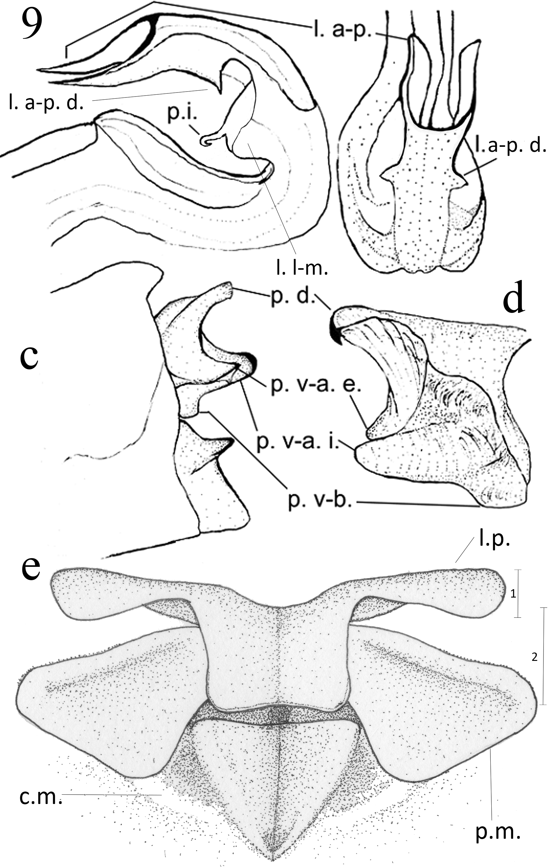
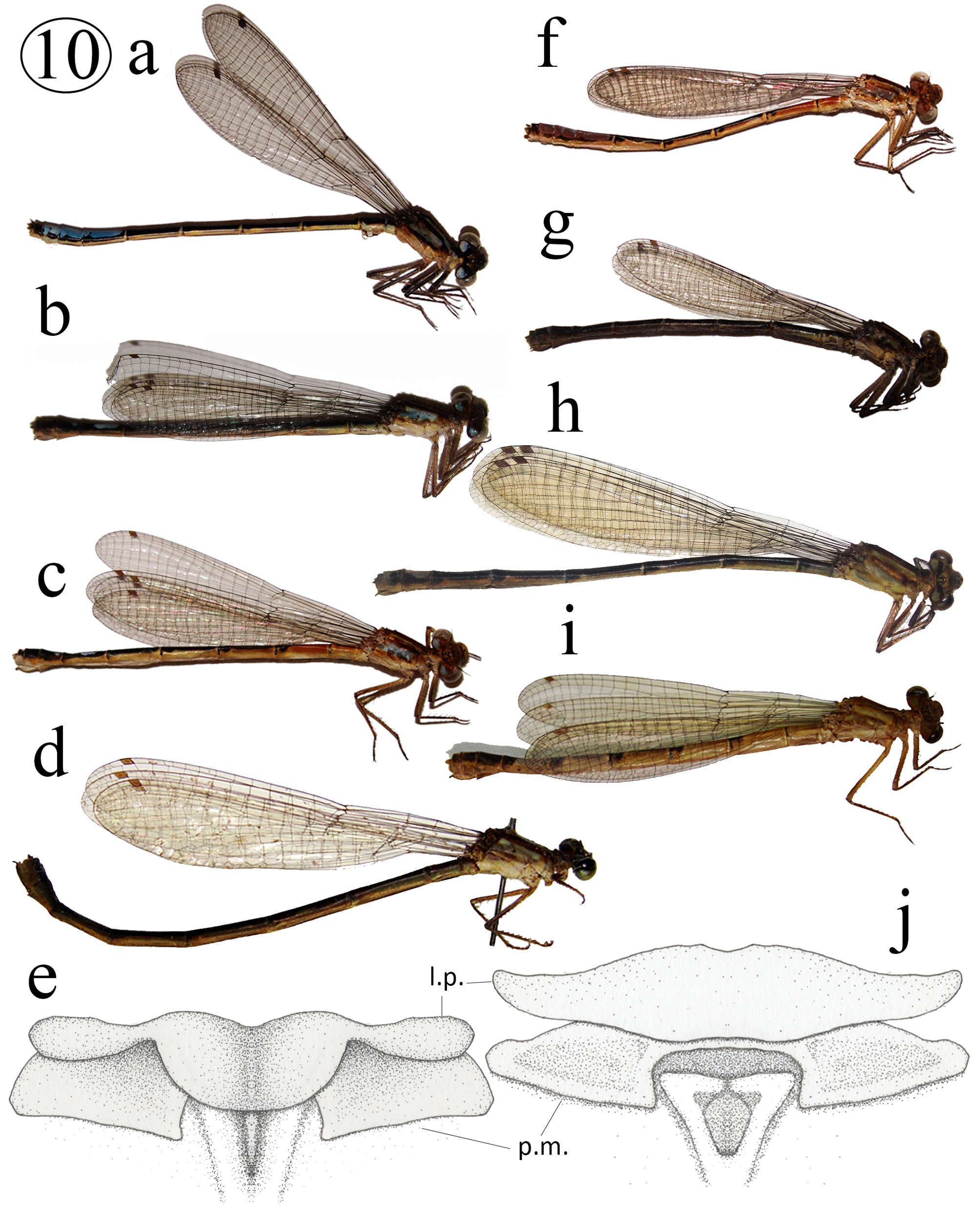
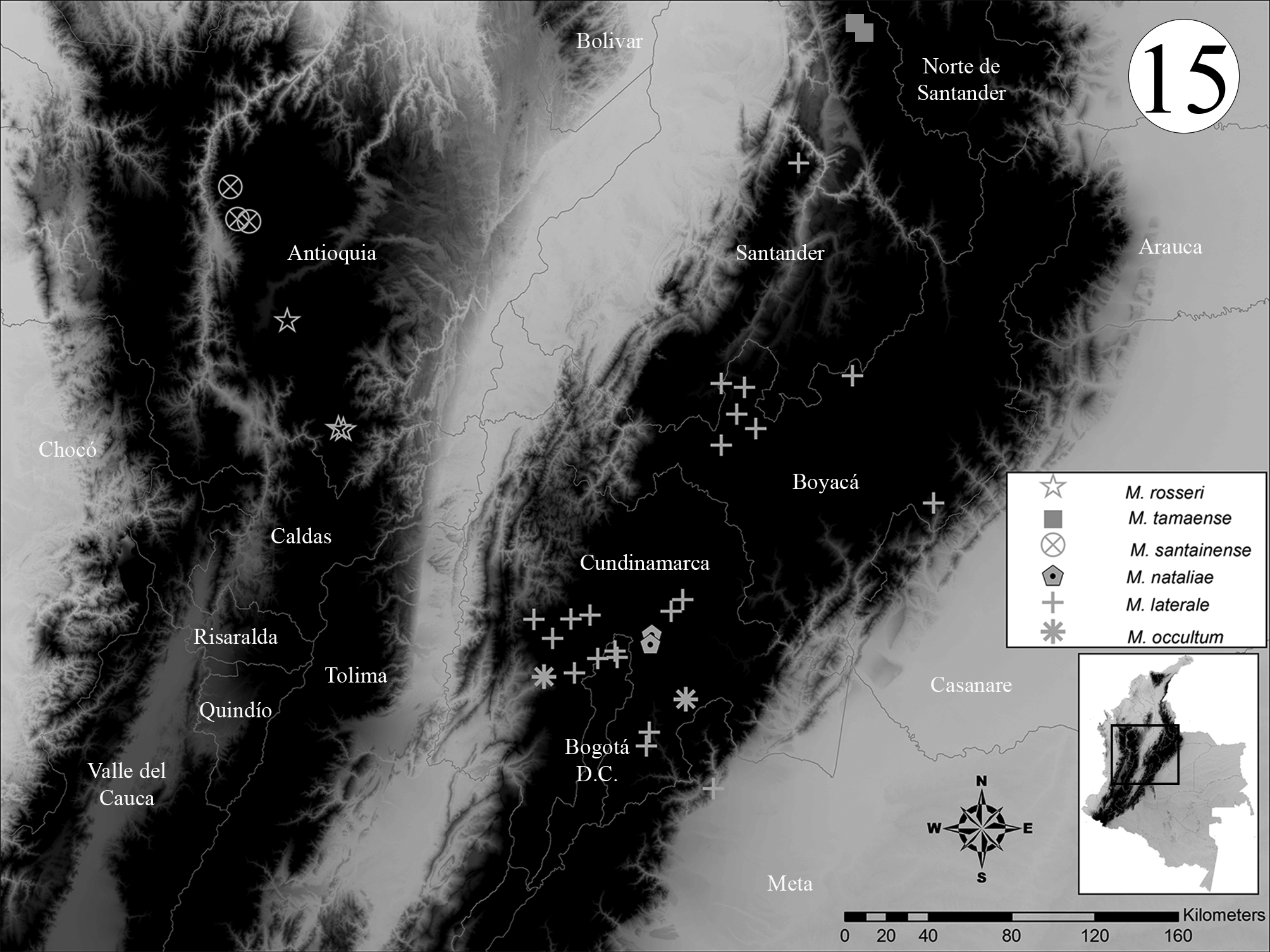
Figs. 9 View FIGURE 9 a–e (diagnostic traits), 10a (habitus ♂), 10b (habitus androchrome ♀), 10c (habitus juvenile androchrome ♀), 10f (habitus juvenile ♂), 10g (habitus gynochrome ♀), 13n (S7– 10 ♂), 14c, f (S7–10 ♀), 15 (map)
Etymology. Named nataliae (noun in the genitive case) after Dr. Natalia von Ellenrieder, whose work, advice, and unconditional help made this work possible.
Specimens examined (24 specimens: male holotype, allotype, and 22 paratypes). Cundinamarca, Municipality Guasca: 9♂ and 3♀, Township El Salitre, 4°50'N 73°55'W, 2700M, 04/02/2006, Leg: E. Realpe & L. Pérez, ANDES. 3♂ and 1♀, same data but CEUA. Holotype, 5♂ and 2♀, Township Pueblo Viejo, Finca El Aji, 4.7934°N 73.923131° W, 3050m, 14/02/2009, Leg: M. Sánchez, ANDES.
Description. Holotype. Head. Labium cream with light blue base. Base of mandible blue. Labrum blue with a basal small black spot and dorsolateral black margins. Gena blue. Anteclypeus blue. Postclypeus black. Frons black with pruinescence, light blue on area surrounding eye, gena, and clypeus, with medial transverse black stripe over clypeus, blue postocular spots reaching eye margin, and cream spot between ocelli and antenna. Antenna black. Back of head cream ( Fig. 10 View FIGURE 10 a).
Thorax. Prothorax black, propleura blue becoming lighter to proximal portion. Medial lobe of posterior prothoracic lobe developed into a caudally projected squarish plate dorsally concave, lateral edges approximately half length of distal edge. Pterothorax black with blue antehumeral stripe, wide blue stripe covering mesepimeron and metepisternum, metepimeron light blue with pruinescence, and cream venter. Meta- and mesocoxae cream, procoxa blue. External side of femur black with small cream spot near base, medial side cream. Tibia external side black and medial side brown. Tarsi and claws brown ( Fig. 10 View FIGURE 10 a). Nine external metafemoral spurs, as long as space between them or shorter, gradually increasing in size toward apex. Six external metatibial spurs, as long as space between them or shorter, gradually decreasing in size toward apex. Tarsal claws with well developed supplementary tooth. Wings hyaline. Pt dark brown, ratio between distal and proximal sides about 1:1. CuP reaching CuPAA slightly distal to confluence of CuPAA with hind margin of wing. 14 Px in right and 12 in left FW, 11 in HW. RP 2 branching between Px 4 and 5 in left FW and under 5 in right FW, between Px 4 and 5 in HW ( Fig. 10 View FIGURE 10 a).
Abdomen. Dorsum black and sides cream except: S1 distal margin and side blue. S2 side bluish. S4–7 with light blue ring on proximal margin. S7 with a dorsal blue spot on distal 9/10. S8–9 with blue dorsum. S10 with pale blue apical projections ( Figs. 10 View FIGURE 10 a, 12n). Genital ligula ( Fig. 9 View FIGURE 9 a–b) with apex concave, with pair of long lateroapical processes extending proximally and terminating in sharp point, also pair of short processes pointing distally about as long as 1/4 length of lateroapical processes and located basal to them, lateromedial lobes hemispherical in lateral view, not visible in ectal view, and inner medial process close to ligula’s flexure. Cercus ( Fig. 9 View FIGURE 9 c–d) external side black, medial side light blue, about 1/2 length of S10, with elongated dorsal process ending in small tooth, external ventroapical process pointed and shorter than dorsal process, inner ventroapical process extending beyond ventroapical external process and terminating in rounded tip which is most distal point of body in lateral view; in posterior view this process is curved outwards and covers the external ventroapical process. Basal process underdeveloped, extending to level of apex of paraproct and ending in blunt tip. Paraproct cream with dorsal process ending in black tip pointing distally, reaching about mid-length of cercus. Total length 31 mm. Abdomen length 25 mm. FW length 18 mm. HW length 17 mm.
Variation in male paratypes. As holotype but seven to nine metafemoral external spurs. Six to eight external metatibial spurs. 12–14 Px in FW, 10–11Px in HW. RP 2 branching between Px 4 and 6 in FW, between Px 3 and 5 in HW. Total length 29–31 mm. Abdomen length 23–25 mm. FW length 17–18 mm. HW length 16–17 mm.
Variation in male juveniles. Head and thorax, light blue instead of blue, brown instead of black ( Fig. 10 View FIGURE 10 f). Coxae and legs cream. Pt brown. Dorsum of S1–3 red, but distal margin of S1 blue, distal margin of S2 black, and distal half of S3 black.
Allotype. Head. As holotype but labium cream. Base of mandible brown. Labrum brown with small black midbasal spot and black dorsolateral margins. Postclypeus and anteclypeus brown, with pruinescence. Frons dark brown with light brown stripe crossing from eye to eye, proximal to vertex and surrounding occipital margin, blue postocular spots not reaching eye margin, approximately hemispherical ( Fig. 10 View FIGURE 10 b).
Thorax. Anterior lobe of pronotum light blue. Medial lobe of posterior prothoracic lobe developed into caudally projected squarish plate concave dorsally, its lateral edge about half as long as distal edge ( Fig. 9 View FIGURE 9 e). Mesepisternal plate brown approximately flat and triangular, carina extending from distal tip of mesostigmal plate to middorsal suture on each side ( Fig. 9 View FIGURE 9 e). Pterothorax brown with pruinescence, middorsal stripe black, mesepisternum with light bluish brown stripe, wide light blue stripe on mesepimeron and distal metepisternum, metepisternum cream ( Fig. 10 View FIGURE 10 b). Coxa cream. External side of remainder of leg brown, medial side cream, femur with a small pale spot near apex. Seven external spurs on right metafemur and six on left. Six external spurs on right metatibia and seven on left. Pt brown. 12 Px in FW, 10 in HW. RP 2 branching between Px 5 and 6 in left FW and under 6 in right FW, between Px 4 and 5 in HW ( Fig. 10 View FIGURE 10 b).
Abdomen. S1–4 with pruinescence, dorsum black and sides greenish cream except: S1 distal margin blue. S4– 7 with incomplete light blue ring on proximal margin, S7 with a blue dorsal spot occupying about 2/3 of dorsum, located proximally and without reaching margins of S7, S8 with pair of dorsal rounded blue spots near proximal margin. S9 with thin pale blue middorsal line that does not reach margins of S9. S10 with medial triangular spot running from proximal edge to middle of segment ( Figs. 10 View FIGURE 10 b, 13c). Vulvar spine cream with black tip. Ovipositor and paraproct cream, cercus brown. Subbasal plate of ovipositor triangular. Ovipositor distal apex reaching level of distal margin of S10. Cercus conical, as long as about half length of S10, its apex representing most distal point of body. Total length 30 mm. Abdomen length 23 mm. FW length 20 mm. HW length 18 mm.
Variation in mature female paratypes. As allotype but entire body can be covered in pruinescence. Head. Base of mandible cream to greenish gray. Labrum cream to gray greenish with small black mediobasal indentation and dorsolateral black margins. Gena cream to gray. Anteclypeus gray and postclypeus brown along apical edge and gray along mediobasal margin. Antefrons dark brown to light green, postfrons with brown stripe from eye to eye proximal to vertex and surrounding it to occipital margin, or completely light brown with dark brown rings surrounding ocelli, postocular spots almost indiscernible, very light blue ( Fig. 10 View FIGURE 10 g).
Thorax. Prothorax brown, anterior lobe of pronotum light blue, propleura blue becoming lighter proximally. Pterothorax light brown with black middorsal stripe, mesepisternal light stripe brown or blue, mesepimeron and distal metepisternum with light green stripe, metepisternum cream ( Fig. 10 View FIGURE 10 g). Six to eight external metafemoral spurs. Six to eight external metatibial spurs. 12–14 Px in FW, 10–11 in HW. RP 2 branching between Px 3 and 5 in HW.
Abdomen. Middorsal light blue line on S9 present or absent, light blue spots on S9–10 present or absent, or dorsum entirely black with only distal margin of segments pale (probably gynochrome females) ( Figs. 10 View FIGURE 10 g, 13a–b). Total length 29–31 mm. Abdomen length 22–24 mm. FW length 18–20 mm. HW length 17–18 mm.
Variation in immature female paratypes. As allotype but light brown instead of dark brown or black, light green mesepisternal stripe, dorsum of S1–3 red except blue distal margin of S1, black distal margin of S2, and black distal half of S3 ( Fig. 10 View FIGURE 10 c).
Diagnosis. The dorsal process of male cercus longer than the ventrobasal external process ( Fig. 9 View FIGURE 9 c–d) groups M. nataliae with M. tamaense , M. demarmelsi , M. gaudiimontanum , M. gaianii , M. laterale , and M. dunklei . The well developed inner ventroapical process of male cercus, curving distally and representing the most distal point of the body ( Fig. 9 View FIGURE 9 c–d), clearly separates this species from M. gaudiimontanum , M. gaianii , and M. laterale . The shape of genital ligula ( Fig. 9 View FIGURE 9 a–b) clearly separates it from all known species in the genus, resembling the ligula of M. demarmelsi but with lateroapical lobes longer and projected parallel to the body axis, versus shorter and projected oblique to the body axis in M. demarmelsi . The medial lobe of posterior prothoracic lobe relatively broad ( Fig. 9 View FIGURE 9 e) separates it from M. demarmelsi . The color pattern of mature males ( Fig. 10 View FIGURE 10 a) is almost identical to that of mature males of M. gaudiimontanum ( Figs. 3 View FIGURE 3 c–d, 5b), but juvenile males of M. nataliae may have red coloration on S2–3 ( Fig. 10 View FIGURE 10 f, but not seen in life), whereas in M. gaudiimontanum there is red on S1–4. The posterior lobe of female pronotum, dorsally concave with distal edge about as long as 1.5x the length of a lateral edge, is unique among species of the genus ( Fig. 9 View FIGURE 9 e). Also unique is the carina extending from distal tip of mesostigmal plate to middorsal suture on each side ( Fig. 9 View FIGURE 9 e). Females present two color patterns: androchrome ( Fig. 13 View FIGURE 13. S 7 – 10 c) with abdominal color pattern similar to females of M. laterale , and gynochrome ( Fig. 13 View FIGURE 13. S 7 – 10 a–b), with abdomen entirely dark, which is shared with other gynochrome females in the genus, i.e. M. occultum ( Fig. 13 View FIGURE 13. S 7 – 10 n), M. tamaense , and M. gaudiimontanum ( Fig. 13 View FIGURE 13. S 7 – 10 l).
Remarks. There are only two known populations of this species, both located in a geographic area relatively well sampled, suggesting that this species may be threatened. The high variability observed for female coloration probably indicates polymorphism. Some specimens were found parasitized by mites.
Distribution. Cerros Orientales of the Sabana de Bogotá, Cordillera Oriental ( Fig.15 View FIGURE 15 ).
Habitat and biology. Adults were found at an artificial reservoir in pasture land; according to the collector "The population was quite large and spiders eat enough of these damselflies, I found a lot trapped in the cobwebs" (Sánchez pers. comm.). Other odonates found at the same locality included (Sánchez pers. comm.) M. laterale , M. demarmelsi , Ischnura chingaza Realpe, 2010 , and Rhionaeschna marchali .


No known copyright restrictions apply. See Agosti, D., Egloff, W., 2009. Taxonomic information exchange and copyright: the Plazi approach. BMC Research Notes 2009, 2:53 for further explanation.