
Microcosmus bitunicatus, Monniot & Monniot, 2001
|
publication ID |
https://doi.org/ 10.5281/zenodo.5391440 |
|
persistent identifier |
https://treatment.plazi.org/id/F57D87A3-FF4C-31AF-E86A-FEE4FD861203 |
|
treatment provided by |
Marcus |
|
scientific name |
Microcosmus bitunicatus |
| status |
sp. nov. |
Microcosmus bitunicatus View in CoL n. sp.
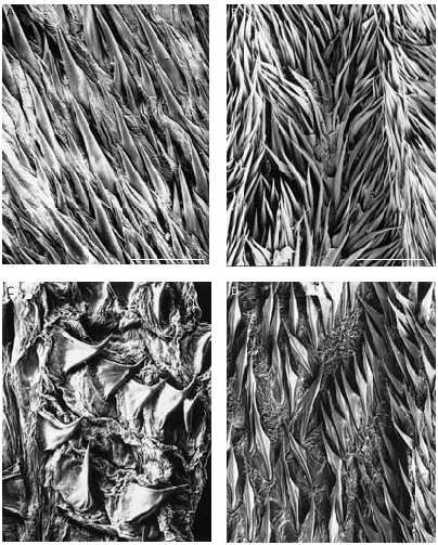
( Figs 108C View FIG ; 130D View FIG )
? Microcosmus manaarensis Herdman, 1906: 311 View in CoL . — Monniot F. & Monniot C. 1996: 265, fig. 59, Philippines.
TYPE MATERIAL. — Philippines. Cebu Straits, Cabilao Island, 9°53.39’N, 123°45.45’E, 10 m, 15.IV.1997 ( MNHN S2 MIC 157).
ETYMOLOGY. — From the Latin tunicatus: dressed in a tunic.
DESCRIPTION
A second specimen has been collected of Microcosmus having two encased tunics, one inside the other. This peculiar structure of the tunic previously seemed a possible artefact, but the collection of another specimen gives this structure a unique significance.
The new specimen ( Fig. 130D View FIG ) is larger, being 10 cm long instead of 5 cm. It was attached in a rocky hollow by a ball of extremely hard compact, tunic. The remainder of the body has developed above the substrate and has a regular shape.
All characters described by Monniot F. & Monniot C. (1996) are confirmed, particularly the independence of the apertures of the external tunic and the siphons of the encased tunic. The spinules ( Fig. 108C View FIG ) covering the external and internal sides of the encased tunic grow from the deepest part of the reflex tunic. As the animal increases in size, the spinules move towards the external rim of the siphon. In some cases they can invade all the body, and this is so for the present specimen.
The absence of spinules on the openings of the external tunic implies that the dissociation between the encased and external tunics occurred at an early stage of the growth of this specimen.
REMARKS
It was with doubt that the first specimen was identified as M. manaarensis . We supposed it to be an ecological form from rocky bottoms of a species normally living on gravel. The internal morphology, the number of branchial folds, and the shape and disposition of the gut and gonads are very similar. On sedimentary bottoms M. manaarensis has a tunic in two parts: one internal which is normal and one external made by a network of ramified hairs agglomerating the sediment. The two tunics are linked by rhizoids. Several Stolidobranchia show a similarly double tunic structure ( Pyura tunica Kott, 1969 ; S. rhizoides Diehl, 1969 ; etc.). But M. bitunicatus n. sp. does not follows this pattern. The external tunic, with its openings corresponding to the siphons, is not in continuity – at least in large specimens – with the functional siphons of the animal. The growth of the two tunics seems to occur independently.
Microcosmus helleri Herdman, 1882 View in CoL possesses a sedimentary form with a thin tunic encrusted with sand, and another form with a thick, clean hard tunic living on rocks, and intermediate forms exist. This is not the case between the typical M. manaarensis View in CoL and M. bitunicatus View in CoL .

No known copyright restrictions apply. See Agosti, D., Egloff, W., 2009. Taxonomic information exchange and copyright: the Plazi approach. BMC Research Notes 2009, 2:53 for further explanation.
|
Kingdom |
|
|
Phylum |
|
|
Class |
|
|
Order |
|
|
Family |
|
|
Genus |
Microcosmus bitunicatus
| Monniot, Françoise & Monniot, Claude 2001 |
Microcosmus manaarensis
| HERDMAN W. A. 1906: 311 |