
Mylonchulus minor (Cobb, 1893) Andrássy, 1958
|
publication ID |
https://doi.org/ 10.11646/zootaxa.3646.4.1 |
|
publication LSID |
lsid:zoobank.org:pub:DDE05DCD-A443-499D-9F38-8C3B43592694 |
|
DOI |
https://doi.org/10.5281/zenodo.5616691 |
|
persistent identifier |
https://treatment.plazi.org/id/03C21B03-FF89-FFC7-FF5B-FDE576DB2D4A |
|
treatment provided by |
Plazi |
|
scientific name |
Mylonchulus minor (Cobb, 1893) Andrássy, 1958 |
| status |
|
Mylonchulus minor (Cobb, 1893) Andrássy, 1958
( Fig. 3 View FIGURE 3 )
= Mononchus (Mylonchulus) minor Cobb, 1893 (Cobb, 1916) = Mononchus (Mylonchulus) brachyuris microdenticulatus minor Cobb, 1893 (Micoletzky, 1922) = Mylonchulus sculentus Jain, Saxena & Sharma, 1993
Measurements. Table 2 View TABLE 2 .
Description. Adult: Body medium-sized, ventrally arcuate, more strongly curved in posterior half after fixation, tapering slightly towards extremities. Cuticle smooth, 1.5–2 µm thick. Body pores indistinct. Lip region truncate to angular, demarcated from adjoining body, about 2.5–3 times as wide as high. Labial sensilla prominent, slightly raised. Amphids cup-shaped with slit-like aperture, 4–7 µm across, located 9–12 µm from the anterior end. Buccal cavity funnel-shaped, about 1.2–1.8 times as long as wide with sclerotized vertical and oblique walls. Dorsal vertical wall bearing anteriorly directed, 8–9 µm long massive tooth, 5–6 µm in thickness with tip located 5–6 μm from anterior end of buccal capsule or at ca 70–80% from its base. Each subventral vertical wall provided anteriorly with 6 transverse rows of rasp-like denticles (n=6) with anterior row relatively more prominent; number of denticulate rows was found to be five in two specimens. A small tooth present on each subventral wall posterior to denticles. Pharyngeal sleeve surrounding buccal cavity at 1/3–1/4 of its length from base. Pharynx cylindroid, muscular, 30–33% of body length. Orifices of the pharyngeal glands, DO located at ca 58–67%, SV1O1 and SV1O2 at ca 69–73%, SV2O1 and SV2O2 at ca 92–93% of pharyngeal length from anterior end. Nerve ring encircling pharynx at ca 27–33% and excretory pore faintly visible at ca 34–39% of pharyngeal length from anterior end. Pharyngo-intestinal junction non-tuberculate with cardial flap of variable length ranging from 9–15 µm. Intestine with polygonal cells. Rectum 0.8–1.1 times as long as anal body diameter. Tail conoid, ventrally bent at half of length in most specimens, tapering into a rounded terminus. Caudal glands well developed, arranged in tandem, opening to exterior through a narrow tubular duct; spinneret terminal.
Female: Reproductive system didelphic, amphidelphic with relatively compact, reflexed ovaries having 8–11 oocytes arranged in single row except the distal proliferation zone; proximal end of ovary containing a large grown oocyte. Oviduct joining ovary sub-terminally, 35–77 µm long. Sphincter not observed at oviduct-uterus junction; uterus devoid of sperms. Vagina extending inward about 1/3–1/4 of the corresponding body diameter with well developed, almond-shaped pars refringens. A smooth-shelled intra-uterine egg observed in one female. Vulva postmedian and transverse.
Male: Not found.
Locality and habitat. Moss containing Mylonchulus minor , scraped from around the roots of Bambusa sp. from Haridwar, Uttar Pradesh, India located at 29°57ʹ22ʺN 78°10ʹ12ʺE coordinates.
Voucher specimens. Seven females on slide Mylonchulus minor (Cobb, 1893) Andrássy, 1958 , no. D20 /1–4 deposited in the Nematode Collection, Department of Zoology, Aligarh Muslim University, Aligarh, Uttar Pradesh, India. One female deposited at USDANC, Beltsville, MD, USA.
Salient characters. Small to medium-sized species with buccal cavity funnel-shaped, about 1.2–1.8 times long as wide; dorsal tooth large, situated in anterior half of buccal cavity; each subventral wall usually bearing 6 transverse rows of denticles; subventral tooth very small; female genital system amphidelphic; tail conoid, ventrally bent at half of its length with rounded terminus; caudal glands arranged in tandem; spinneret terminal.
Remarks. This is a widely distributed species of Mylonchulus , and has been reported from different parts of the world (Ahmad & Jairajpuri, 2010). The present specimens showed morphological as well as morphometric characteristics similar to M. minor (Cobb, 1893) Andrássy, 1958 . The typical features of the individuals as stated by Cobb (1916) were the funnel-shaped buccal cavity with a prominent dorsal tooth but very short or relatively inconspicuous subventral teeth, likely to be overlooked. Intrapopulation variations were observed in the size of subventral tooth which ranged from a slight protuberance ( Fig. 3 View FIGURE 3 B, C) to a prominent triangular tooth ( Fig. 3 View FIGURE 3 G), subventral rasp-like denticles being either regularly arranged in rows or slightly dispersed, the cardial flap ranging from insignificant ( Fig. 3 View FIGURE 3 L) to visibly longer ( Fig. 3 View FIGURE 3 I); and the ovaries slender ( Fig. 3 View FIGURE 3 M) to prominently broad ( Fig. 3 View FIGURE 3 Q). Contrary to the description of the species apud Cobb (1916) as having a conoid, arcuate tail bent near the middle, the shape of tail in the present specimens varied from cylindroid ( Fig. 3 View FIGURE 3 U), slightly bent to strongly bent ( Fig 3 View FIGURE 3 X).
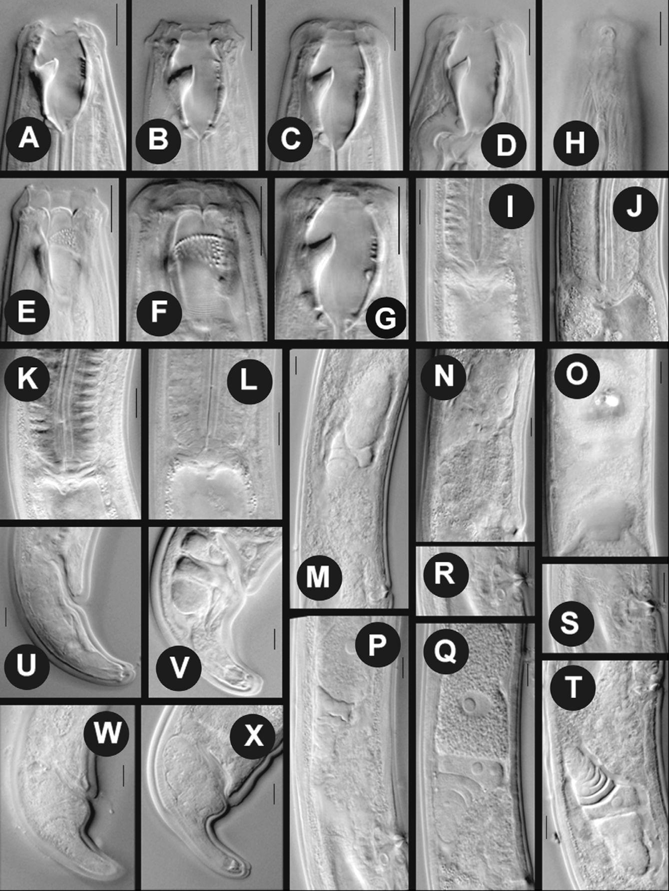
TABLE 2. Morphometrics of Mylonchulus minor (Cobb, 1893) Andrássy, 1958, M. obtusicaudatus (Daday, 1899) Andrássy, 1958, M. vasis Yeates, 1992 and M. contractus Jairajpuri, 1970 b. Measurements are in Μm and in the form: mean ± SD (range).
| Characters M. minor female (n) (8) Body length 1010±60 (930–1140) | M. obtusicaudatus female (6) 1400.3±90.7 (1280–1620) | M. vasis female (6) 916.6±65.8 (839–1014) | M. contractus female M. contractus male (10) (1) 762.6±43.6 (696– 822) 801 |
|---|---|---|---|
| Body diameter at vulva 48.2±3.6 (43–53) | 56.2±6.5 (46–70) | 43.3±4.0 (38–50) | 31.6±0.9 (30–33) 28 |
| a 21.1±1.6 (19.0–24.0) b 3.4±0.1(3.2–3.5) | 25.1±2.2 (20.6–28.1) 3.9±0.2 (3.7–4.6) | 21.1±1.4 (19–23.3) 3.1±0.1(3–3.3) | 24.1±1.5 (21.6–26.4) 28.6 3±0.1(2.9–3.1) 3 |
| c 25.1±1.7 (22.6–27.1) c' 1.4±0.9 (1.2–1.5) | 24.9±1.2(22.3–26.8) 1.6±0.1(1.3–1.9) | 26.5±2 (24.2–29.8) 1.1±0.1(1.0–1.3) | 27±2.5 (22.8–31.4) 24.2 1.2±0.1(1.1–1.4) 1.1 |
| V /T 58.2±1.1(56.7–60.1) | 62.0±0.8 (60.7–63.1) | 61.8±3.2 (58.7–66.5) | 59.7±1.1 (57.7–61.0) 45.5 |
| G1 15.5±1.9 (12.0–18.1) | 12.3±1.2 (10.8–14.9) | 15.0±2.1 (13.3–18.2) | 12.3±1.1 (11.3–13.5) – |
| G2 14±1.8 (12.2–17.7) | 11.9±1.3 (9.1–13.7) | 12.3±0.8 (11.4–13.5) | 10.4±0.5(9.7–11.2) – |
| Lip height 9.8±0.3 (9–10) | 10.9±0.5 (10–12) | 9.5±0.5 (9–10) | 7.7±0.4 (7–8) 7 |
| Lip diameter 24.8±1.2 (23–27) Stoma length 25±0.5 (24–26) Stoma diameter 15.2±1.1(13–17) | 28±0.7 (27–29) 31.2±0.9 (30–33) 19.2±0.7(18–20) | 24.1±1.1 (23–26) 30.6±0.8 (30–32) 16.0±0.8 (15–17) | 18.5±0.9 (17–20) 18 24.7±0.8 (23–26) 25 12.4±0.5 (12–13) 12 |
| Dorsal tooth position 5.7±0.5 (5–6) | 8.6±0.5 (8–9) | 6.6±0.5 (6–7) | 4.6±0.5 (4–5) 10 |
| Pharynx length 294.5±11.2 (280–318) | 355.8±15.9 (335–390) | 290.8±12.9 (276–312) | 248.5±13.3 (233–268) 263 |
| Nerve ring from ant. end 89.8±7.2 (80–100) | 99.7±5.8 (85–110) | 94±3.5 (90–99) | 82±4.3 (75–90) 85 |
| Excretory pore from ant. end 109.2±8.6 (96–125) | 125.3±17.1 (108–173) | 98±1.6 (96–100) | 92.6±3.8 (89–95) 93 |
| Rectum length 24.6±1.3 (23–27) | 28.7±2.9 (24–33) | 23±1.8 (21–25) | 19.7±1.4 (18–22) 24 |
| Anal body diameter 29.1±1 (28–31) | 34.9±1.9 (32–38) | 28.1±2.3 (25–31) | 21.9±0.7 (21–23) 28 |
| Tail length 40.7±3 (37–47) | 56.4±4.5 (50–62) | 34.5±1.7 (32–37) | 28.4±3.1 (24–34) 33 |
| Vulva–anus distance 345.2±21.3 (307–398) | 426.7±28.9 (388–476) | 323.1±18.6 (300–345) | 277.9±17.3 (250–295) – |
| Spiculelength – | – | – | – 36 |
| Gubernaculumlength – | – | – | – 23 |
No known copyright restrictions apply. See Agosti, D., Egloff, W., 2009. Taxonomic information exchange and copyright: the Plazi approach. BMC Research Notes 2009, 2:53 for further explanation.