
Neogreenia zeylanica ( Green, 1896 )
|
publication ID |
https://doi.org/ 10.11646/zootaxa.5418.5.1 |
|
publication LSID |
lsid:zoobank.org:pub:0DDD7278-0E9C-4979-ACB9-1915D4209282 |
|
DOI |
https://doi.org/10.5281/zenodo.10787187 |
|
persistent identifier |
https://treatment.plazi.org/id/03C24061-FFC4-D274-FF0F-242AFF29FD8D |
|
treatment provided by |
Plazi |
|
scientific name |
Neogreenia zeylanica ( Green, 1896 ) |
| status |
|
Neogreenia zeylanica ( Green, 1896) View in CoL
Monophlebus zeylanicus Green 1896: 6 View in CoL . Type data: SRI LANKA: Pundaluoya, on stems of Antidesma bunius View in CoL . Syntypes, female, type depository: Natural History Museum , London, UK .
Kuwania zeylanica ( Green, 1896) View in CoL ; Green 1922: 425.
Neogreenia zeylanica ( Green, 1896) View in CoL ; Foldi 2001: 211; Varshney 1992: 11.
Host plants: Antidesma bunius (L.) Spreng ( Phyllanthaceae ).
Distribution: India, Sri Lanka.
Remarks. Green (1922) described and illustrated the adult female and male, young larva and nymph, and provided brief descriptions of a later larva (probably a male) and male nymph. According to these descriptions and the syntype material examined, it can be inferred that the “young larva” is a first-instar nymph, the “nymph” is a third-instar female nymph, the “later larva” is a third-instar male nymph, and the “male nymph” is a male pupa. Varshney (1992) recorded this species in the list of scale insects in India. Here N. zeylanica is redescribed based on photographs of some syntype specimens and the description in Green (1922).
Material examined. Syntypes, all from SRI LANKA: collection data as above: 1 ♀, mounted with 1 first-instar nymph and another ♀ together on 1 slide; ♂, mounted singly on a slide; 1 third-instar ♀, mounted singly on a slide; 1 ♂ pupa, mounted singly on a slide ( NHM) .
Adult female
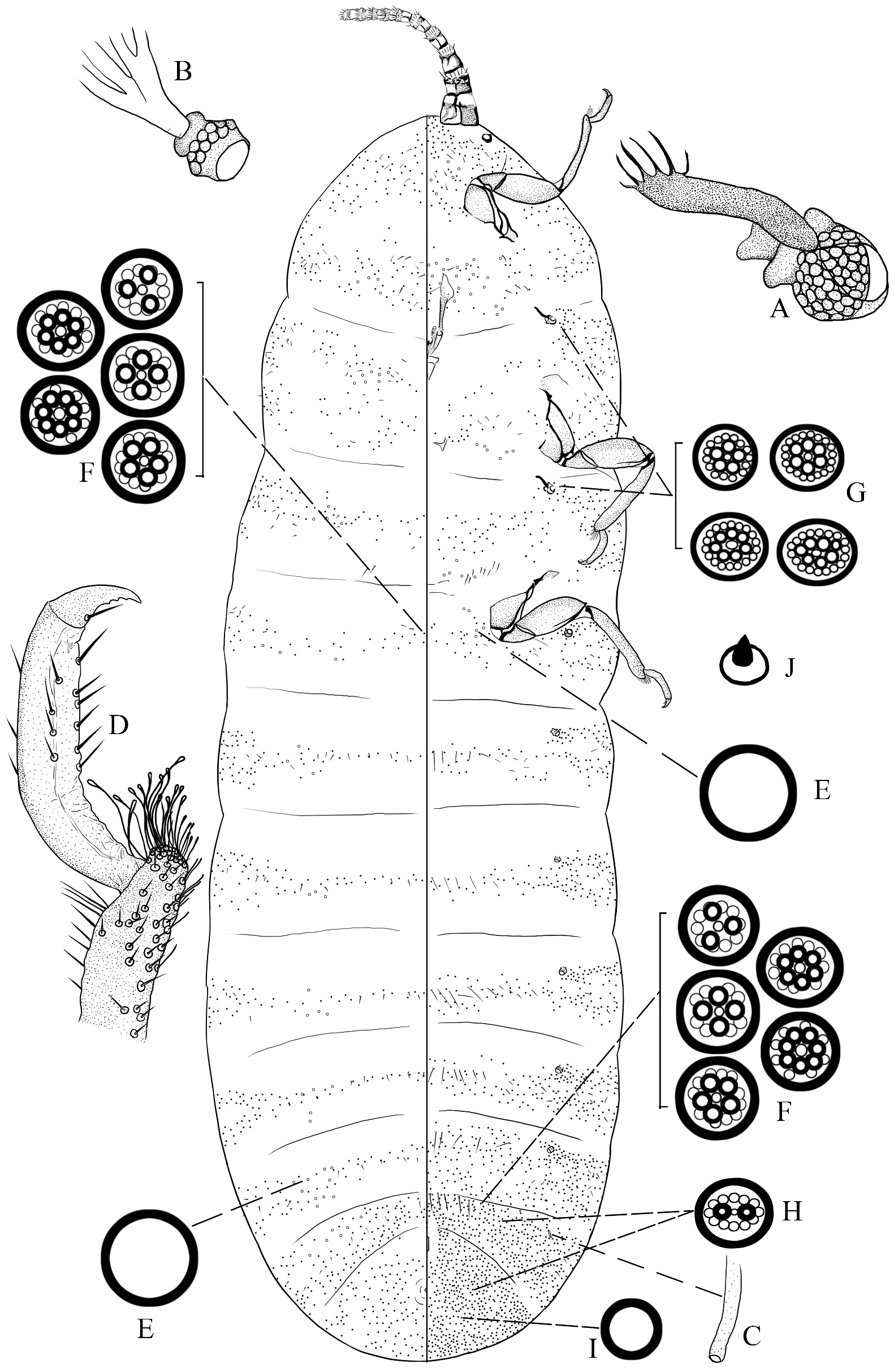
Slide-mounted material (n=1) ( Fig. 12 View FIGURE 12 ). Body elongate, about 7.7 mm long and 2.5 mm wide, head narrow and abdominal apex rounded. Derm membranous but margins of posterior abdomen slightly sclerotized. Antennae each 10 segmented, situated close to each other; total length of each about 1,000 μm; each segment with apex membranous, basally and subdistally sclerotized, also with a sclerotized bar on most segments; scape (segment I) largest; proximal 3 segments each almost cylindrical; other segments each more-or-less bowl-like; segments III–IX each with a ring of hair-like setae. Eyes each with sclerotized margin and membranous centre, situated lateral to antennal base. Mouthparts present: clypeolabral shield longer than labium, labium 2 segmented. Thoracic spiracles ( Fig. 12A View FIGURE 12 ) each with a sclerotized bar, and a dense group of sieve-like disc-pores at inner end of atrium. Abdominal spiracles with anterior 6 pairs ( Fig. 12B View FIGURE 12 ) developed, each with a group of sieve-like disc-pores at inner end of atrium; smaller, tube-like spiracles ( Fig. 12C View FIGURE 12 ) present on abdominal segment VII at least. Legs developed; hind leg trochanter + femur about as long as tibia + tarsus, tibia about 2.2 times as long as tarsus. Tibiae ( Fig. 12D View FIGURE 12 ) each with about 15–20 capitate digitules; tarsi each with few setae on distal half; claw with 3 small denticles and a pair of pointed digitules, shorter than claw. Anal opening simple, without pores or setae, located dorsally on medial area of abdominal segment VIII. Vulva opening longitudinal and slit-like, situated on venter of abdominal segment VIII.
Dorsum. With disc-pores of 2 types: (i) large simple pores ( Fig. 12E View FIGURE 12 ), each about 13 μm in diameter, probably present from thorax to abdominal segment VII; (ii) compound multilocular disc-pores ( Fig. 12F View FIGURE 12 ), each about 10 μm in diameter, with 3–7 subcentral loculi and a ring of indistinct peripheral loculi, scattered on head and posteriormost abdominal segment, forming transverse bands across other segments. Setae of only 1 type, short, nearly spine-like, each about 25 μm long, few, distribution same as that of compound multilocular disc-pores, except the last segment indistinct.
Venter. With disc-pores of 5 types: (i) large simple pores ( Fig. 12E View FIGURE 12 ), same size and structure as on dorsum but fewer, present near each coxa; (ii) compound multilocular disc-pores ( Fig. 12F View FIGURE 12 ), same structure, size and distribution as on dorsum; also pores ( Fig. 12G View FIGURE 12 ) each about 9–10 μm in diameter, with 5–7 subcentral loculi and an outer ring of distinct peripheral loculi, or some pores with irregularly distributed loculi, sometimes oval, these pores present around each thoracic spiracle and some abdominal spiracles; (iii) bilocular compound multilocular disc-pores ( Fig. 12H View FIGURE 12 ), each broadly oval, about 10 μm wide, with 2 subcentral loculi and a ring of indistinct peripheral loculi, present on 2 posteriormost segments at least; (iv) thick-rimmed simple pores ( Fig. 12I View FIGURE 12 ), each about 7.5 μm in diameter, present on abdominal segment VIII around vulva; (v) sieve-like disc-pores, each about 8 μm in diameter, some slightly polygonal, with many irregularly distributed loculi, forming dense group in atrium of each thoracic spiracle, and a few present in atria of anterior 6 pairs of abdominal spiracles. Setae of 2 types: (i) short setae, same size as on dorsum, present on submargins and margins of each segment and scattered on head and abdominal segment VIII; longer setae, almost hair-like, present on median areas of venter; and (ii) short conical spine-like setae ( Fig. 12J View FIGURE 12 ), each about 5 μm long; present around thoracic spiracles and abdominal spiracles.
Adult male
Slide-mounted material (n=1). Body about 2.2 mm long and 0.6 mm wide across prealare. Setae spine-like, each 5–10 µm long, with a large convex socket (5–9 µm wide), numerous on posterior part of abdomen. Loculate pores, each 7–8 µm in diameter, with 2–4 (mostly 4) loculi, few, present dorsally on submedian areas of abdominal segment VII at least. Venter of head with a subpentagonal sclerotization; a pair of compound eyes present. Antennae damaged, basal 3 segments remaining. Post-tergite shaped seemingly like inverted “π”. Fore wings large and well developed, each about 1.8 mm long; subcosta (Sc) present along CT from the wing base toward the apex; radius (R) present posterior to Sc, with a line of circular sensoria; pterostigma (ptst) probably present. Legs slender, one fore leg and one middle leg remaining. Thoracic spiracles present but abdominal spiracles not detected. Abdominal segments VI and VII each with a row of large dorsal tubular ducts medially, with 8 ducts on segment VI and 9 on segment VII; and thin-rimmed simple pores on tergites VII at least; penial sheath situated terminally, with anterior area broad and subpentagonal, narrowing posteriorly to an acute apex. Aedeagus (aed) emerging from ventral slit in penial sheath. Eversible endophallus absent.
Third-instar female nymph (cyst)
Slide-mounted material (n=1). Body oval, about 2.8 mm long and 1.9 mm wide; head broad and abdomen slightly narrower; derm membranous medially, but posterior abdomen sclerotized marginally. Antennae reduced. Eyes absent. Labium and clypeolabral shield present. Legs absent. Thoracic spiracles each with a sclerotized bar. Abdominal spiracles with anterior 6 pairs developed, each with a group of sieve-like disc-pores at inner end of atrium. Anal opening with U-shaped sclerotization, located medially on posteriormost dorsal segment. Cicatrices absent. Compound multilocular disc-pores, each 9–10 µm in diameter, with 5–7 subcentral loculi (mostly 6 and 7 loculi) surrounded by outer ring of peripheral loculi, present on margins of each segment. Large simple pores forming group on submargins on each side of each segment.
Male pupa
Slide-mounted material (n=1). Body elongate, about 2.1 mm long and 0.58 mm wide. Antennae each 10 segmented, total length about 1,120 µm; segments almost cylindrical, with similar widths, becoming slightly narrower towards apical segment. Mouthparts absent. Legs long and smooth; without claw. Fore wing bud broad, about 520 μm long and about 260 μm wide; hind wing bud small, nearly round. Thoracic spiracles developed, each with a sclerotized bar. Abdominal spiracles present. Sub-apex of abdomen with conical projection, formed of rudiment of sclerotized penial sheath. Compound multilocular disc-pores each 7–8 µm in diameter, with 3 or 4 subcentral loculi surrounded by an outer ring of indistinct peripheral loculi. Spine-like setae each about 25 µm long.
| NHM |
University of Nottingham |
| VI |
Mykotektet, National Veterinary Institute |
No known copyright restrictions apply. See Agosti, D., Egloff, W., 2009. Taxonomic information exchange and copyright: the Plazi approach. BMC Research Notes 2009, 2:53 for further explanation.
|
Kingdom |
|
|
Phylum |
|
|
Class |
|
|
Order |
|
|
Family |
|
|
Genus |
Neogreenia zeylanica ( Green, 1896 )
| Zheng, Xinyi, Watson, Gillian W., Zhang, Jiangtao, Tan, Zhixiang & Wu, San’An 2024 |
Neogreenia zeylanica ( Green, 1896 )
| Foldi, I. 2001: 211 |
| Varshney, R. K. 1992: 11 |
Kuwania zeylanica ( Green, 1896 )
| Green, E. E. 1922: 425 |
Monophlebus zeylanicus
| Green, E. E. 1896: 6 |